
ブロックテスト対策(26th Nov)
・解剖学:①アセス出題内容(50問)、②過去問(44問)
・生理学:①Ch 30,31(63問),Ch32(59問)の教科書自作問題、②アセス出題内容(50問)、③過去問(76問)
・組織学:①アセス出題内容(50問)、②炎症レクチャー自作問題(60問)、③過去問(47問)
・生化学:①アセス出題内容(40問)、②過去問(36問)、③自作問題(73問)
| 科目 | 内容 | 問題数 | URL |
|---|---|---|---|
| 解剖学 | アセス出題内容 | 50問 | アセス解剖学リンク |
| 過去問 | 44問 | 解剖学過去問リンク | |
| 生理学 | Ch 30,31(教科書自作問題) | 63問 | 生理学30,31リンク |
| Ch 32(教科書自作問題) | 59問 | 生理学32リンク | |
| アセス出題内容 | 50問 | 生理学アセスリンク | |
| 過去問 | 76問 | 生理学過去問リンク | |
| 組織学 | アセス出題内容 | 50問 | 組織学アセスリンク |
| 炎症レクチャー自作問題 | 60問 | 炎症レクチャーリンク | |
| 過去問 | 47問 | 組織学過去問リンク | |
| 生化学 | アセス出題内容 | 40問 | 生化学アセスリンク |
| 過去問 | 36問 | 生化学過去問リンク | |
| 自作問題 | 73問 | 生化学自作リンク | |
| 倫理 | 過去問(Ch6,7) | 33問 | 倫理過去問リンク |
| 教科書(Ch11) | 倫理PPTリンク | ||
| PCM | 過去問(Outbreak) | 50問 | PCM過去問リンク |
Contents
アセスメント出題内容(12th Nov 2024)
Anatomy
| What part of the male urethra where urinary and reproductive functions met | B. Prostatic urethra 男性の尿道には、尿路系(urinary system)と生殖器系(reproductive system)の機能が交差する特定の部分があります。この部分は「前立腺部尿道(prostatic urethra)」と呼ばれます。 解説: 男性の尿道は、以下の4つの部分に分けられます: **膀胱頸部(bladder neck)**から始まる「膀胱部尿道(pre-prostatic urethra)」 前立腺(prostate)を貫く部分である「前立腺部尿道(prostatic urethra)」 骨盤の筋肉を通る「膜性尿道(membranous urethra)」 陰茎内部を通る「海綿体尿道(spongy urethraまたはpenile urethra)」 特に「前立腺部尿道(prostatic urethra)」は重要な役割を果たします。この部分には、**射精管(ejaculatory ducts)**が開口しており、**尿(urine)と精液(semen)が同じ通路を通るようになります。そのため、この部位で排尿(urination)と射精(ejaculation)**という2つの異なる機能が交差します。 他の尿道の部分(例:膜性尿道や海綿体尿道)は、主に尿の排出に関与しており、生殖機能との直接的な関連性はありません。 |
| The left gonadal (testicular and ovarian) vein drains into what structure? The renal vein lies posterior to what A. Left renal vein B. Abdominal aorta C. Inferior Vena Cava D. Left common iliac vein | A. Left renal vein |
| What structure is flattened and is the superior part of the ureter? Renal pelvis Minor calyx Renal sinus | a. Renal pelvis |
| GS Resident reviewed the kidney’s anatomy prior to contemplated surgery. The main arterial blood supply of the kidney arises from what structure? a. Renal Artery b. Abdominal Aorta | Abdominal Aorta |
| During exploratory surgery, the surgeon is trying to identify what structure surrounding the right kidney. What structure of the right renal artery crosses posteriorly? a. right renal vein b. abdominal aorta c. right common iliac artery d. Inferior vena cava | d. Inferior vena cava |
| A medical senior clerk was asked to perform urethral catheterization. The medical clerk must be careful on what urethral segment of the male urethra because it is thin and less distensible. A. Intermediate segment B. Intramural segment C. Prosthetic urethra D. Internal urethra | A. Intermediate segment |
| During a CT scan of the ureter, ureteric colic were found lodged at one of the normal constriction points. What is the likely site of this? a) Ureterovesical junction b) Renal pelvis c) Ureteropelvic junction d) Mid-ureter | c) Ureteropelvic 尿管結石(ureteric colic)は、尿管の自然な生理的狭窄部(normal constriction points)に引っかかることがよくあります。これらの狭窄部は結石が停滞しやすい部位として知られています。CTスキャンで見つかった場合、最も可能性の高い部位は以下の3つの狭窄部のいずれかです: 尿管の自然な狭窄部: 腎盂尿管移行部(ureteropelvic junction, UPJ) 腎盂(renal pelvis)から尿管(ureter)が始まる部分。最も上部に位置します。 総腸骨動脈交差部(crossing of the iliac vessels) 尿管が骨盤腔に入る際、総腸骨動脈・静脈(iliac vessels)と交差する部分。この部分で圧迫を受けることが多いです。 膀胱壁貫通部(ureterovesical junction, UVJ) 尿管が膀胱(bladder)に入る部分。最も下部に位置します。 |
| What is the most consistent blood supply of the abdominal ureter? Renal artery Common iliac Internal iliac Abdominal aorta | a. Renal artery 腹部尿管(abdominal ureter)の血液供給は複数の動脈から供給されますが、その中で最も一貫した供給源は**腎動脈(renal artery)**です。 解説: 尿管の血液供給は、**尿管動脈叢(ureteric arterial plexus)**として知られる網状の構造を通じて供給されます。この動脈叢は、尿管の周囲に形成され、複数の動脈から枝を受けています。供給源は尿管の部位ごとに異なりますが、腹部尿管の主要な供給源として以下が挙げられます: 腎動脈(renal artery) 腎臓に血液を供給する主要な動脈であり、その枝の一部が腹部尿管に直接供給します。これは、腹部尿管に最も一貫して血液を供給する動脈です。 精巣動脈または卵巣動脈(testicular or ovarian artery) 腹部尿管に補助的な血液供給を提供しますが、腎動脈ほどの一貫性はありません。 腹部大動脈の枝(branches of the abdominal aorta) 特に腹部尿管の上部に小さな枝を供給します。 |
| A surgeon was doing exploratory procedure, and was examining the left and right renal vein. The left renal vein was longer because it passes to what structure posteriorly? A. Abdominal aorta B. IVC C. Left renal artery D. Left common iliac artery | ans. A abdominal aorta |
| Ureteric colic referred pain; responsible for the referred pain a. sympathetic b. parasympathetic c. visceral afferent d. visceral efferent | c. Visceral afferent **尿管疝痛(ureteric colic)**は、尿管に存在する結石や他の障害物が尿管を通過または詰まることによって引き起こされる激しい痛みです。この痛みは、**内臓求心性神経(visceral afferent nerves)**によって脳に伝達されるため、尿管の痛みがどのように感じられるか、そして関連痛(referred pain)として現れる理由を理解するために重要です。 尿管疝痛と内臓求心性神経の関係: 尿管の痛みの伝達: 尿管内で結石が詰まったり、尿管が伸展(膨らみ)することによって引き起こされる痛みは、内臓求心性神経を通じて脊髄に伝達されます。この神経は、主に**交感神経(sympathetic nervous system)に関連しており、痛みの信号が脊髄のT11からL2(主にT10~L2)**の神経節に伝わります。 尿管の異常(結石、尿流の阻害など)はこの神経系を刺激し、脳に痛みを感じさせます。 |
| What is the most common arterial blood supply of the female pelvic ureters? a. Uterine Arteries b. Inferior vesical arteries c. Internal Iliac Arteries d. Ovarian Arteries | a. Uterine Arteries 女性の骨盤尿管(pelvic ureter)の最も一般的な動脈血供給源は、**卵巣動脈(ovarian artery)および子宮動脈(uterine artery)**です。 解説: 女性の骨盤尿管の血液供給は複数の動脈からなりますが、主に以下の動脈から血液を供給されます: 卵巣動脈(Ovarian artery): 卵巣動脈は、腹部大動脈から分岐して卵巣に血液を供給する動脈であり、その枝が骨盤尿管にも血液を供給します。特に尿管の上部と中部に関連しています。 子宮動脈(Uterine artery): 子宮動脈は、内腸骨動脈(internal iliac artery)から分岐し、子宮に血液を供給する動脈ですが、その枝が骨盤尿管に血液を供給します。尿管の下部に関連する部分に血液を供給することが多いです。 内腸骨動脈(Internal iliac artery): 内腸骨動脈から直接分岐する動脈も骨盤尿管の下部に血液を供給します。特に**膀胱動脈(vesical artery)や直腸動脈(rectal artery)**の枝が関連しています。 |
| Motor action to detrusor muscle of the urinary bladder | parasympathetic 排尿筋の運動制御のメカニズム: 副交感神経(Parasympathetic Nervous System): 排尿筋の収縮は主に副交感神経(特に仙髄神経(S2~S4))からの信号によって引き起こされます。 副交感神経の求心性神経(afferent fibers)は膀胱の壁にある筋層に作用し、排尿筋を収縮させます。これにより、膀胱内の圧力が増加し、尿が尿道に向かって押し出されます。 主に、**アセチルコリン(acetylcholine)という神経伝達物質がムスカリン受容体(muscarinic receptors)**に結合することで、排尿筋が収縮します。 交感神経(Sympathetic Nervous System): 交感神経は膀胱の**β3受容体(β3 receptors)**を刺激し、排尿筋の弛緩を促進します。これにより、膀胱が尿を貯めることができる状態が維持されます。 交感神経は主にT11~L2の脊髄から起こり、**内腸骨動脈(internal iliac artery)**の枝を介して膀胱の筋肉に伝達されます。 尿意(Bladder sensation)と脳の役割: 膀胱が満杯になると、**求心性神経(afferent nerves)**が脳に尿意を伝えます。これにより、排尿の準備が整い、脳からの指令で副交感神経が活性化され、排尿筋の収縮が促進されます。 排尿の過程: 膀胱が満杯になると、**仙髄神経(S2~S4)**からの副交感神経が刺激され、排尿筋を収縮させます。同時に、尿道括約筋(external urethral sphincter)が弛緩(Pudendal nerveにより外尿道括約筋を随意的に弛緩させる)し、尿道が開きます。これにより、尿が体外に排出されます。 |
| What branch does the anterior artery of kidney supply? A. Apical segment B. Posterior segment C. Anterosuperior segment D. Apicoposterior segment | C. Anterosuperior segment |
| What structure separates bladder from the Pubic bones? | Retropubic space of retzius 場所と構造: Retropubic spaceは、**恥骨(pubis)**の上、膀胱の前面と、前立腺(男性の場合)または子宮(女性の場合)の後面との間に位置しています。 この空間は、膀胱や前立腺、尿道など、泌尿器系の重要な構造物に近接しています。 空間内には、**脂肪組織(fatty tissue)**が豊富に含まれており、これが膀胱や前立腺を支持する役割を果たします。 解剖学的構造物: **前立腺(男性)または子宮(女性)**の前面 膀胱の前面 恥骨の上部 恥骨筋(pubic muscles)や膀胱の筋肉(detusor muscle)も関連します。 |
| In females, what structure is directly related to the posterior wall of the bladder A. Retropubic space of Retzius B. Anterior wall of vagina C. Posterior wall of vagina D. Anterior wall of bladder | B. Anterior wall of vagina |
| What surface of the bladder covers the peritoneum? Superior Inferolateral Posterior | a. Superior 膀胱の**表面(surface of the bladder)**のうち、**上面(superior surface)が腹膜(peritoneum)**に覆われています。 解説: 膀胱の構造: 膀胱は、前面(anterior)、後面(posterior)、下部(inferior)、**上面(superior)**という4つの主な表面を持っています。 **上面(superior surface)**は、膀胱の上部であり、腹腔内に向かって突出しています。 腹膜との関係: 膀胱の上面は腹腔内に位置し、ここで膀胱は腹膜によって覆われています。 膀胱の上面は、腹膜の一部である**前腹膜(anterior peritoneum)**によって覆われ、膀胱を支持しています。 しかし、膀胱の**前面(anterior surface)や下部(inferior surface)は腹膜に覆われていません。下部は骨盤腔(pelvic cavity)**内であり、直接骨盤内臓器と接しています。 |
| This sphincter contracts during ejaculation to prevent retrograde ejaculation of semen into the bladder A. Common iliac B. Internal urethral sphincter C. (Forgot here) D. External urethral sphincter | B. Internal urethral sphincter |
| The main arteries of the bladder are coming from what? A. Abdominal aorta B. External Iliac Arteries C. Common Iliac Arteries D. Internal Iliac Arteries | D. Internal Iliac arteries 膀胱の血液供給は主に内腸骨動脈から供給され、いくつかの枝を通じて膀胱に血液を供給します。以下がその主な動脈です: 膀胱動脈(Vesical arteries): 上膀胱動脈(superior vesical artery): 内腸骨動脈から分岐し、膀胱の上部と上面を供給します。この動脈は、膀胱の前面や頂部(上部)に血液を供給します。 下膀胱動脈(inferior vesical artery): 男性では前立腺と精嚢に血液を供給するほか、膀胱の下部にも血液を供給します。女性では、子宮や膣に血液を供給する枝が含まれます。 内腸骨動脈(Internal iliac artery): 内腸骨動脈は膀胱に血液を供給する主要な動脈であり、膀胱動脈はその分岐枝です。 その他の動脈供給: 子宮動脈(uterine artery)や卵巣動脈(ovarian artery)(女性の場合)も膀胱に関与することがありますが、これらは膀胱動脈から分岐して供給されることが一般的です。 |
| Where does the left renal vein drain? | IVC |
| in the male urethra, what is the most distensible part | prostatic urethra |
| This structure runs parallel to the female urethra | Vagina |
| A vascular surgeon is repairing the vessels that provide the main arterial supply of the kidney. Which structure is being repaired? Abdominal Aorta Renal Artery IVC Common Iliac Artery | B. Renal Artery |
Histology
| This is the dark stained region of the kidney that contains the renal corpuscles and tubule cross sections. A. Renal Medulla B. Renal Hilum C. Renal Cortex D. Renal Pelvis | C. Renal Cortex |
| What is the functional unit of the Kidney? A. Glomerulus B. Renal Pyramid C. Nephron D. Loop of Henle | C. Nephron |
| Which part of the kidney is Loop Henle located? | Renal Medulla |
| These are the vessels that enter the glomerulus and divides into loops of capillaries | Afferent arteriole |
| Blood filtration occurs in a. Proximal Convoluted Tubule b. Glomerulus c. Distal Convoluted Tubule d. Collecting System | Glomerulus |
| Which of the following is NOT part of the juxtaglomerular apparatus? a) Juxtaglomerular Granular Cells b) Lacis Cells c) Macula Densa d) Bowman’s Capsule | d) Bowman’s Capsule 1. 傍糸球体細胞(Juxtaglomerular cells): 位置: これらの細胞は、糸球体の近くにある**入球小動脈(afferent arteriole)**の壁に存在します。 機能: 傍糸球体細胞は、血圧を調整するホルモンである**レニン(renin)**を分泌します。レニンは、血液中のアンジオテンシンをアンジオテンシンIに変換し、その後アンジオテンシンIIを生成することにより、血圧を上昇させる役割を果たします。 2. マクラデンサ(Macula densa): 位置: マクラデンサは、**遠位曲尿細管(distal convoluted tubule)**の上皮細胞で、糸球体の近くに位置します。 機能: マクラデンサは、尿中のナトリウム濃度を感知し、その情報を基に入球小動脈の拡張や収縮を調整します。ナトリウム濃度が低いと、レニンの分泌を促進し、腎臓からの血液流量を調整して血圧を保つ役割を果たします。 3. 入球小動脈(Afferent arteriole)と出球小動脈(Efferent arteriole): 位置: 傍糸球体細胞とマクラデンサは、入球小動脈と出球小動脈の間に位置しています。 機能: 入球小動脈と出球小動脈は、糸球体内の血流量と圧力を調整します。これにより、腎臓のろ過機能を制御することができます。 Lacis Cells(ラシス細胞)についての解説: 位置: Lacis cellsは、糸球体と**マクラデンサ(Macula densa)**の間に位置し、**入球小動脈(afferent arteriole)と出球小動脈(efferent arteriole)**の間に存在します。 特徴: Lacis cellsは、間質細胞であり、構造的に支持的な役割を果たします。 これらの細胞は、**平滑筋細胞(smooth muscle cells)**とは異なり、血流の調整に直接関与するわけではありませんが、JGAの機能を補完する役割を持っています。 機能: Lacis cellsは、**細胞外マトリックス(extracellular matrix)**を分泌し、周囲の構造をサポートする役割を持っています。これにより、JGA全体が機能するための基盤を提供しています。 また、Lacis cellsは、レニン分泌を調節するためにマクラデンサと相互作用する可能性があるとも考えられていますが、その正確な役割は完全には解明されていません。 |
| Which of the ff. is not part of the filter barrier? A. Glomerular Basement Membrane B. Capillary endothelial lining C. Podocytes D. Mesangial cells | D. Mesangial cells Glomerular Basement Membrane(糸球体基底膜): 糸球体の毛細血管と**ポドサイト(podocytes)**の間に位置する膜です。この基底膜はろ過のための物理的バリアを提供し、大きな分子や血球を血液からろ過して尿に通さないようにします。 Capillary Endothelial Lining(毛細血管内皮細胞層): 糸球体毛細血管の内側を覆っている内皮細胞です。これらの細胞はろ過の選択性を高め、特に血液の成分がろ過される際に重要です。内皮細胞には孔(ポア)があり、これがろ過の一部を担います。Fenestrated Podocytes(ポドサイト): ポドサイトは、糸球体基底膜の上に配置された特殊な足状突起(foot processes)を持つ細胞です。これらは、基底膜と接触して、さらにろ過バリアを強化します。ポドサイトの突起はスリット膜(slit diaphragm)を形成し、ろ過される物質を制御します。 Mesangial cells(メサンギウム細胞): メサンギウム細胞は、糸球体内のメサンギウム領域に存在し、毛細血管の間に位置しています。これらの細胞は、糸球体の支持、構造的な安定性の維持、および免疫応答に関与しますが、フィルター障壁の一部とは見なされません。メサンギウム細胞はろ過バリアそのものに直接関与するわけではなく、むしろ糸球体内の血流の調整や細胞間の支持を担当します。 |
| These cells have contractile properties and provide physical support for capillaries within the glomerulus. | mesangial cells 収縮能(Contractile properties): メサンギウム細胞は平滑筋細胞(smooth muscle cells)のような収縮能を持ちます。これにより、糸球体の血管の口径(血流量)を調整することができます。血流が多すぎる場合、これらの細胞が収縮して血流を制限し、逆に血流が少ない場合には、拡張して血流を増加させることができます。 物理的支持(Physical support): メサンギウム細胞は、糸球体の毛細血管の間で構造的なサポートを提供します。これにより、糸球体の安定性が保たれ、ろ過過程が正常に機能します。 免疫機能: メサンギウム細胞はまた、免疫応答にも関与しています。例えば、糸球体の病変や炎症に対して反応することがあり、免疫細胞の浸潤を促進することがあります。 血流の調整: メサンギウム細胞の収縮によって、糸球体内の血圧を調整することができます。この機能は、腎臓が効率的に血液をろ過するために重要です。 |
| In which portion of the nephron would you find intercalated cells? a. Proximal Convoluted Tubule b. Distal Convoluted Tubule c. Collecting Ducts | Collecting Ducts 位置: インターカレーテッド細胞は、腎臓の集合管と遠位曲尿細管(distal convoluted tubule)の間にある集合管内に位置します。 種類: インターカレーテッド細胞は、**A型インターカレーテッド細胞(Type A intercalated cells)とB型インターカレーテッド細胞(Type B intercalated cells)**の2つのタイプに分類されます。それぞれが異なる役割を持ち、酸塩基平衡を調整します。 A型インターカレーテッド細胞: 主に酸の排泄に関与しており、**プロトン(H⁺)**を尿中に分泌します。この過程は、体内のpHを低下させ、血液のpHを正常範囲に保つために重要です。 B型インターカレーテッド細胞: 主に重炭酸イオン(HCO₃⁻)を分泌して、血液中のpHを上げる役割を持ちます。これにより、体内の酸塩基バランスを調整します。 酸塩基平衡の維持: A型細胞は、体内で生成された過剰な酸を排出し、B型細胞は、逆に体内でのアルカリ(重炭酸イオン)の排泄を助けることで、血液と尿のpHを維持します。 水分と電解質の調整: インターカレーテッド細胞は、ナトリウムやカリウム、カルシウムなどのイオンの輸送にも関与しています。これにより、腎臓は体内の水分や電解質のバランスを調整します。 |
| Which of the ff are more characteristic of proximal convoluted tubules than distal convoluted tubules. A. Simple squamous epithelium B. Stained more with longer microvilli C. Simple cuboidal epithelium | B. Stained more with longer microvilli |
| Which part of the nephron is most of the glucose absorbed? | PCT |
| The rate of Na+ absorption here is regulated by | Aldosterone Na⁺吸収の調節因子: アルドステロン(Aldosterone): アルドステロンは、腎臓の遠位曲尿細管および集合管におけるNa⁺の再吸収を促進するホルモンです。アルドステロンは、**副腎皮質(adrenal cortex)**から分泌され、ナトリウムチャネル(Na⁺チャンネル)の数を増加させることにより、Na⁺の再吸収を促進します。これにより、ナトリウムの体内保持が増加します。 アンジオテンシンII(Angiotensin II): アンジオテンシンIIは、レニン-アンジオテンシン系(Renin-Angiotensin System)の一部で、血圧を調整します。アンジオテンシンIIはアルドステロンの分泌を刺激し、また、ナトリウムの再吸収を増加させることにより、血圧を上昇させます。特に近位尿細管(proximal tubule)および遠位曲尿細管でのNa⁺吸収に関与します。 ナトリウム-カリウムATPアーゼ(Na⁺-K⁺ ATPase): Na⁺-K⁺ ATPaseポンプは、腎臓の尿細管細胞の基底側に存在し、Na⁺を細胞外に、K⁺を細胞内に輸送する役割を果たします。このポンプは、Na⁺吸収の駆動力を提供し、Na⁺の体内再吸収を効率的に行わせます。 バソプレッシン(Vasopressin): **バソプレッシン(ADH)**は、主に水の再吸収を促進しますが、ナトリウムの再吸収にも影響を与えることがあります。バソプレッシンは、腎臓の集合管に作用し、水と一緒にナトリウムの再吸収を増加させることが知られています。 ANP(心房性ナトリウム利尿ペプチド、Atrial Natriuretic Peptide): ANPは、心臓の右心房から分泌され、ナトリウムの排泄を促進する役割を持っています。ANPは、ナトリウムの再吸収を抑制し、尿中へのナトリウム排泄を促進します。 |
| What cell is responsible in producing renin? a. Lacis cells b. Juxtaglomerular granular cells | Juxtaglomerular cells ジュクスタグロメルラー細胞は、糸球体の周囲、特に入球小動脈(afferent arteriole)の壁に位置しています。これらの細胞は、入球小動脈と出球小動脈の接合部付近にあり、特に糸球体の**マクラデンサ(macula densa)**と密接に連携しています。 |
| What enzyme cleaves angiotensinogen to angiotensinogen I? | Renin |
| This cell is rich with aquaporins and function as channels for water molecules A. Podocytes B. Lacis cell C. Principal cells D. Intercalated cells | C. Principal Cells Principal Cells(プリンシパル細胞) プリンシパル細胞は、腎臓の集合管に存在する主要な細胞で、主にナトリウム(Na⁺)の再吸収とカリウム(K⁺)の排泄を担当しています。また、これらの細胞は水の再吸収にも重要な役割を果たします。 プリンシパル細胞の役割: ナトリウム再吸収: プリンシパル細胞は、**ナトリウムチャネル(ENaC: Epithelial Sodium Channels)**を通じてナトリウムを再吸収します。このプロセスは、アルドステロン(Aldosterone)によって調節され、ナトリウムの体内保持を増加させ、血圧の上昇を促進します。 カリウム排泄: 逆に、ナトリウムが再吸収される過程で、カリウムは細胞内から尿中に排泄されます。この過程は、アルドステロンによって調節されます。アルドステロンはカリウムの排泄を促進し、ナトリウムの再吸収を増加させます。 水分調整: プリンシパル細胞は、アクアポリン(Aquaporin)の活性化を介して、集合管での水の再吸収にも関与します。 |
| What is the approximate length of the penile urethra? | 15 cm |
| Muscle layer of urinary bladder forms what muscles | – Detrusor |
| What is the lining epithelium of the ureter? SSKE SSNKE Transitional Simple Cuboidal | C. Transitional |
| Which is NOT a part of juxtaglomerular complex? Podocytes JG cells Lacis cells Macula densa | a. Podocytes |
| What part of Nephron is involved in the countercurrent multiplier system? A. Collecting System B. Loop of henle | B. Loop of henle |
Physiology
| What occurs in the thick ascending limb of henle? Production of erythropoietin Uptake of water Active uptake of sodium Passive uptake of sodium | c. Active uptake of sodium **ヘンレの太い上行脚(Thick Ascending Limb of Henle’s Loop, TAL)**は、腎臓における重要な構造の一部で、水の再吸収を行わないが、ナトリウム(Na⁺)、カリウム(K⁺)、塩化物(Cl⁻)の再吸収に関与しています。この部分は、腎臓の濃縮・希釈機能において重要な役割を果たしています。 ヘンレの太い上行脚で起こること: ナトリウム・カリウム・塩化物の再吸収(Na⁺-K⁺-2Cl⁻共輸送): ヘンレの太い上行脚では、ナトリウム(Na⁺)、カリウム(K⁺)、**塩化物(Cl⁻)**が共同で再吸収されます。これには、**Na⁺-K⁺-2Cl⁻共輸送体(NKCC2)**が関与しています。この輸送体は、細胞内にこれらのイオンを取り込み、血液へと再吸収します。 水の再吸収がない: ヘンレの太い上行脚は、水に対して不透過性を持っています。つまり、この部分では水は再吸収されません。そのため、ここでは水分の濃縮や希釈は行われず、イオン(Na⁺、K⁺、Cl⁻)のみが再吸収されます。 電位差の生成: Na⁺-K⁺-2Cl⁻共輸送体の働きにより、細胞内に取り込まれたイオンが基底側で再吸収されると、腎小管内腔における陽イオンの過剰が生じます。このため、上行脚内腔と周囲の間に**電位差(+)**が生じ、これが他のイオンや水分の移動に影響を与えることがあります。 カルシウム(Ca²⁺)とマグネシウム(Mg²⁺)の再吸収: ヘンレの太い上行脚では、Na⁺やCl⁻の再吸収とともに、カルシウム(Ca²⁺)とマグネシウム(Mg²⁺)も再吸収されます。特に、電位差が生じることによって、これらの二価の陽イオンの再吸収が促進されます。 |
| What is the primary function of juxtamedullary nephrons in the Kidney? | Concentration of urine that extends loop of Henle deep to the medulla ジュクスタメデュラ腎単位の主な機能は、長いヘンレのループを利用して逆行性髄質濃縮を行い、濃縮尿の生成をサポートすることです。これにより、腎臓は体内の水分と塩分を効率よく調整し、浸透圧の違いを利用して尿の濃縮を実現します。 |
| What is the function of erythropoietin | B. stimulates red blood cell production in response to low partial pressure of oxygen |
| In the kidneys, where does high hydrostatic pressure drive filtration into Bowman’s capsule? A.Peritubular Capillaries B. C. Vasa recta D. Glomerular Capillaries | D. Glomerular Capillaries |
| Best describes the effect of angiotensin II on kidneys | constrict the efferent arterioles, increase GFR 輸出細動脈(Efférent Arteriole)の収縮: アンジオテンシンIIは、特に糸球体輸出細動脈(efferent arteriole)に強い収縮作用を持っています。この作用によって、糸球体内圧が上昇し、結果として**糸球体濾過率(GFR)**が増加します。 通常、糸球体における濾過は、**動脈血の流入(輸入細動脈)と流出(輸出細動脈)**のバランスによって調整されますが、アンジオテンシンIIが輸出細動脈を収縮させると、糸球体内で血液の滞留時間が長くなり、濾過圧が高まるため、GFRが一時的に増加します。 |
| Which describes the function of macula densa in regulation of GFR? A. It senses change in blood pressure and constricts afferent arterioles B. It detects sodium chloride concentration and adjusts GFR (kani ans?) C. It releases renin to glomerular capillaries D. It (i forgot unsa dire na word) collecting tubules to release ADH | B. It detects sodium chloride concentration and adjusts GFR (kani ans?) 位置: マクラデンサは、遠位尿細管が糸球体の近くを通過する部位に存在します。具体的には、近位尿細管と遠位尿細管が接しているところの遠位尿細管の上皮細胞の一部です。 1. ナトリウムの感知と調節 ナトリウム濃度の監視: マクラデンサは、通過する尿(遠位尿細管内の尿)のナトリウム濃度を感知します。もしナトリウム濃度が低下している場合(例えば、血圧が低い、またはGFRが低い場合)、マクラデンサはこれを感知して信号を出します。 ナトリウム濃度が低い場合: 低ナトリウム濃度を感知すると、マクラデンサはレニンの分泌を促進します。これにより、**レニン-アンジオテンシン-アルドステロン系(RAAS)**が活性化し、アンジオテンシンIIが生成されます。アンジオテンシンIIは、**輸出細動脈(efferent arteriole)**を収縮させることで、GFRを増加させます。 ナトリウム濃度が高い場合: 逆に、ナトリウム濃度が高い場合には、マクラデンサはレニン分泌を抑制します。これにより、RAASが抑制され、血流が適切に維持され、GFRが安定します。 2. 血液の流れとGFRの調整 GFRの調節: マクラデンサが感知するもう一つの重要な因子は、糸球体への血流(血圧)です。血流が多すぎる場合や、逆に少なすぎる場合、マクラデンサはこれを感知して糸球体の血管を調整します。例えば、血流が少ない場合、レニンの分泌を促進し、血流を回復させ、GFRを安定させます。 3. 近位尿細管とのフィードバック機構(TGF) 近位尿細管の調節: マクラデンサは、近位尿細管とともに**腎臓のトランスミッションフィードバック機構(TGF)**を形成します。この機構により、糸球体での濾過速度が過剰または不足している場合に、それを補正します。例えば、GFRが急激に増加した場合、マクラデンサが反応して尿の流れを減らし、濾過速度を適正に保とうとします。 |
| which best describes the role of juxtamedullary nephrons? a. short loops of henle,reabsorption of electrolytes? b. c. Long loops of henle, diving deep? into the medulla, aiding in the concentration of urine d. | c.Long loops of henle, diving deep? into the medulla, aiding in the concentration of urine |
| Which of the following is most likely to occur if the efferent arteriole is constricted and the afferent arteriole is unchanged? A. Decrease in Glomerular filtration rate B. Increase in Glomerular filtration rate, promotes filtration C. Reduced Renal blood flow without affecting Glomerular filtration rate D. Decrease in Peritubular capillary hydrostatic pressure, reduced reabsorption | B. Increase in Glomerular filtration rate, promotes filtration 輸出細動脈が収縮し、輸入細動脈が変化しない場合、糸球体内圧が上昇し、これにより一時的に**糸球体濾過率(GFR)**が増加する可能性があります。この現象は、腎臓が血液を効率的に濾過するために重要ですが、長期間にわたる圧力の増加は腎臓に負担をかける可能性があるため、注意が必要です。 |
| Determines the urinary filtration rate like creatinine | Creatinine Clearance |
| Enzyme release by kidney in response to blood pressure, activating the RAAS. | Renin |
| Which factor primarily determines GFR by influencing glomerular hydrostatic pressure? | Afferent arteriolar resistance 主な要因: 輸入細動脈(afferent arteriole)と輸出細動脈(efferent arteriole)の口径 輸入細動脈の口径(afferent arteriole diameter): 輸入細動脈は、血液が腎臓の糸球体に流れ込む入口です。この口径が広いと、より多くの血液が糸球体に流れ込むため、糸球体内圧(glomerular hydrostatic pressure)が上昇します。逆に、輸入細動脈が収縮すると、流入する血液量が減少し、糸球体内圧が低下します。 輸出細動脈の口径(efferent arteriole diameter): 輸出細動脈は、糸球体を通過した後の血液が流れ出る出口です。輸出細動脈が収縮すると、血液が糸球体を出る際の抵抗が増加し、糸球体内に血液が滞留しやすくなります。これにより、糸球体内圧が上昇し、GFRが増加します。反対に、輸出細動脈が広がると、血液の滞留が少なくなり、糸球体内圧が低下します。 |
| What is the role of the macula densa in relationship to decrease sodium chloride concentration in the distal convoluted tubule? | It stimulates renin release, which increases efferent arteriolar resistance マクラデンサ(Macula Densa)は、腎臓において重要な役割を果たす細胞群で、特に遠位曲尿細管(distal convoluted tubule)におけるナトリウムクロライド(NaCl)濃度の低下を感知し、その情報を基に腎機能の調整を行います。具体的には、ナトリウムクロライド濃度が低下した場合、マクラデンサはその変化を感知し、レニンの分泌を刺激することで、**輸出細動脈(efferent arteriole)**の抵抗を増加させ、**糸球体濾過率(GFR)**を調節します。 |
| In condition where stong sympathetic nervous system is activated, how does RBF and and GRF change a.Both increase b. Both decrease c.RBF decrease,GRF remain unchanged d.RBF increase, GRF decrease | B. Both decrease 交感神経系が強く活性化された場合、腎血流(RBF)および糸球体濾過率(GFR)はどのように変化するかについて説明します。 交感神経系の影響 交感神経系が活性化されると、ノルアドレナリン(noradrenaline)やアドレナリン(adrenaline)が分泌され、これが腎臓の血管に作用します。特に、交感神経の刺激はα1アドレナリン受容体に作用し、血管収縮を引き起こします。 腎血流(RBF)の変化 交感神経系が強く活性化されると、腎臓の血管収縮が引き起こされます。これにより、腎臓に供給される血液量(腎血流、RBF)は次第に減少します。特に**輸入細動脈(afferent arteriole)と輸出細動脈(efferent arteriole)**が収縮するため、腎血流が減少します。 輸入細動脈が収縮すると、血液が腎臓に流れ込む量が減少し、腎血流が減少します。 輸出細動脈の収縮は、糸球体内圧の上昇を引き起こし、一時的に糸球体濾過率(GFR)を維持しますが、長期的には腎血流の低下がGFRに影響を与えることがあります。 糸球体濾過率(GFR)の変化 交感神経が強く活性化されると、初期段階では糸球体内圧が維持され、GFR(糸球体濾過率)は比較的安定していることが多いです。しかし、長期間にわたる交感神経系の強い刺激は、腎血流の低下により、最終的にはGFRの低下を引き起こすことがあります。 初期段階では、輸出細動脈の収縮によって糸球体内圧が上昇し、GFRを維持することがあります。 しかし、長期的に交感神経が活性化され続けると、腎血流が減少し、最終的にはGFRが低下する可能性があります。 |
| Changes likely to occur with severe constriction of efferent arteriole? | 糸球体濾過率(GFR)の一時的な増加: 糸球体内圧が上昇すると、濾過圧も高くなり、**糸球体濾過率(GFR)**が増加する可能性があります。 これは、血液が糸球体に滞留し、より多くの液体と溶質が濾過されるためです。 腎血流(RBF)の減少: 輸出細動脈の収縮は、血液が腎臓を通過する際の抵抗を増加させるため、腎血流(RBF)が減少します。これにより、腎臓全体の血液供給が減少し、腎臓の他の部分への血流も影響を受けます。 |
| What effect does angiotensin II have on low blood pressure? | It preferentially constricts efferent arterioles |
| How does the tubuloglomerular feedback mechanism stabilize GFR in response to a increase sodium chloride concentration at macula densa? A. By causing afferrent arteriole dilation and increasing GFR B. By signaling afferent arteriole constriction, hence decreases GFR C. By Directly increasing renin release, therefore constrict afferent arterioles D. By stimulating aldosterone release, hence enhances sodium reabsorption | B. By signaling afferent arteriole constriction, hence decreases GFR チューブ-糸球体フィードバック(tubuloglomerular feedback, TGF)機構は、腎臓が糸球体濾過率(GFR)を安定させるために重要な役割を果たします。このメカニズムは、特にナトリウムクロライド(NaCl)濃度の変化を感知し、それに応じて腎血流やGFRを調節します。 ナトリウムクロライド濃度の増加とマクラデンサの役割 ナトリウムクロライド濃度の増加: 遠位曲尿細管におけるナトリウムクロライド(NaCl)濃度が増加すると、これが**マクラデンサ(macula densa)**という細胞群によって感知されます。マクラデンサは、**傍糸球体装置(juxtaglomerular apparatus, JGA)**の一部であり、尿細管の最上部に位置しています。 マクラデンサによる信号伝達: ナトリウムクロライド濃度の増加は、マクラデンサの細胞に取り込まれたNaCl濃度が高いため、マクラデンサから信号が発生します。これにより、マクラデンサは**近くの傍糸球体細胞(juxtaglomerular cells)**にシグナルを送ります。 TGFメカニズムによるGFRの調整 レニン分泌の抑制: マクラデンサからの信号は、傍糸球体細胞に対してレニンの分泌を抑制する効果を持ちます。レニンの分泌が減少することで、アンジオテンシンIIの生成が減少し、結果として血管の収縮が減少します。 輸入細動脈の拡張: レニンの分泌が抑制されることにより、**輸入細動脈(afferent arteriole)が拡張します。輸入細動脈の拡張は、腎臓への血流を増加させ、糸球体内圧と糸球体濾過率(GFR)**の増加を抑制します。これにより、GFRの過度な上昇が防がれます。 NaCl濃度の正常化: 最終的に、この調整によってNaCl濃度が正常化し、糸球体濾過率(GFR)は安定します。もしNaCl濃度が高すぎる場合、TGFメカニズムによりGFRが減少し、濾過されるナトリウム量も調整されます。 |
| What condition leads to an increase in bowman’s capsule hydrostatic pressure and decrease in GRF A. Increased glomerulus hydrostatic pressure B. Renal Artery constriction C. Urinary tract obstruction (ex. Kidney stones) D. Increase plasma proteins concentration | ボウマン嚢(Bowman’s capsule)内圧の上昇は、糸球体濾過率(GFR)を減少させる原因となります。以下のような状態がこのような圧力上昇を引き起こし、結果としてGFRを低下させることがあります。 ボウマン嚢内圧の上昇を引き起こす条件 尿路閉塞(Urinary Obstruction): 尿路閉塞(たとえば、尿管結石や腫瘍などによる閉塞)が起こると、尿の排出が阻害されます。尿がボウマン嚢に蓄積するため、ボウマン嚢内の圧力(ボウマン嚢内圧)が上昇します。この圧力の上昇は、糸球体の濾過を逆に押し戻すため、糸球体濾過率(GFR)が低下します。 腎盂腎炎(Pyelonephritis): 腎盂腎炎のような腎臓の感染症や炎症によっても、ボウマン嚢内の圧力が上昇することがあります。炎症が尿の流れに影響を与え、尿がボウマン嚢に戻ることで内圧が上昇します。このため、GFRは低下します。 腎不全(Acute Kidney Injury or Chronic Kidney Disease): **急性腎障害(AKI)や慢性腎疾患(CKD)**においても、腎臓の機能障害が進行するとボウマン嚢内圧の上昇が見られることがあります。これがGFR低下の一因となります。 腎浮腫(Renal Edema): 腎臓に浮腫(腫れ)が発生すると、腎組織や尿の流れに影響を与え、ボウマン嚢内圧が上昇することがあります。浮腫は腎臓の血液供給や尿の排出に影響を及ぼし、結果としてGFRが低下します。 |
| If a pt has low blood flow, what happens to Pg? | COP will increase, GFR decrease. 血流が低下した場合、糸球体内圧(Pg)にどのような影響があるかについて説明します。また、**コロンイド浸透圧(COP)**の変化や、**糸球体濾過率(GFR)**の低下についても詳しく見ていきます。 血流低下と糸球体内圧(Pg)の関係 血流の低下がPgに与える影響: 血流(腎血流、RBF)が低下すると、糸球体に供給される血液量が減少します。これにより、糸球体内の**糸球体内圧(Pg)**が低下します。Pgの低下は濾過の圧力を減少させるため、糸球体での濾過が減少します。 コロンイド浸透圧(COP)の増加: 血流が低下すると、腎臓の濾過が減少し、糸球体での血液の流れが遅くなります。この結果、糸球体内で水分が濃縮され、コロンイド浸透圧(COP)が増加します。コロンイド浸透圧とは、血液中のタンパク質(主にアルブミン)によって引き起こされる浸透圧で、血液中に水分を引き寄せる力です。 血流が低下し、尿の生成が少なくなると、血液中のタンパク質濃度が相対的に高くなり、これがCOPの増加を引き起こします。 GFR(糸球体濾過率)の低下: 糸球体内圧(Pg)が低下し、同時にコロンイド浸透圧(COP)が増加すると、GFR(糸球体濾過率)が低下します。GFRは糸球体内圧とコロンイド浸透圧の差によって決まるため、COPが増加することで濾過圧が減少し、濾過が抑制されることになります。 血流が低下すると、糸球体での濾過が困難になり、最終的にGFRが減少します。 |
| How does Nitric Oxide (NO) impact the renal blood flow and GFR? | NO acts as a vasodilator, increasing renal blood flow and GFR |
| Which mechanism is primarily responsible for autoregulation of Renal Blood Flow and GFR in response to increased Arterial Blood Pressure? A: Increase Renin release and Angiotensin II production B: Myogenic mechanism causing vascular smooth muscle to contract C: Decreased Sympathetic Nitric Oxide Activation D: Increase afferent arteriole dilation | B. Myogenic mechanism causing vascular smooth muscle to contract 1. 血管平滑筋反射(Myogenic Mechanism) 血管平滑筋反射は、血管の壁にある平滑筋が血流の変化に反応して収縮または拡張する現象です。 血圧が上昇すると、腎臓の**輸入細動脈(afferent arteriole)**の壁が引き伸ばされます。この引き伸ばしに反応して、血管平滑筋が収縮し、血流の過剰な増加を防ぎます。これにより、腎血流が安定し、GFRが急激に上昇しないように調節されます。 この反射的な収縮は、血流が速すぎるときに腎臓内の血圧を調整し、濾過率を一定に保つ役割を果たします。 2. チューブ-糸球体フィードバック(Tubuloglomerular Feedback, TGF) **チューブ-糸球体フィードバック(TGF)**は、尿細管内のナトリウムクロライド(NaCl)濃度を感知することで、腎臓の血流と濾過率を調整するメカニズムです。 血圧が上昇すると、腎臓内の血流が増加し、糸球体濾過率(GFR)が一時的に増加します。これにより、**遠位曲尿細管(distal convoluted tubule)のマクラデンサ(macula densa)**が感知するナトリウムクロライド濃度も増加します。 マクラデンサが高いNaCl濃度を感知すると、レニン分泌を抑制し、輸入細動脈の収縮を引き起こします。この収縮により、腎血流が減少し、GFRが安定します。 自己調節機構の効果 血圧の上昇に対して、これらのメカニズムが協力して腎血流を調整し、腎臓の濾過機能が過度に変化しないように維持します。血管平滑筋反射は即時的な反応を提供し、輸入細動脈を収縮させて血流を安定化します。 チューブ-糸球体フィードバックは、ナトリウムクロライド濃度の変化を感知し、腎血流と濾過率を長期的に調整します。 |
Biochemistry
| What can be a base and an acid? A. Amphipathic B. Ampotheric C. Allosteric D. All of the Above | A. Ampotheric **Amphipathic(アンフィパシック)**は、分子の中に親水性と疎水性の部分が存在する特性を指し、分子が両方の性質を持つことが特徴です。主に脂質や界面活性剤に見られます。 **Amphoteric(アンフォテリック)**は、物質が酸性または塩基性として機能する特性を指し、pHによって酸や塩基として作用することが特徴です。水やアミノ酸などがその例です。 |
| Which of the following is true of acids? a. Proton acceptors b. Proton donors c. Neutral substances d. Electron donors | Proton Donors 酸 (acids) は化学的性質の一つとして「プロトン(H⁺)供与体」であることが知られています。これは、酸が溶液中で水素イオン(プロトン)を放出する能力を持っているからです。例えば、塩酸 (HCl) は水中で以下のように解離します: HCl→H⁺+Cl⁻ この反応では、H⁺(プロトン)が供与されています。 他の選択肢の誤りは以下の通りです: a. Proton acceptors: プロトンを受け取る物質は塩基 (bases) に該当します。酸はプロトンを供与するため、この選択肢は正しくありません。 c. Neutral substances: 中性物質(neutral substances)は酸や塩基ではなく、pHがほぼ7の物質です(例: 純水)。酸は通常、pHが7未満の物質を指します。 d. Electron donors: 電子供与体は酸ではなく、通常、還元剤や特定の化学種(例: Lewis塩基)に関連付けられます。酸は電子を受け取る場合もありますが、主にプロトン供与体として定義されます。 |
| The capacity of a substance to withstand rupture when placed under stress. | surface tension |
| What term describes the interactions that arise from transient dipoles between molecules? a. Covalent bonding b. Ionic bonding c. Van der Waals forces d. Hydrogen bonding | Van der waals Explanation: ファンデルワールス力(Van der Waals forces)は、分子間の一時的な双極子(瞬間的な電荷の偏り)から生じる弱い引力の総称です。この力は、分子が互いに接近した際に電子の分布が変化し、瞬間的な双極子が誘発されることにより発生します。以下の他の選択肢と比較して、この力の特徴を確認しましょう: a. Covalent bonding (共有結合): 原子間で電子を共有する強い結合であり、分子内での結合に関連します。分子間力とは異なります。 b. Ionic bonding (イオン結合): 正と負のイオン間の静電引力による強い結合であり、ファンデルワールス力よりも強力で特定のイオン間に限定されます。 d. Hydrogen bonding (水素結合): 水素原子と電気陰性の高い原子(例: 酸素、窒素、フッ素)間の特殊な分子間力を指し、ファンデルワールス力より特異性が高い力です。 |
| Which of the following is true? a. Oxygen atom is capable of forming two single bonds with hydrogen b.. Water molecule bind to other molecules with “hydrogen bonds” c. Hydrogen bonds are strong d. Water is from hydrogen bonds. | b a. Oxygen atom is capable of forming two single bonds with hydrogen (酸素原子は水素と2つの単結合を形成できる): この選択肢は一部正しいように思えますが、正確ではありません。水(H₂O)の分子構造では、酸素原子は2つの水素原子と共有結合を形成します。ただし、「酸素原子が単独で2つの結合を形成する」と誤解される可能性があるため、この選択肢は適切ではありません。 b. Water molecule bind to other molecules with “hydrogen bonds” (水分子は他の分子と「水素結合」で結合する): 正しい記述です。水分子は極性を持つため、酸素原子の負電荷側と他の水分子の水素原子の正電荷側が引き合い、「水素結合」を形成します。この結合により、水の表面張力や高い沸点などの特性が生じます。 c. Hydrogen bonds are strong (水素結合は強い): 誤りです。水素結合は共有結合やイオン結合よりもはるかに弱い結合です。ただし、水分子のように多くの水素結合が集まると、全体として強い影響を及ぼすことがあります。 d. Water is from hydrogen bonds (水は水素結合によって形成されている): 誤りです。水分子そのものは、酸素原子と水素原子の共有結合によって形成されています。水素結合は、個々の水分子間の相互作用であり、水分子そのものを構成する結合ではありません。 |
| What bond in water allows it to dissolve other molecules? (non verbatim) | Hydrogen bonds 水は極性分子であり、酸素原子は部分的な負電荷、そして水素原子は部分的な正電荷を持っています。このため、他の極性分子やイオン性の溶質(例えば塩や砂糖)の正負の電荷に引き寄せられ、水素結合を形成します。これにより溶質が溶解します。 |
| What are the difference of hydrogen bond and salt bridge? A.Salt bridge is stronger than hydrogen bond B.Salt bridge do not facilitate binding of molecule and ion C.Salt bridge act over larger distance D.Salt bridge is an electrostatistic | C.Salt bridge act over larger distance 塩橋とは、主に生化学や分子生物学の分野で使用される用語で、正電荷を持つ分子(カチオン)と負電荷を持つ分子(アニオン)間で形成される静電的相互作用のことを指します。この相互作用は、イオン性グループ間に働く引力によるものであり、特にタンパク質の構造や酵素の機能、分子間の安定性において重要な役割を果たします。 |
| Interactions that arise from the transient dipoles | van der Waals |
| Involved in the process of hydrolysis A.Nucleophilic attack cleavage of Amide, Glucoside, Esterbonds B. Involves enzyme that catalyzes the formation of Glucosyl to Phosphate D.Bipolymer are formed | A 加水分解は、**親核攻撃(Nucleophilic attack)**に基づきます。水分子が電気陰性な原子(通常は酸素や窒素)に親核性を示し、共有結合を切断します。 この反応は、以下の結合で特に見られます:アミド結合(Amide bond): タンパク質のペプチド結合。 グルコシド結合(Glucoside bond): 多糖類(例: デンプン、セルロース)。 エステル結合(Ester bond): 脂質やヌクレオチドの一部。 これらの加水分解反応は、通常、酵素(例: プロテアーゼ、アミラーゼ、リパーゼ)によって促進されます。 |
アセスメント出題内容(19th Nov 2024)
解剖
| The renal vein lies posterior to what | 腎静脈(renal vein)の位置関係を解説すると、腎静脈は通常、腎動脈(renal artery)の後方に位置します。この配置は、腎臓から血液を心臓に戻すための構造的な配置として非常に重要です。 以下に、腎静脈の位置関係の解説を示します: 腎動脈との位置関係 腎静脈は腎動脈の後方にあり、腎臓から心臓へ血液を戻します。この配置は腎門(renal hilum)の解剖学的構造の中で見られ、通常「静脈が一番前、動脈がその後ろ、尿管がさらに後ろ」という順序で並びます。 左腎静脈の特別な位置 左腎静脈は、腹部大動脈(aorta)の前を横切り、さらに膵臓の尾部や十二指腸の一部と関係するため、これらの構造の背後に位置することがあります。 |
| What structures form the angle of trigone of the bladder? A. Internal urethral orifice and intramural urethra B. Ureteric orifices and external urethral sphincter C. Ureteric orifices and internal urethral sphincter D. Ureteric orifices and internal urethral orifice | D. Ureteric orifices and internal urethral orifice 膀胱のトライゴン (trigone) は、以下の3つの構造によって形成されています。 尿管口 (ureteral orifices) これは、尿管(腎臓から尿を膀胱に運ぶ管)の2つの開口部で、膀胱の上部に位置しています。尿管が膀胱に入る場所です。 内尿道口 (internal urethral orifice) これは、膀胱の下部にある開口部で、尿が膀胱から尿道に排出される場所です。 これらの3つの点が三角形を形成し、膀胱の底部に位置するトライゴンを構成します。 |
| Physician prescribes a medication that inhibits the motor function of the internal urethral sphincter. a. Sympathetic fiber agonist b. Parasympathetic fiber agonist c. Visceral afferent fiber agonist d. Visceral efferent fiber agonist | b Explanation (解説): 内尿道括約筋(internal urethral sphincter)の運動機能は、自律神経系によって制御されています。 交感神経(Sympathetic fibers) 内尿道括約筋の収縮を引き起こします。したがって、交感神経作動薬(sympathetic fiber agonist)は括約筋の弛緩を抑制するため、正解ではありません。 副交感神経(Parasympathetic fibers) 内尿道括約筋を弛緩させる作用があります。副交感神経作動薬(parasympathetic fiber agonist)は括約筋の運動機能を抑制するため、正解です。 内臓求心線維(Visceral afferent fibers) 内臓求心線維は感覚信号を伝達しますが、運動機能の抑制には直接関与しません。よって、不正解です。 内臓遠心線維(Visceral efferent fibers) 運動機能を制御しますが、選択肢において特定の自律神経系(交感神経か副交感神経)が明確に示されていないため、不適切です。 |
| What is the longest and most mobile segment of a male urethra? A. Prostatic urethra B. Intramural segment C. Intermediate segment D. Spongy urethra | D. Spongy urethra |
| The female external urethral orifice is located where? A. Vestibule B. Between the labia majora C. Posterior to the vagina D. Anterior to the rectum | A. Vestibule |
| At what level of the vertebra are the kidneys located? | T12-L3 |
| A surgeon studies the structures at the renal hilum prior to surgery. Which of the following is the correct arrangement of these structures from anterior to posterior? A. Renal pelvis, renal vein, renal artery B. Renal artery, renal vein, renal pelvis C. Renal vein, renal artery, renal pelvis D. Renal vein, renal pelvis, renal artery | c. Renal vein, renal artery, renal pelvis |
| The male urinary fundus is separated from the rectum by what structure? a. Retrovesical pouch b. Retropubic space (of Retzius) c. Retrovesical septum d. Retrorectal septum | C. Retrovesical Septum |
| ALBAREZ | |
| prevents the reflux of urine for the urinary bladder to the ureters? A. Contraction of detrusor muscle B. Internal urethral sphincter C. Sympathetic fibers D. External urethral sphincter | A. Contraction of detrusor muscle Explanation: 尿が膀胱から尿管に逆流するのを防ぐ主な構造は、膀胱の膀胱平滑筋 (detrusor muscle) の収縮です。膀胱が収縮することで、膀胱内の圧力が高まり、尿が膀胱から尿道に流れることを助け、尿管に逆流しないようにします。この機能は尿管膀胱弁 (ureterovesical valve) によって補強されます。 他の選択肢の説明: b. Internal urethral sphincter (内尿道括約筋) は尿道の入り口にあり、尿の排出を調整する役割を持ちますが、尿管に逆流するのを防ぐ役割はありません。 c. Sympathetic fibers (交感神経繊維) は膀胱の弛緩や排尿の調整に関与しますが、逆流防止の直接的な役割はありません。 d. External urethral sphincter (外尿道括約筋) は排尿を制御する筋肉ですが、尿管への逆流を防ぐことはありません。 |
| During surgery, the surgeon examines the right gonadal vein. Where does it drain to? Right renal vein Right internal iliac vein Common iliac vein D. IVC | D. IVC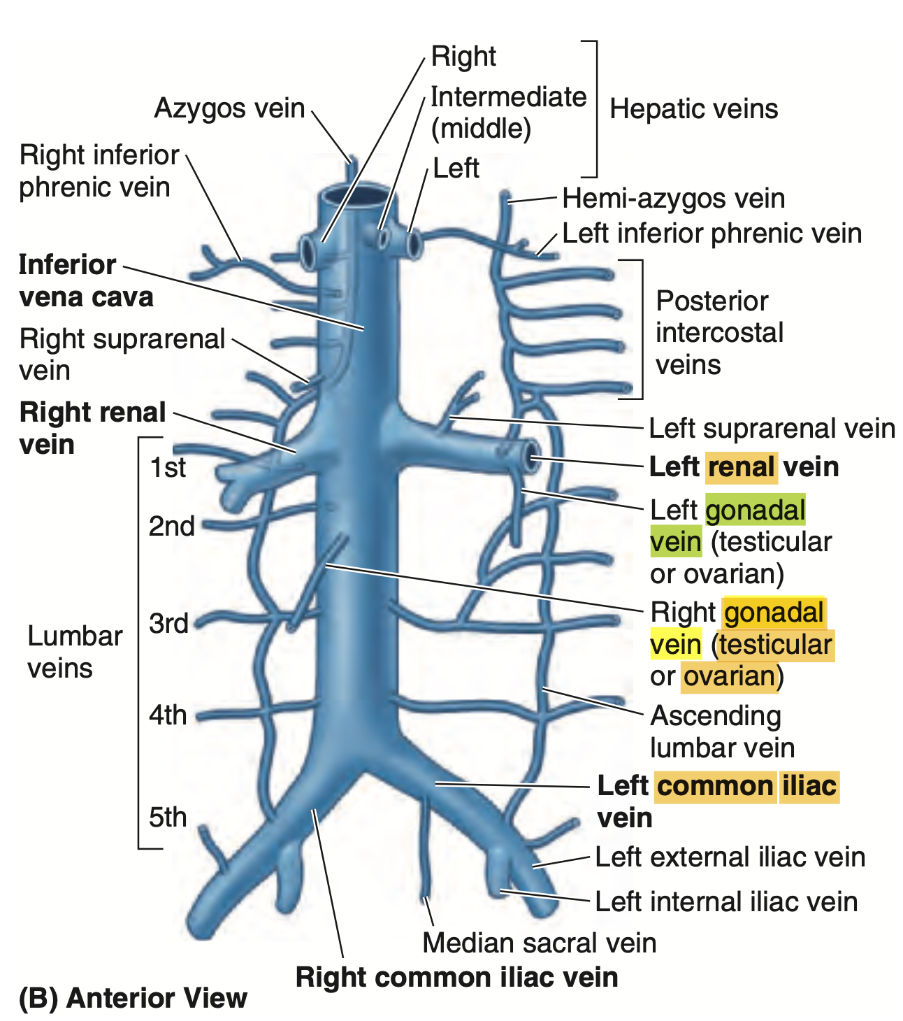 |
| Q: The duct of the bulbourethral gland opens into which structure? a. Prostatic urethra b. Membranous urethra c. Spongy (penile) urethra d. Ejaculatory duct | Spongy urethra Explanation: 球状尿道腺 (bulbourethral gland) の導管は、海綿体尿道 (spongy or penile urethra) に開口します。球状尿道腺は、精液の前駆体である粘液分泌物を分泌し、性交時に尿道を潤滑する役割を果たします。その分泌物は、尿道の海綿体部分(陰茎の内部を通る尿道)に開口します。 他の選択肢の説明: a. Prostatic urethra (前立腺尿道): これは、尿道が前立腺を通過する部分で、球状尿道腺の導管は開口しません。 b. Membranous urethra (膜性尿道): これは前立腺尿道から続く、骨盤内に位置する短い部分で、球状尿道腺の導管は開口しません。 d. Ejaculatory duct (射精管): これは前立腺内で、精液と尿道の交差点付近に開口しますが、球状尿道腺の導管の開口部ではありません。 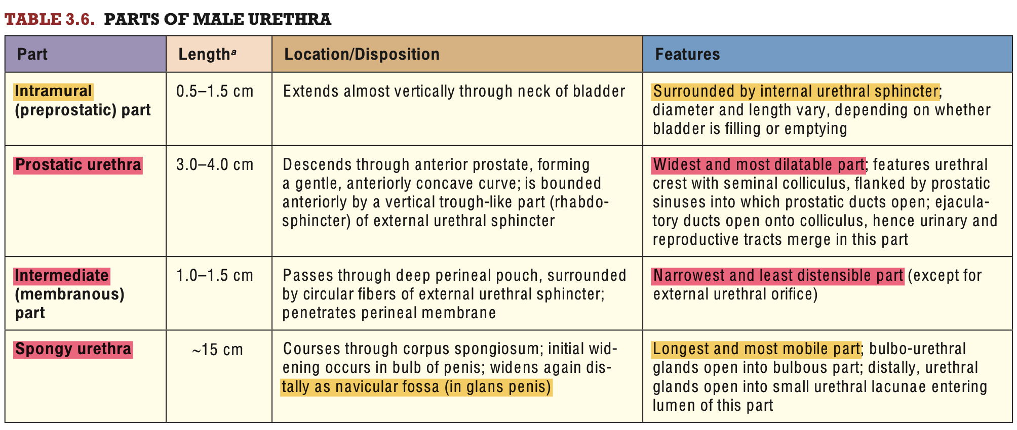 |
| What segment of the male urethra is surrounded by the external urethral sphincter? Spongy Intramural Prostatic intermediate | D. intermediate Spongy urethra(海綿体部尿道) 陰茎内にある尿道部分で、陰茎海綿体に囲まれています。この部分は外尿道括約筋に囲まれていません。 Intramural urethra(壁内部尿道) 膀胱の出口にある短い尿道部分で、膀胱括約筋に関連しますが、外尿道括約筋には囲まれていません。 Prostatic urethra(前立腺部尿道) 前立腺を貫く尿道部分で、前立腺によって囲まれていますが、外尿道括約筋には関係がありません。 Intermediate urethra(中間部尿道) 骨盤膜(pelvic diaphragm)を通る部分で、外尿道括約筋に囲まれています。この筋肉は随意的に尿流を制御する役割を持っています。したがって、正解です。 |
| What is the arterial supply of the male distal urethra? Prostatic branch of inferior vesical Middle rectal artery Dorsal Artery | c. Dorsal artery 男性尿道の動脈供給は部位によって異なります。遠位尿道(spongy urethra)は特に陰茎海綿体に囲まれており、血液供給は以下のように分類されます。 Prostatic branch of inferior vesical artery この動脈は前立腺部尿道(prostatic urethra)や膀胱の下部を供給しますが、遠位尿道には関与していません。不正解です。 Middle rectal artery この動脈は主に直腸や周辺の組織を供給します。尿道には関与しないため、不正解です。 Dorsal artery of the penis 陰茎の背部動脈(dorsal artery of the penis)は陰茎全体の主要な血液供給源であり、遠位尿道を供給します。したがって、正解です。 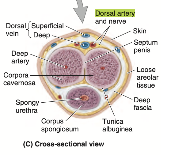 |
| Consistent arterial blood supply of male pelvic ureter? A.Uterine artery B. Inferior vesical artery C. Internal iliac artery D. Ovarian artery | B. Inferior vesical artery 男性の骨盤部尿管 (pelvic ureter) の主な血流供給は、下膀胱動脈 (inferior vesical artery) から供給されます。この動脈は、内腸骨動脈(internal iliac artery)の枝であり、骨盤内の膀胱や尿管に血液を供給します。下膀胱動脈は特に男性の尿管の下部 a. Uterine artery (子宮動脈): これは女性の子宮に血液を供給する動脈で、男性の尿管には関与しません。 c. Internal iliac artery (内腸骨動脈): 内腸骨動脈は骨盤の多くの臓器に血液を供給しますが、尿管への供給は、下膀胱動脈や他の枝を通じて行われます。 d. Ovarian artery (卵巣動脈): 卵巣動脈は女性の卵巣に血液を供給する動脈で、男性の尿管には関与しません。 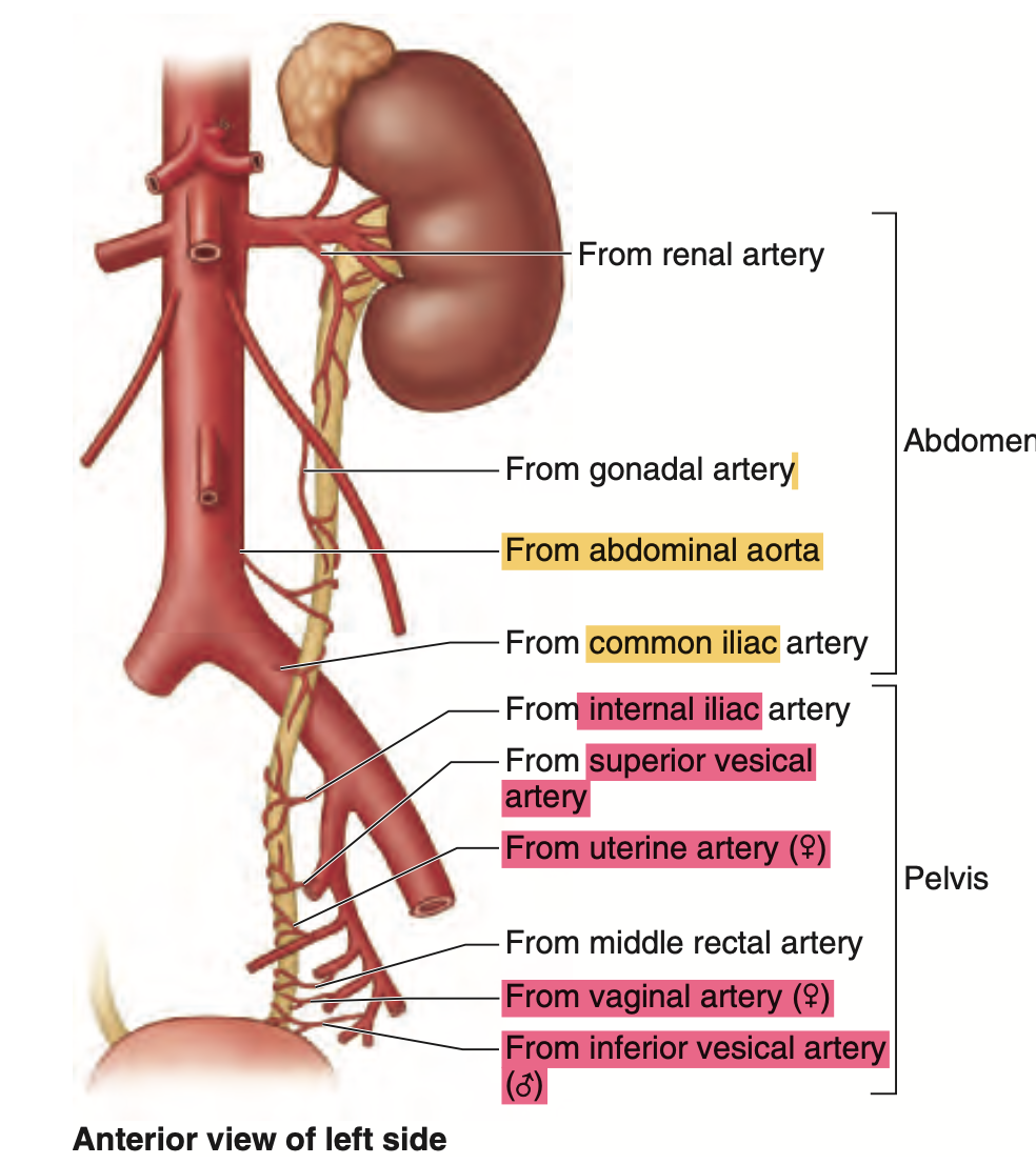 |
| What segment of the male urethra is the ducts of the bulbourethral gland seen? Intramural Prostatic Intermediate Spongy | Spongy Urethra 球尿道腺(bulbourethral gland)は、陰茎尿道(spongy urethra)に粘液を分泌する小さな腺で、その管は尿道の特定のセグメントに開口します。 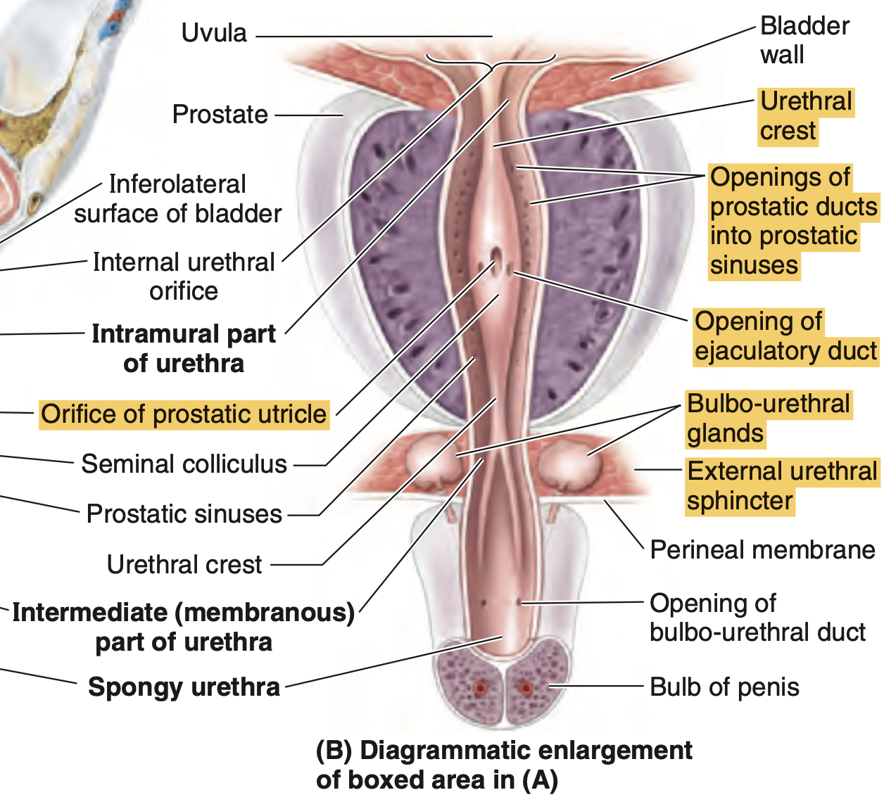 |
| Cause of renal vein entrapment syndrome A. Compression of the right renal vein between the abdominal aorta and the superior mesenteric artery B. Compression of the left renal vein between the abdominal aorta and the superior mesenteric artery C. Compression of the left renal vein between the abdominal aorta and the inferior mesenteric artery | B. Compression of the left renal vein between the abdominal aorta and the superior mesenteric artery 左腎静脈は、通常腹部大動脈と上腸間膜動脈の間を通過しますが、これらの血管によって圧迫されると、腎臓から心臓への血流が制限されます。 圧迫が続くと、左腎静脈高圧症や血栓が形成され、腎機能に影響を与えることがあります。 |
| USMAN, SUSAN | |
| Which of the following structures in females is considered the anatomical equivalent of the male bulbourethral gland? A) Skene’s glands B) Bartholin’s glands C) Ovaries D) Clitoris | Bartholin’s glands バルトリン腺(Bartholin’s glands)は膣前庭の粘液分泌腺で、男性の球尿道腺と同じく潤滑のための粘液を分泌します。この腺は胚発生学的にも機能的にも球尿道腺の女性版と考えられています。正解です。 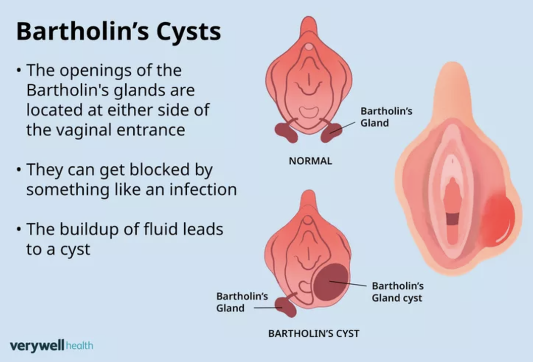 |
| Which part of empty bladder points to the superior edge of the pubic symphysis? a. Body b. Fundus C. Apex d. Neck | APEX 空の膀胱 (empty bladder) の頂点 (apex) は、恥骨結合 (pubic symphysis) の上縁に向かって位置しています。膀胱が空であるとき、頂点は恥骨結合のすぐ上にあり、通常は前方に向かっています。膀胱が満杯になると、その形が変わり、頂点は上向きに移動します。 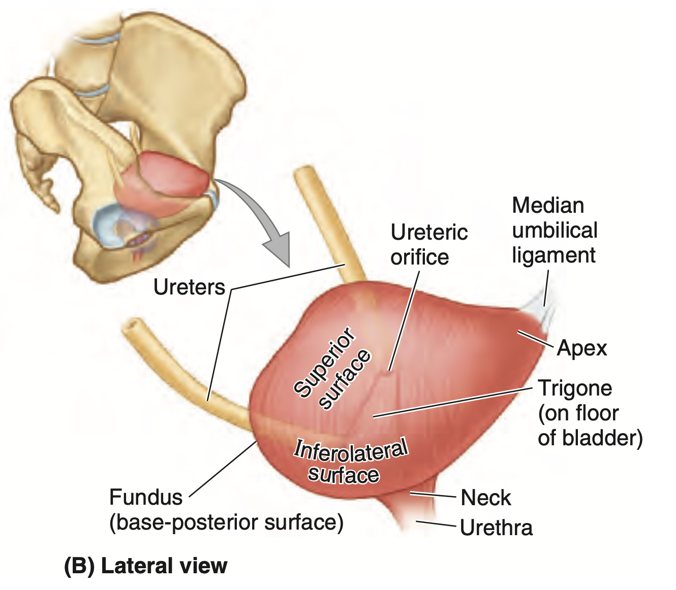 |
| In the male urinary bladder, what is formed by the muscle fibers in the bladder neck? a. Perineal membrane b. External urethral sphincter c. Internal urethral sphincter d. Rectovesical septum | b. internal urethral sphincter |
| The bulborethral gland is found in which segment of the male urethra A Prostatic B intramural C intermediate D spongy | C. Intermediate |
| Q: What is the condensed, membranous layer that covers the kidney and surrounding fat? a. Renal fascia b. Perinephric fat c. Gerota’s fascia d. Pararenal fat | Renal fascia Moore The kidneys, suprarenal glands, and the fat surrounding them are enclosed (except inferiorly) by a condensed, membranous layer of renal fascia. 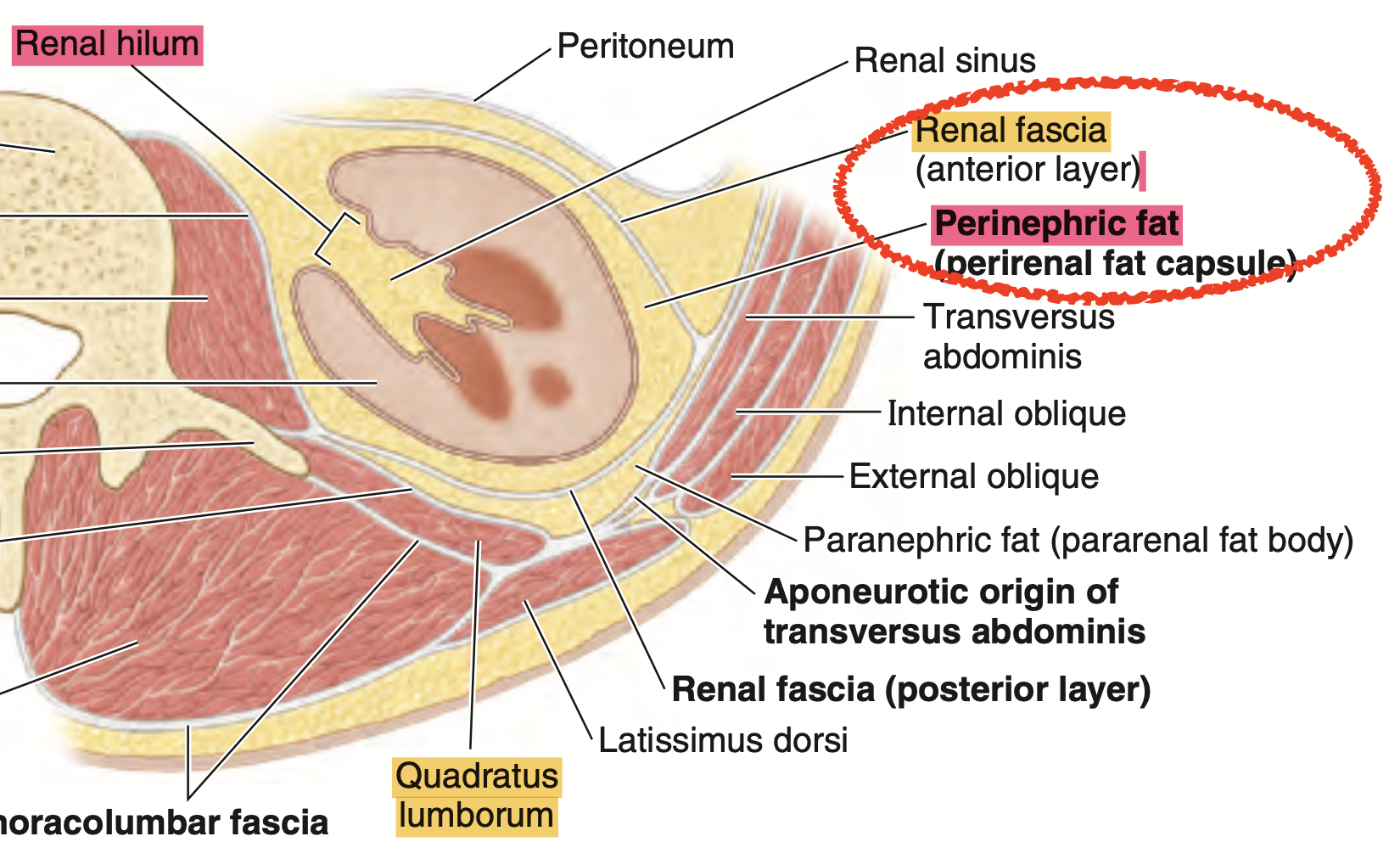 |
| A horseshoe kidney typically results from the fusion of what structures? A. Fusion of the superior poles B. Fusion of the inferior poles C. Fusion of the medial poles D. Fusion of the lateral poles | B. Inferior poles Moore The kidneys are close together in the embryonic pelvis. In approximately 1 in 600 fetuses, the inferior poles (rarely, the superior poles) of the kidneys fuse to form a horseshoe kidney (Fig. B2.32D). This U-shaped kidney usually lies at the level of L3–L5 vertebrae because the root of the inferior mesenteric artery prevented normal relocation of the kidneys. 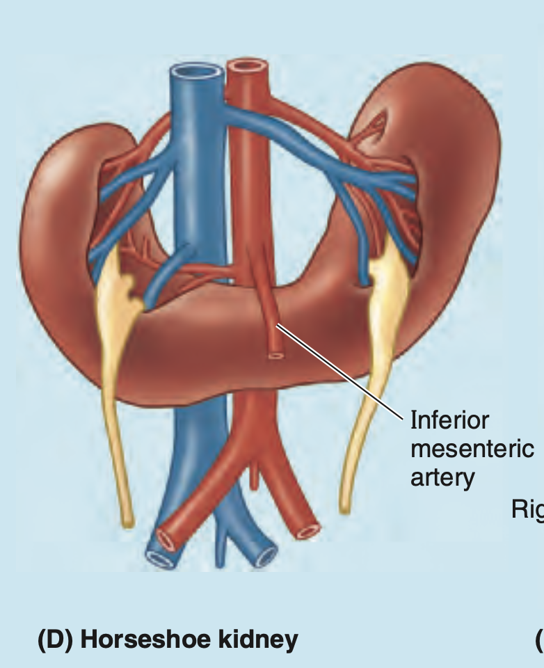 |
| SOLIS Segment surrounding internal urethral sphincter A.intramural urethra B,prostatic urethra C.intermediate urethra D.spongy urethra | A. intramural urethra |
組織
| How many nephrons are there in each kidney A.100 B.1,000 C 1.25millions D. 1.25billions | C 1.25millions ネフロン(nephron)は腎臓の基本的な機能単位であり、尿生成や老廃物の濾過を行います。それぞれの腎臓には約 1百万から1.5百万個 のネフロンが含まれているとされています。 |
| Type of nephron that has longer loop of henle that extends deep into medulla Juxtamedullary nephron Cortical nephron Medullary nephron | A. Juxtamedullary nephron ジュクスタメデュラ腎単位 (juxtamedullary nephron) は、長いヘンレループ (long loop of Henle) を持ち、そのループは腎髄質 (medulla) に深くまで伸びています。このタイプの腎単位は、尿の濃縮に重要な役割を果たし、腎髄質内での水分再吸収を促進します。ジュクスタメデュラ腎単位は、腎臓全体の約15%しか占めていませんが、尿の濃縮能力を高めるために特化しています。 |
| This is when the substances move from the epithelial cells into the tubular lumen. A. Tubular absorption B. Filtration C. Crossing over D. Tubular secretion | D. Tubular secretion Explanation: 尿細管分泌 (tubular secretion) は、上皮細胞 (epithelial cells) から尿細管腔 (tubular lumen) へ物質が移動する過程を指します。このプロセスでは、血液から尿へ物質が移動し、尿の成分が調節されます。腎臓では、尿細管で不要な物質や過剰なイオンが血液から分泌され、最終的に尿として排泄されます。 |
| H. OMAR Which of the following statements about the nephrons is correct? A. Nephrons increase in number as we age. B. Nephrons decrease in number as we age. C. The number of nephrons remains constant as we age. D. Nephrons can easily regenerate if there is an injury or death to it. | B. Nephrons decrease in number as we age |
| Q: What is the most substantial part of the filtration process in the kidneys? a. Capillary endothelial cells b. Glomerular basement membrane c. Slit diaphragm | Glomerular basement membrane 糸球体基底膜 (glomerular basement membrane) は、腎臓における濾過 (filtration) プロセスで最も重要な役割を担う部分です。この基底膜は、糸球体内の血液とボーマン嚢の間の物質のろ過を制御します。基底膜は、サイズと電荷に基づいて物質を選択的にろ過し、血液中の不要な物質が尿として排泄される過程を助けます。特に、血漿タンパク質や大きな分子を通さない役割が重要です。 |
| RELATOR | |
| 32. Which of the following is NOT FOUND in the renal corpuscle A. Bowman’s capsule B. Glomerolus C. Proximal convoluted tubule D. Mesangium | C. |
| From the urinary space, the filtrate of the blood flows into what part of the nephron? Distal convoluted tubules Proximal convoluted tubules Collecting duct Loop of henle | proximal ct(? ) 血液から濾過されたフィルトレートは、尿間隙 (urinary space) から近位曲尿細管 (proximal convoluted tubules) に流れ込みます。ここで濾過された物質が再吸収される過程が始まります。近位曲尿細管は、腎臓の主要な再吸収部位であり、グルコース、アミノ酸、ナトリウム、カルシウム、ビタミンなどの多くの重要な物質を血液に再吸収します。 The urinary space (U) between the parietal layer and the glomerulus (G) drains into the lumen of the proximal tubule. |
| OKPE | |
| These cells are secreted in response to ADH Principal cells Intercalated cells Pedicel (?) Lacis cells | A. Principal cells ? 主細胞 (Principal cells) は、腎臓の集合管 (collecting ducts) と後部尿細管に存在し、抗利尿ホルモン (ADH, antidiuretic hormone) に応答して水の再吸収を調節します。ADH が主細胞に作用すると、アクアポリン-2 (aquaporin-2) と呼ばれる水チャネルが細胞膜に挿入され、水が再吸収されるようになります。このメカニズムにより、体内の水分バランスが維持されます。 他の選択肢の説明: b. Intercalated cells (介在細胞): 介在細胞は、酸塩基平衡の調節に関与し、プロトン (H⁺) や重炭酸イオン (HCO₃⁻) を分泌または再吸収しますが、ADH への応答はしません。 c. Pedicels (足突起): これはポドサイト (糸球体の足細胞) の構造の一部であり、濾過機能に関与しますが、ADH には関与しません。 d. Lacis cells (ラクシス細胞): これらは糸球体付近の間質細胞であり、レニン分泌や血流調節に関与しますが、ADH への応答はありません。 |
| What is the brush border of the proximal convoluted tubule composed of ? A. Cilia B. Microvilli C. Stereocillia D. Flagella | B. Microvilli |
| What is the leading cause of end stage kidney disease in the US Diabetes. Hypertension Autoimmune Cancer | a. Diabetes |
| PEGARIT | |
| Patient has Renal Artery Stenosis (decrease size of renal arteries), what happens if left untreated? Kidney will secrete renin results Hypertension No change BP decreased BP due to constriction decreased GFR | a. Kidney will secrete renin results Hypertension 腎動脈狭窄 (renal artery stenosis) による腎臓への血流低下は、腎臓の糸球体近接装置 (juxtaglomerular apparatus) が感知します。この状態では、腎臓は低血圧と誤解し、レニン (renin) を分泌します。レニンはアンギオテンシノーゲンをアンギオテンシンIに変換し、その後アンギオテンシンIIに変わることで、血管収縮とアルドステロン分泌を促進します。これにより血圧が上昇し、持続的な高血圧 (hypertension) が発生します。 |
| This cell in collecting duct system is eosinophilic and helps in maintaining acid base balance by secreting bicarbonate a.intercalated A cell b.intercaleted B CELLS C. Principal cells D principle vells | Answer: b. Intercalated B cell Explanation: 介在細胞B型 (Intercalated B cells) は、腎集合管 (collecting ducts) に存在し、主に重炭酸イオン (HCO₃⁻) を分泌することで酸塩基平衡を維持します。この細胞は酸性環境を改善する役割を果たし、体液のpHが正常に保たれるよう調整します。 A型間在細胞(Type A Intercalated cells):主に水素イオンを分泌して、血液をアルカリ性に調整します。このタイプは血液が酸性に傾いたときに活動し、尿に水素イオンを排泄して酸性物質を排出します。 B型間在細胞(Type B Intercalated cells):主に重炭酸イオンを分泌して、血液がアルカリ性になりすぎるのを防ぎます。このタイプは、血液がアルカリ性に傾いたときに働き、重炭酸イオンを排泄することで酸塩基バランスを保ちます。 |
| TAN, JOAN | |
| Transport of urine from renal pelvis to urinary bladder -Ureter -Urethra -Hilum -Renal medulla | URETER |
| GONZAGA This is pain in urination. A. Dysuria B. Hematuria C. Oliguria D. Anuria | Dysuria |
| What is the lining epithelium of the urinary bladder? A. Urothelium B. Nonkeratinized stratified squamous C. Keratinized stratified squamous D. Simple squamous | Urothelium |
| Lining epithelium of distal part of female urethra | Nonkeratinized Stratified Squamous |
生理
| What is the lowest osmolarity of diluted urine that the kidney can produce in response to reduced body fluid osmolarity? A. 100 mOsm/L B. 50 mOsm/L C. 300 mOsm/L D. 1200 mOsm/L | B. 50 mOsm/L 腎臓の希釈能力 腎臓の髄質(medulla)と集合管(collecting ducts)の調節により、尿を希釈する能力が最大限発揮されると、尿浸透圧は約 50 mOsm/L まで低下します。 抗利尿ホルモン(ADH)の抑制 抗利尿ホルモン(ADH)の分泌が抑制されることで、集合管の水透過性が低下し、水が再吸収されにくくなります。その結果、希釈尿が生成されます。 生理的限界 正常な腎機能では、尿浸透圧は 50~1200 mOsm/L の範囲で変動します。この範囲の下限が 50 mOsm/L です。 |
| What triggers ADH release from posterior pituitary gland? Decreased Plasma Osmolarity Increased Plasma Osmolarity I forgot I also forgot lmao | B. Increased Plasma Osmolarity ADHの分泌を引き起こす主な要因は以下の通りです: 血漿浸透圧の上昇(最も感受性の高い刺激) 血液量や血圧の低下 アンジオテンシンIIの作用 ストレスや痛みなどの追加刺激 |
| GURUNG | |
| Obligatory Urine volume in a 70-kg person A. 1.0L B. 0.5L C. 1.5L D. 😉 | 1. 腎臓の濃縮能力 腎臓の尿濃縮能力により、尿浸透圧を最大1200 mOsm/L程度に達することができます。 70kgの成人では、1日に約600-800 mOsmの溶質(老廃物や電解質)が排泄される必要があります。 2. 最大濃縮尿浸透圧を考慮した計算 最大濃縮尿浸透圧が1200 mOsm/Lの場合: 必須尿量=排泄する溶質量(約600-800 mOsm) /最大尿浸透圧(1200 mOsm/L) 計算すると、約 500 mL/日 となります。 |
| How is mechanisms Hyperosmolarity in renal medulla achieved? A. Through Filtration process in the glomerulus B. By countercurrent multiplier mechanism involving loop of Henle and vasa recta C. Via increase water permeability in the collecting tubule D. By Active water transport in the collecting duct | Answer: b. 1. Countercurrent Multiplier Mechanism 腎髄質の高浸透圧は、主にヘンレのループ(loop of Henle)の働きによって達成されます。 下行脚(descending limb)は水に透過性が高く、溶質にはほとんど透過性がありません。一方、上行脚(ascending limb)は水に不透過性ですが、ナトリウムやクロライドなどの溶質を能動的に輸送します。 この対向流の働きにより、腎髄質の浸透圧が増加し、深部(内髄)に向かって浸透圧勾配が形成されます。 |
| What happens to the permeability of the CCT to water when ADH is low or absent? a. it becomes highly permeable to water and promotes water reabsorption (?) b. it remains nearly impermeable to water c. (?) d. promotes transport of water to interstitial fluid (?) | b. it remains nearly impermeable to water |
| PADILLOS | |
| What % of renal blood flow is directed to the medulla 50% 15% 5% 25% | C. 5% (closest) |
| Tubular segment where water is permeable, but impermeable to sodium and urea. A pct B descending loop of henle C ascending loop of henle E dct | b 特徴:水には高い透過性がありますが、ナトリウムや尿素には不透過性です。そのため、周囲の腎髄質の高浸透圧により、水が受動的に再吸収されます。 役割:尿が濃縮される過程に寄与し、髄質の浸透圧勾配を助けます。 |
| Which of the following best describes the role of urea recirculation in maintaining the hyperosmolarity of the renal medulla? A. Urea is actively transported from the proximal tubule into the interstitial fluid. B. Urea diffuses out of the medullary collecting duct into the interstitial fluid and re-enters the thin descending loop of Henle. C. Urea is entirely reabsorbed in the proximal tubule, preventing it from contributing to medullary osmolarity. D. Urea is secreted into the distal tubule and excreted without recirculation. | B 集合管での尿素の拡散: 腎髄質の集合管では、尿素が尿細管から間質液(interstitial fluid)に拡散します。このプロセスは、特に抗利尿ホルモン(ADH)が作用しているときに促進されます。 ヘンレのループへの再取り込み: 拡散した尿素は、ヘンレのループの薄い下行脚(thin descending limb)および一部の薄い上行脚(thin ascending limb)に再び入ります。 浸透圧勾配の維持: この循環によって、腎髄質の高浸透圧勾配が増強され、集合管を通過する水の再吸収が促進されます。 |
| GAYDA | |
| What triggers the osmoticreceptor-ADH feedback system in response to a water deficit? | 水分不足に応答して浸透圧受容体-ADHフィードバックシステムを作動させる主な要因は、血漿浸透圧の上昇です。この変化が視床下部の浸透圧受容体によって感知され、ADHが分泌されて水分再吸収が促進されます。 |
| CAPANGPANGAN | |
| What is a condition, excessive thirst without physiologic stimulus Diabetes Mellitus Diabetes Insipidus Psychogenic thirst Hypodipsia | Psychogenic thirst 生理的刺激がないにもかかわらず、異常な渇き(過剰な水分摂取欲求)を感じる精神的な状態です。 心理的要因や強迫的行動が関与していることが多いです。 この状態は、腎臓やホルモンの異常によるものではなく、精神的ストレスや精神疾患(例:強迫性障害、統合失調症)に関連している場合があります。 |
| At what threshold level in urine is triggered in response to an increased plasma sodium concentration? | 血漿ナトリウム濃度の基準値 正常値: 血漿ナトリウム濃度は 135–145 mEq/L に維持されます。 閾値: 血漿ナトリウム濃度が 145 mEq/L を超えると、浸透圧が増加し、体液の恒常性を調整するためのフィードバックシステムが作動します。 |
| What drives the passive reabsorption of chloride ions in the renal tubular lumen? a. A positive electrical potential created by active sodium reabsorption b. A negative electrical potential created by active sodium reabsorption c. active transport of K+ d. increase secretion of water into the tubular lumen | B. A negative electrical potential created by active sodium reabsorption メカニズム:ナトリウムイオン(Na⁺)が腎尿細管上皮細胞を通じて能動的に再吸収されると、細胞外に正電荷が移動します。 これにより、管腔内が相対的に負電位となります(negative electrical potential)。 影響:この負電位が、塩化物イオン(Cl⁻)を管腔内から細胞へ受動的に引き込む駆動力となります。 |
| What part of the Nephron is very permeable to water owing to the large presence of aquaporins–1(AQP1). a. thick ascending limb b. Proximal Convulated tubule c. Distal convulated tubule d. collecting ducts | B. Proximal Convulated Tbule 特徴:近位尿細管には大量のアクアポリン-1(AQP1)が存在し、水に対する透過性が非常に高いです。 水は溶質(ナトリウム、グルコースなど)の再吸収に伴い、浸透圧勾配に従って受動的に再吸収されます。 近位尿細管では、ろ過された水の約 65–70% が再吸収されます。 役割:大量の水と溶質が同時に再吸収されるため、腎臓の水バランス調整の第一段階を担います。 |
| Which of the following best describes solvent drag? Active transport of water carrying solutes through aquaporins A: movement of solutes along with water across tight junctions by osmosis B: Diffusion of water and solute through impermeable membranes C: Passive diffusion of solutes across the proximal tubules | B. Movement of solutes along with water across tight junctions by osmosis 溶媒ドラッグ(Solvent drag)とは、水が浸透圧勾配に従って移動する際に、水分子と一緒に溶質(ナトリウム、カリウム、カルシウムなど)が引きずられる現象を指します。 この現象は、特に近位尿細管や腸管などの**タイトジャンクション(tight junctions)**を介した水の再吸収中に顕著に見られます。 |
| Does not require energy directly from ATP or other high-energy phosphate | Secondary Active Transport 2次能動輸送は、直接ATPのエネルギーを使用しないが、一次能動輸送によって形成されたイオン勾配をエネルギー源として利用します。 ナトリウムイオン(Na⁺)の濃度勾配や電気化学的勾配がよく使われ、これが他の物質(例えば、グルコースやアミノ酸)の輸送を駆動します。 |
| What process is used for protein reabsorption in the proximal tubule? | Pinocytosis |
| What is the transport maximum for glucose on healthy individual a. 125 b. 100 c. 375 d.500mg/min | c. 375 mg/min |
| What drives the secondary active transport of glucose at the luminal membrane in the proximal tubule A. Hydrostatic Pressure gradient B. Sodium gradient by Na-K ATPase pump C. Passive diffusion of glucose D. hydrogen ion secretion in the luminal membrane | B. Sodium gradient by Na-K ATPase pump |
| Which of the following is a correct match between the proximal convoluted tubule and its glucose transporter. Upper GLUT1 Later GLUT 2 SGLT2 and GLUT2 SGLT2 and GLUT 1 | c.SGLT2 and GLUT2 SGLT2(Sodium-Glucose Co-Transporter 2) 位置: 近位尿細管の**早期部分(S1セグメント)**に存在。 機能: ナトリウムとグルコースを共輸送する(1:1比率)。 役割: グルコースの約 90% を再吸収。 GLUT2(Glucose Transporter 2) 位置: 近位尿細管の基底側膜に存在。 機能: 再吸収されたグルコースを細胞から血液に輸送。 役割: SGLT2によって取り込まれたグルコースを循環系に戻します。 SGLT1(Sodium-Glucose Co-Transporter 1) 位置: 近位尿細管の後半部分(S3セグメント)。 役割: 残りの約10%のグルコースを高効率(2:1比率でNa⁺と共輸送)で再吸収。 GLUT1(Glucose Transporter 1) 位置: 基底側膜。 役割: 再吸収の補助的役割を果たすが、SGLT2と主に関係するのはGLUT2。 |
| MICULOB | |
| What is the effect of loop diuretics like furosemide in a thick ascending loop of henle? | フロセミドのようなループ利尿薬は、ヘンレの上行脚においてNa⁺/K⁺/2Cl⁻共輸送体を阻害することで、ナトリウムや水の再吸収を減少させ、尿量を増加させます。この効果により、浮腫や高血圧の治療に利用されますが、電解質異常や脱水などの副作用に注意が必要です。 |
| KAPUNAN | |
| How do potassium-sparing diuretics like spironolactone act on principal cells? | Answer: C. They block mineralocorticoids receptors, inhibits aldosterone reabsorption and potassium secretion. スピロノラクトンはミネラルコルチコイド受容体(mineralocorticoid receptors)をブロックし、アルドステロンの作用を抑制することで、ナトリウムの再吸収を阻害し、カリウムの排泄(分泌)を抑制します。 |
| What is the diuretic that directly blocks sodium channels in the luminal membrane of principal cells? | Amilorid **アミロライド(Amiloride)やトリアムテレン(Triamterene)**が該当します。これらはカリウム保持性利尿薬の一種で、集合管の主細胞の管腔側膜に存在するナトリウムチャネル(ENaC: Epithelial Sodium Channel)を直接阻害します。 |
| what is the role of type b interlacted B cells? Secrete h+ions and absorb hco3- ions secrete hco3-ions and absorb h+ions | b Type B介在細胞:アルカリ性化(アルカローシス)の際に活性化され、HCO₃⁻を尿中に分泌し、H⁺を血液中に再吸収します。 |
| Which of the ff explains the pressure natriuresis? A. Dec sodium excretion bc of B. Inc water reabs bc of dec GFR C. Dec sodium secretion bc of N/K ATPase pump D. Inc sodium excretion bc of elevated arterial pressure | D. 1. 圧利尿のメカニズム 動脈圧が上昇すると、腎臓に流入する血液量が増加し、腎間質(interstitial space)の圧力が高まります。 腎臓の血管床の圧力が上昇することで、以下の現象が起こります:尿細管でのナトリウム再吸収の抑制: 高い腎間質圧が尿細管周囲の毛細血管の圧力を増加させ、ナトリウム再吸収が低下します。 ナトリウム排泄の増加: 尿中にナトリウムがより多く排泄されます(ナトリウム利尿)。 |
生化
| hydroxylase enzymes catalyze hydroxyl groups into lysine and proline, what vitamin act as a cofactor? | Vitamin C ヒドロキシラーゼ酵素の役割 ヒドロキシラーゼ酵素は、コラーゲンの合成過程で重要な働きをします。 プロリンおよびリシン残基にヒドロキシル基(-OH)を付加して、それぞれヒドロキシプロリンおよびヒドロキシリシンを生成します。 この修飾は、コラーゲン分子の安定性を高め、三重らせん構造を形成するために必須です。 ビタミンCの役割 ビタミンCは、プロリルヒドロキシラーゼ(prolyl hydroxylase)およびリシルヒドロキシラーゼ(lysyl hydroxylase)の**補酵素(cofactor)**として働きます。 酵素反応には**鉄(Fe²⁺)**が必要であり、ビタミンCは鉄を還元型(Fe²⁺)に保つことで酵素の活性を維持します。 酵素反応の結果、ヒドロキシル基がプロリンやリシンに付加されます。 |
| A 28 year old woman cane to her obstetrician when she missed her period and her pregnancy kit turned out positive. This is an example of what Nitrogen Balance? a. Positive Nitrogen Balance b. Negative Nitrogen Balance c. Both d. Neither | Positive Nitrogen Balance 28歳の女性が妊娠検査で陽性となった場合、その体内では胎児の成長や組織形成のためにタンパク質合成が増加しています。これは正の窒素平衡(Positive Nitrogen Balance)の一例です。 その他の例: 成長期の子供。 筋肉をつけるために運動している人。 回復期の患者(傷の治癒や組織再生)。 |
| Process that transfers a-amino nitrogen to a-ketoglutarate to form glutamate a. hydroxylation b. transamination c. transdeamination d. oxidation | b. transamination 1. Transamination(アミノ基転移) 概要: トランスアミネーションは、アミノ酸のα-アミノ基をα-ケトグルタル酸に移動させる反応です。このプロセスで、元のアミノ酸はα-ケト酸になり、α-ケトグルタル酸は**グルタミン酸(Glutamate)**に変換されます。 触媒酵素: トランスアミナーゼ(アミノ基転移酵素、Transaminase)。 ピリドキサールリン酸(PLP; ビタミンB6誘導体)を補酵素として必要とします。 主要な例: Amino Acid+α-Ketoglutarate ⟶α-Keto Acid+Glutamate 重要性: トランスアミネーションは、アミノ酸代謝や窒素の代謝で中心的な役割を果たします。 |
| 79. Vitamin B derivative that serve as a cofactor for transamination processnin amino acid metabolism. A. Pyridoxal phosphate B. Cobalamin C. Tetrahydropterin D. N-acetyl d-glutamate | A Pyridoxal Phosphate (PLP): Pyridoxal phosphate (PLP) はビタミンB6(ピリドキシン)の活性型で、アミノ酸代謝における重要な補酵素(cofactor)です。 トランスアミネーション (transamination) は、アミノ酸のアミノ基(-NH₂)をケト酸(α-keto acid)に移す反応で、これによりアミノ酸とケト酸の相互変換が行われます。PLPはこの反応の補酵素として作用します。 PLPは、**アミノ基転移酵素(トランスアミナーゼ)**と結合し、反応の触媒を助けます。 |
| 80. The posttranslational modification of preprocollagen occurs where ER Nucleus Mitochondria Cytosol | Endoplasmic Reticulum Preprocollagenの概要: コラーゲンは細胞外マトリックスの主要構成要素であり、線維形成に重要な役割を果たします。 初期には、コラーゲンはpreprocollagenとして合成されます。この段階では、まだ完全な構造を持っていません。 翻訳後修飾 (Posttranslational Modifications): 翻訳後修飾は、preprocollagenが成熟したコラーゲンに変換される重要なプロセスです。 主な修飾プロセスは以下の通りです: シグナルペプチドの除去: シグナルペプチドが除去され、procollagenになります。この反応は、粗面小胞体 (ER) 内で行われます。 水酸化反応 (Hydroxylation): プロリンやリシンの残基が水酸化されます。この修飾はER内で行われる必要があり、酵素(プロリルヒドロキシラーゼ、リシルヒドロキシラーゼ)とビタミンCが必要です。 糖鎖付加 (Glycosylation): 水酸化されたリシンに糖鎖が付加されるプロセスもERで進行します。 三重らせん構造の形成 (Triple Helix Formation): プロコラーゲンが三重らせん構造を形成します。このプロセスもERで行われます。 修飾の場としてのER (Endoplasmic Reticulum): 粗面小胞体(ER)は、タンパク質の合成後修飾と折り畳みの場として機能します。 Preprocollagenの翻訳後修飾は、特にERの管腔内で進行し、その後ゴルジ体を経て細胞外に分泌されます。 |
| What is the function of prolyl hydroxylase and lysyl hydroxylase | Hydroxylation **Prolyl Hydroxylase(プロリルヒドロキシラーゼ)とLysyl Hydroxylase(リシルヒドロキシラーゼ)**は、コラーゲンの合成過程で重要な酵素です。この酵素はプロリン(Proline)およびリシン(Lysine)残基を修飾し、コラーゲンの安定性と機能に寄与します。 |
| SAPLOT | |
| enzyme that converts phenylalanine to tyrosine a. tyrosine aminotransferase b. alanine aminotransferase c. phenylalanine hydroxylase | C. Phenylalanine Hydroxylase Phenylalanine Hydroxylaseは、フェニルアラニンをチロシンに変換する酵素で、正常なアミノ酸代謝と神経伝達物質の合成に欠かせません。この酵素の欠乏や機能不全は、フェニルケトン尿症(PKU)という重大な代謝異常症を引き起こします。 |
| SABATE | |
| Young child experienced an automobile accident. For surgery. Diet consulted with a dietitian to provide a diet containing an non-essential amino acid. Which of the following is an essential amino acid for faster growth and recovery? a. Arginine b. Tyrosine c. Alanine d. Glycine | a. Arginine(アルギニン) 機能と役割:**準必須アミノ酸(conditionally essential amino acid)**として分類され、通常は体内で合成されますが、成長期やストレス下、外傷、手術後の回復など特定の状況では外部からの摂取が必要になります。 |
| Most abundant protein in the body? Myoglobin Albumin Collagen | Collagen is the most abundant protein in the human body. It accounts for roughly 30% of the total protein content. Myoglobin: While myoglobin is an important protein found in muscle tissue, its primary function is to store oxygen for use by muscle cells during exercise. It’s not as abundant in the body as collagen. Albumin: Albumin is the most abundant protein in blood plasma. Its primary function is to maintain osmotic pressure in the blood and transport various substances, such as hormones and drugs. While it’s a significant protein, it’s not as widely distributed in the body as collagen. |
| Identify which is not a precursor of essential amino acid a ketoglutarate 3 phosphoglycerate glutamate aspartate | a ketoglutarate |
| Identify which is not a precursor of essential amino acid A.Aspartate B.Alpha keto glutarate C.Glutamate D.Histamine | Histamine |
| Water and hydrogen bonding are important for all of the following properties EXCEPT: a. Surface tension b. High heat capacity c. High heat of vaporization d. Ability to dissolve hydrocarbons | answer: d. Ability to dissolve hydrocarbons a. Surface tension(表面張力) 水素結合の影響: 水分子同士が水素結合で強く結びつくため、表面での分子間力が強くなり、表面張力が高くなります。 例: 小さな昆虫が水面を歩けるのは、この表面張力のおかげです。 b. High heat capacity(高比熱) 水素結合の影響: 水分子間の水素結合を切るのに多くのエネルギーが必要なため、水は高い比熱を持っています。 重要性: この性質により、環境や生物の温度が急激に変化しにくくなっています。 c. High heat of vaporization(高蒸発熱) 水素結合の影響: 水が蒸発する際、水分子間の水素結合を切るために大量のエネルギーが必要です。 重要性: 体温調節(汗をかいて蒸発させる)に役立ちます。 |
| SY, SITTI | |
| What is binded by a Covalent Bond? a. Hydrophobic b. Hydrogen c. Disulfide d. Van Der Waals | answer: c a. Hydrophobic(疎水性) 説明:疎水性相互作用は、水分子との相互作用を避ける非極性分子(例: 炭化水素)の間で起こる弱い力です。 共有結合ではなく、分子間力(非共有結合)による相互作用です。 b. Hydrogen(水素結合) 説明:水素結合は、水素原子が電気陰性度の高い原子(例: 酸素や窒素)と弱い相互作用を持つことで形成されます。 これは分子間または分子内の弱い結合であり、共有結合ではありません。 c. Disulfide(ジスルフィド結合) 説明:ジスルフィド結合は、2つのシステイン残基の硫黄原子(-SH基)が共有結合を形成することで生じます。 共有結合の一種で、タンパク質の三次構造や四次構造を安定化させる重要な役割を果たします。 例: インスリンの構造やケラチンの安定化。 d. Van Der Waals(ファンデルワールス力) 説明:分子間で弱い引力を生じる相互作用であり、共有結合ではなく物理的な力に分類されます。 例: 分子間の一時的な双極子相互作用。 |
| IRABON | |
| Patient falls and has a fracture. Also has blue sclera and aortic murmur. A. Fibrillin B. Type 1 C. Type IV D. a-trypsin | answer: B. Type 1 青色強膜(blue sclera) 青色強膜は、強膜が薄く、下の脈絡膜が透けて見えるために生じる。これはコラーゲン異常(特にType Iコラーゲン)による結合組織の脆弱性に起因します。 骨折 転倒による骨折は、骨が非常に脆弱である**骨形成不全症(Osteogenesis Imperfecta, OI)**に典型的です。OIの主な原因は、Type Iコラーゲンの産生や構造異常です。 大動脈雑音(aortic murmur) 結合組織の異常により、心血管系(特に弁や血管壁)に影響が出る場合があります。Type Iコラーゲンの欠損は、弁膜疾患や大動脈の異常を引き起こすことがあります。 a. Fibrillin フィブリリンはマルファン症候群(Marfan Syndrome)に関連。マルファン症候群では、関節の過伸展、大動脈解離、レンズ脱臼が主な症状です。 c. Type IV(Type IV Collagen) Type IVコラーゲンは主に基底膜に存在し、アルポート症候群(Alport Syndrome)など腎疾患に関連。 d. α-Trypsin(α1-Antitrypsin) α1-アンチトリプシン欠乏症は、主に肺気腫や肝疾患に関連。 |
| A 67 yr old male, has a history of heavy smoking, lost significant weight for the past month. Chest x-ray showed suspicious mass on the right lobe of his lung. His condition indicates what type of nitrogen balance? Positive nitrogen balance Negative nitrogen balance Both Neither | Negative nitrogen balance |
| Positive Nitrogen Balance happens when: There is a balance between excretion and ingestion of nitrogen There is an ingestion over excretion of nitrogen There is an excretion over ingestion of nitrogen No significant changes | B. There is an excess ingestion over excretion of nitrogen |
| A 3 months old infant was brought to the ER due to seizure. Patient was noted to have pale skin with blue eyes due to a lack ……… of this. A. Phenylalanine hydroxylase B. Phosphatase C. Tyrosinase D. Melanin | C : Tyrosinase a. Phenylalanine hydroxylase 役割:フェニルアラニンをチロシンに変換する酵素。 欠乏するとフェニルケトン尿症(PKU)を引き起こし、知的障害や発育遅滞の原因となります。 b. Phosphatase 役割:様々な細胞代謝プロセスでのリン酸基除去に関与。 メラニン合成には直接的な関与はありません。 c. Tyrosinase 役割:チロシンを酸化してドーパ(DOPA)に変換し、メラニン産生を開始する酵素。 チロシナーゼ欠乏症は、**眼皮膚型アルビニズム(Oculocutaneous Albinism, OCA1)**の原因となり、皮膚の色素減少や青い目が見られる。 d. Melanin 役割:皮膚、毛髪、眼の色素を形成する物質。 欠乏が症状の直接原因ですが、これは結果であり、原因ではありません。 |
| In alkaptonuria, what compound accumulates a) Melanin b) tyrosine c) d) homogentisic acid | D. Homogentisic Acid 1. アルカプトン尿症(Alkaptonuria)の概要 アルカプトン尿症は、**ホモゲンチジン酸酸化酵素(homogentisate oxidase)**の欠損によって引き起こされる遺伝性代謝異常症です。 フェニルアラニンとチロシンの代謝経路における異常が原因で、ホモゲンチジン酸が代謝されずに蓄積します。 蓄積したホモゲンチジン酸の特徴: 尿の黒変: ホモゲンチジン酸は空気に触れると酸化され、尿が黒くなります。 結合組織への沈着(Ochronosis): ホモゲンチジン酸が長期間蓄積すると、関節や軟骨に沈着し、黒色化や関節炎を引き起こします。 |
| Amino acid that is not transaminated A. Tyrosine B. Lysine C. Glutamine D. Serine | B. Lysine トランスアミネーション(Transamination)とは? トランスアミネーションは、アミノ基がアミノ酸からα-ケト酸に転移する反応です。 この反応は、アミノ酸の分解や合成において中心的な役割を果たします。 主な触媒酵素は、**アミノトランスフェラーゼ(transaminase, aminotransferase)で、補酵素としてピリドキサールリン酸(PLP, ビタミンB6)**が必要です。 Threonineも |
| infant presents with lethargy, irritability, and poor feeding. noted to have spasticity. The parents report that the baby has a sweet smelling urine | answer : a. Valine, leucine, and isoleucine 主訴: 乳児が嗜眠(lethargy)、興奮性(irritability)、摂食不良(poor feeding)を呈し、筋緊張亢進(spasticity)を伴う。 特徴的所見: 尿が甘い香り(「メープルシロップ」のような匂い)を放つ。 原因 酵素欠損: 分岐鎖α-ケト酸デヒドロゲナーゼ複合体(Branched-Chain α-Keto Acid Dehydrogenase Complex, BCKDC)の欠損。この酵素は分岐鎖アミノ酸(ロイシン、イソロイシン、バリン)の代謝に必須です。 遺伝形式: 常染色体劣性遺伝(Autosomal Recessive)。 |
| Enzyme responsible for rate limiting step urea cycle A. Carbamoyl phosphate synthetase I B. Carbamoyl phosphate synthetase II C D | A. carbamoyl phosphate synthetase I 律速段階(rate limiting)の酵素: Carbamoyl Phosphate Synthetase I(CPS I) CPS Iの機能: 尿素回路の最初のステップを触媒します。 ミトコンドリア内で、アンモニア(NH₄⁺)と二酸化炭素(CO₂)を結合させ、カルバモイルリン酸(Carbamoyl Phosphate)を生成します。 この反応にはATPが必要です。 律速段階の理由: 尿素回路全体の速度を制御する重要なステップであり、CPS Iが調節されます。 **N-アセチルグルタミン酸(N-acetylglutamate, NAG)**がCPS Iを活性化する補因子として機能します。 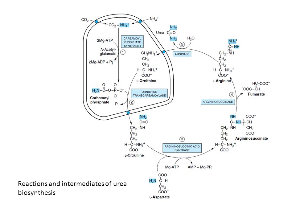 |
| S/SX: vomiting, irritability, lethargy, hyperargininemia. def of what enzyme | arginase 症状(嘔吐、興奮性、嗜眠、血中アルギニン濃度の上昇)は、尿素回路障害に関連しています。特に、アルギナーゼ(ARG1)の欠損症が疑われます。この酵素は尿素回路の最終段階でアルギニンを分解する役割を担っており、その欠損により高アルギニン血症が生じます。他の尿素回路酵素欠損症(OTCやCPS1欠損症)ではアルギニンの蓄積は見られません。正しい答えは Arginase (ARG1) の欠損です。 |

コメント