
Contents
- 1 サマリー(教科書から)
- 2 自作(教科書ベース)
- 2.1 Question 1:腎小体
- 2.2 Question 2:Renin
- 2.3 Question 3:Loop of Henle
- 2.4 Question 4:Proximal tubule
- 2.5 Question 5:多発性嚢胞腎
- 2.6 Question 6:腎臓の血液供給経路
- 2.7 Question 7:Calcitriol
- 2.8 Question 8:皮質と髄質
- 2.9 Question 9:糸球体腎炎
- 2.10 Question 10:片方腎の摘出
- 2.11 Question 11:濾過障壁
- 2.12 Question 12:Podocytes
- 2.13 Question 13:Proximal convoluted tubule
- 2.14 Question 14:Diabetic glomerulosclerosis
- 2.15 Question 15:Mesangial cells
- 2.16 Question 16:Bowmanカプセル(糸球体小体)
- 2.17 Question 17:Slit diaphragm
- 2.18 Question 18:Proximal convoluted tubule
- 2.19 Question 19:mesangial cells
- 2.20 Question 20:糸球体基底膜(GBM)
- 2.21 Question 21:PCTのendocytosis
- 2.22 Question 22:ヘンレのループの対向流増幅システム
- 2.23 Question 23:遠位尿細管の緻密斑(Macula densa)
- 2.24 Question 24:傍糸球体装置(JGA)
- 2.25 Question 25:薬物や有機イオンのクリアランス
- 2.26 Question 26:Sickle cell nephropathy
- 2.27 Question 27:抗利尿ホルモン(ADH)
- 2.28 Question 28:介在細胞(Intercalated cells)
- 2.29 Question 29:Aquaporins
- 2.30 Question 30:Lithotripsy
- 2.31 Question 31:尿管と膀胱の移行上皮
- 2.32 Question 32:尿運搬経路
- 2.33 Question 33:尿管の壁構造
- 2.34 Question 34:Umbrella cells
- 2.35 Question 35:Pyelonephritis
- 3 教科書の問題(10)
- 4 過去問
- 4.1 Question 1:尿道の長さ
- 4.2 Question 2:間在細胞
- 4.3 Question 3:末期腎疾患
- 4.4 Question 4:Macula Densa
- 4.5 Question 5:ボウマン嚢の内臓層(visceral layer)
- 4.6 Question 6:近位尿細管
- 4.7 Question 7:間質細胞(Mesangial cells)
- 4.8 Question 8:Aldosterone
- 4.9 Question 9:有窓型毛細血管
- 4.10 Question 10:PCT
- 4.11 Question 11:傍糸球体装置
- 4.12 Question 12:移行上皮
- 4.13 Question 13:腎杯
- 4.14 Question 14:排尿困難
- 4.15 Question 15:集合管主細胞
- 4.16 Question 16:傍糸球体細胞
- 4.17 Question 17:太い上行脚
- 4.18 Question 18:糸球体のろ過障壁
- 4.19 Question 19:膀胱後部
- 4.20 Question 20:葉間動脈
- 5 Quiz
- 5.1 Question
- 5.2 Question
- 5.3 Question
- 5.4 Question
- 5.5 Question
- 5.6 Question
- 5.7 Question
- 5.8 Question
- 5.9 Question
- 5.10 Question
- 5.11 Question
- 5.12 Question
- 5.13 Question
- 5.14 Question
- 5.15 Question
- 5.16 Question
- 5.17 Question
- 5.18 Question
- 5.19 Question
- 5.20 Question
- 5.21 Question
- 5.22 Question
- 5.23 Question
- 5.24 Question
- 5.25 Question
- 5.26 Question
- 5.27 Question
サマリー(教科書から)
腎臓 (Kidney)
- 各腎臓には、外側に厚い皮質 (cortex) があり、内部には8〜12個の腎錐体 (renal pyramids) に分かれた髄質 (medulla) が存在し、各錐体とその周囲の皮質組織が腎葉 (renal lobe) を構成します。
- 各腎錐体の頂部乳頭 (apical papilla) は、小腎杯 (minor calyx) に接続し、これは2〜3個の大腎杯 (major calyces) によって腎盂 (renal pelvis) に連結されています。
- 尿管 (ureter) は腎盂から尿を運び出し、腎門 (renal hilum) から腎動脈 (renal artery) と腎静脈 (renal vein) も位置しています。
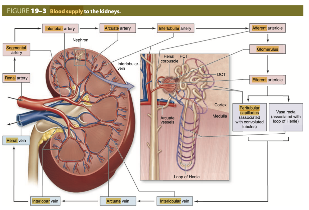
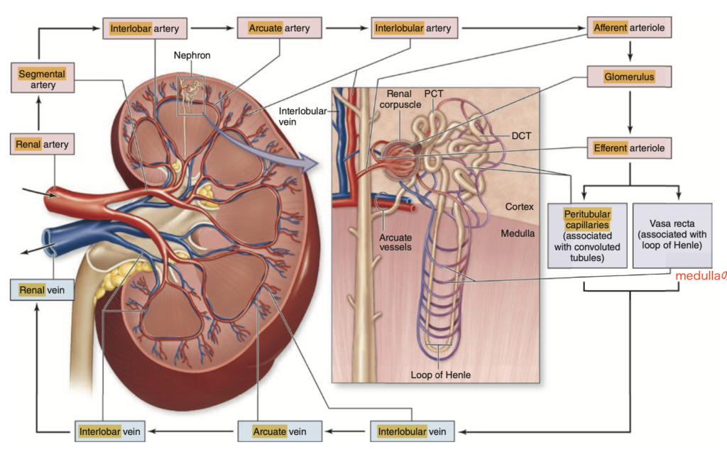
腎血管系 (Renal Vasculature)
- 腎動脈 (renal arteries) は腎葉間動脈 (interlobar arteries) に分岐し、さらに皮質内に入り微小血管を形成します。静脈分枝は動脈供給に並行しています。
- 皮質内では、輸入細動脈 (afferent arterioles) が糸球体 (glomeruli) という毛細血管群に入り、これらは細静脈ではなく輸出細動脈 (efferent arterioles) によって排出され、毛細血管内での高い静水圧を可能にします。
- 皮質糸球体からの輸出細動脈は広範に分岐し、周囲の毛細血管 (peritubular capillaries) となりますが、髄質糸球体からの輸出細動脈は髄質内で長い微小血管ループ(ヴァサ・レクタ (vasa recta))を形成します。
ネフロン (Nephrons)
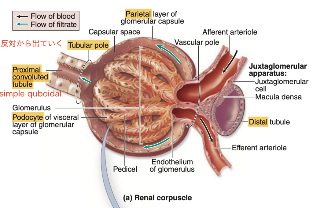
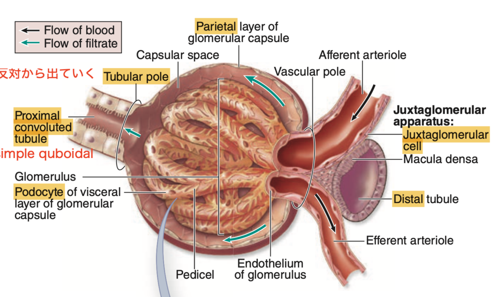
- 腎臓の機能単位であるネフロンは約100万個存在し、腎小体 (renal corpuscle) と長い尿細管 (renal tubule)、集合管系 (collecting ducts) から成ります。
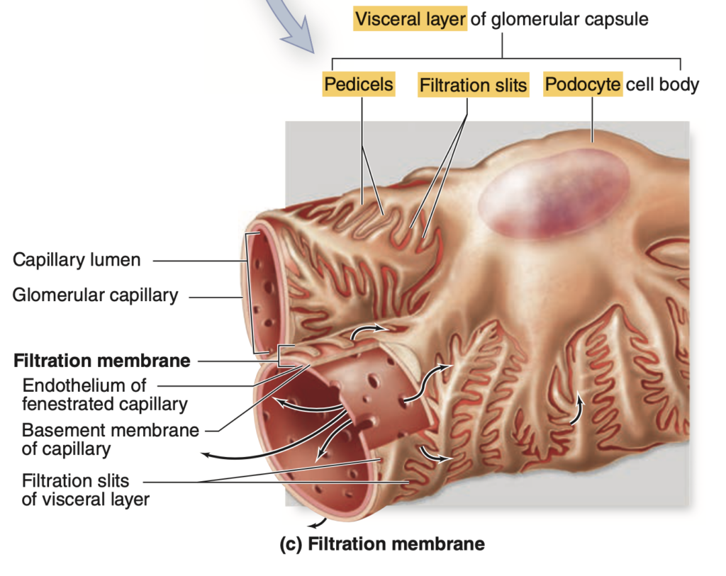
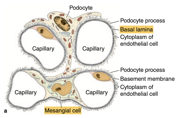
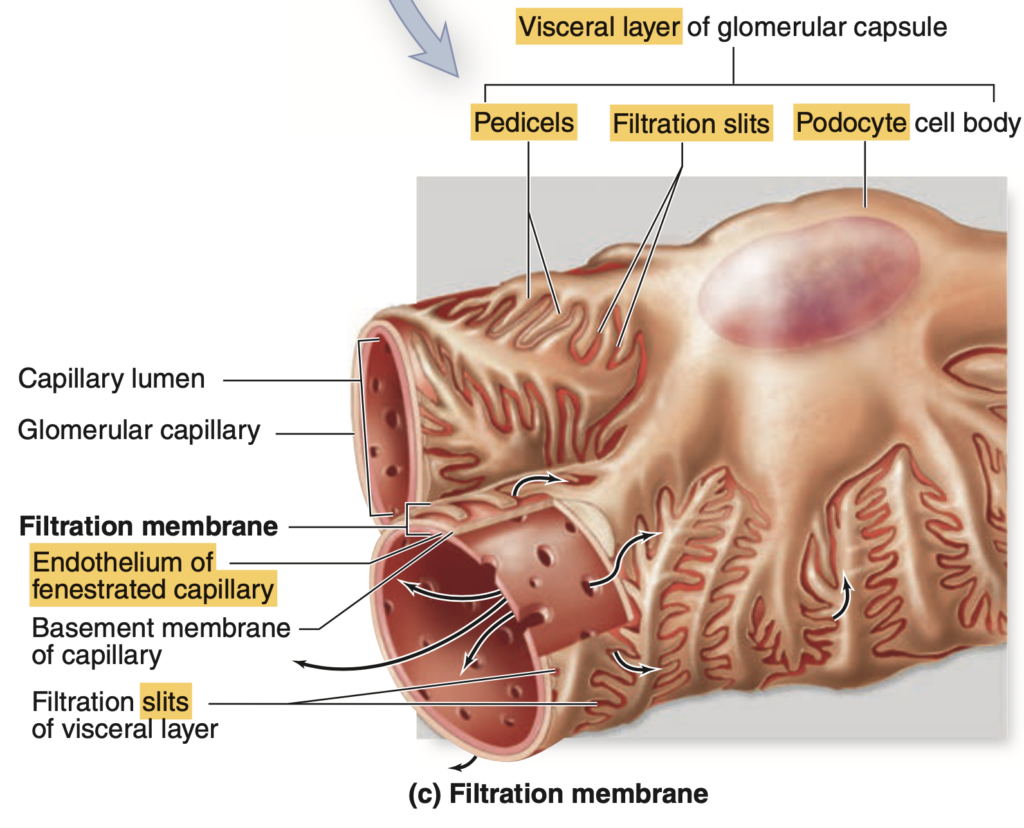
- 腎小体には、単層扁平上皮の壁側層 (parietal layer) を持つボーマン嚢 (Bowman capsule) と、糸球体毛細血管を覆う足細胞 (podocytes) の特殊な内臓層 (visceral layer) があります。
- 足細胞は、毛細血管を囲む大きな一次突起 (primary processes) を延ばし、二次突起 (pedicels) の間に狭いスリット孔 (slit pores) を形成します。
- 糸球体の高い静水圧により、血漿中の水と小さな溶質が糸球体ろ過膜を通過し、ボーマン嚢内腔 (capsular space) に移動します。
- 糸球体のろ過膜は、窓のある毛細血管内皮、タイプIVコラーゲンなどからなる厚い基底膜 (basal lamina)、および二次突起の間にあるスリット膜 (slit diaphragm) から成ります。
- 腎小体からろ液は皮質と髄質を通る尿細管に入り、再吸収および分泌が行われます。
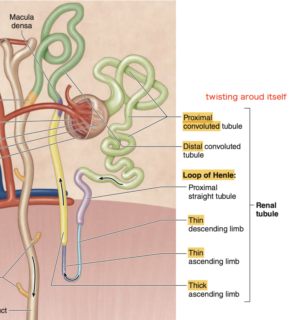
尿細管 (Tubules)
- 尿細管の最初の部分である近位尿細管 (PCT) は主に皮質に位置し、長い微絨毛と多数のミトコンドリア、大きな基底側突起 (basolateral folds) を持つ単層立方上皮で構成されます。
- 近位尿細管では、すべてのグルコース、他の有機栄養素、小さなタンパク質、ペプチド、そして大量の水と電解質がろ液から再吸収され、周囲の毛細血管に移動します。
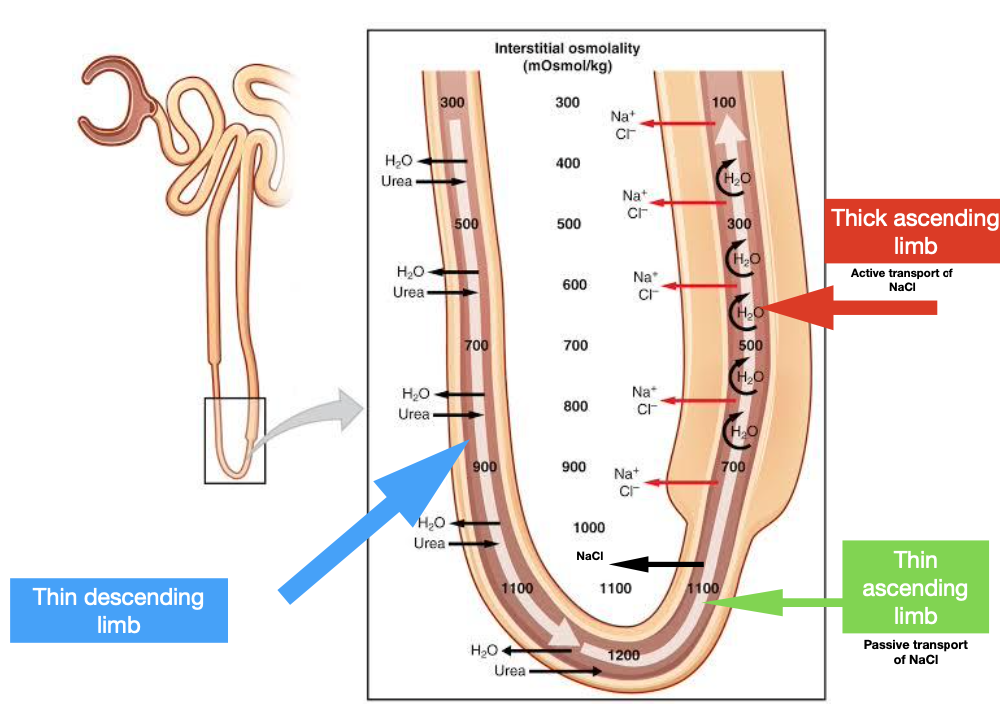
- 近位尿細管からのろ液はヘンレのループ (Loop of Henle) に入り、これは髄質に位置し、薄い降下脚と上行脚があり、後者は厚い上行脚 (TAL) として皮質に戻ります。
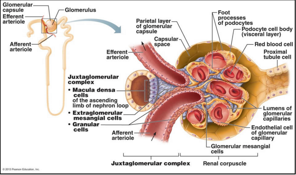
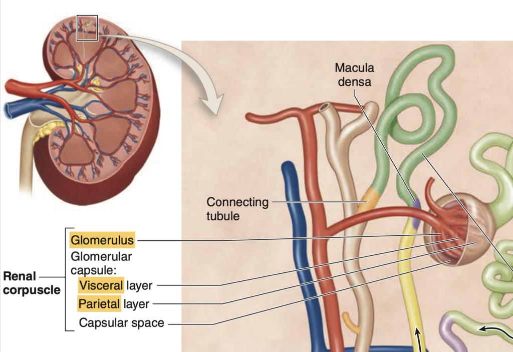
- TAL(または遠位直尿細管)は、糸球体の血管極 (vascular pole) で動脈と接触し、マクラデンサ (macula densa) として局所的に肥厚します。
- マクラデンサの高い上皮細胞と、隣接する輸入細動脈内の特殊な平滑筋細胞である傍糸球体細胞 (juxtaglomerular cells) が、血圧の調整に重要な傍糸球体装置 (JGA) を形成します。
- マクラデンサ以降、尿細管は遠位尿細管 (DCT) となり、ろ液の電解質濃度がさらに調整され、短い接続尿細管へと続きます。
- 複数のネフロンからの接続尿細管は集合管 (collecting ducts) に結合し、単層立方上皮で構成され、髄質に向かって並行に進み、徐々に大きくなり、柱状上皮になります。
尿路 (Urinary Tract)
- 集合管の主細胞 (Principal cells) は淡染し、ミトコンドリアが少なく、アクアポリン (aquaporins) が豊富な細胞膜を持ち、受動的な水の再吸収を行います。
- 最大の集合管はろ液を小腎杯 (minor calyces) に運び、ここで最終的な調整が行われ、尿と呼ばれます。
- 小腎杯、大腎杯、腎盂、尿管、膀胱は尿路上皮 (urothelium, transitional epithelium) で覆われ、尿の高浸透圧や毒性から細胞を保護します。
- 尿路上皮の表面には傘細胞 (umbrella cells) と呼ばれる大きく膨らんだ細胞があり、ウロプラキン (uroplakin) タンパク質が密集した膜構造により細胞質が保護されます。
- 膀胱が膨らむと、折りたたまれていた粘膜が広がり、尿路上皮がやや薄くなり、傘細胞のヒンジ膜が部分的に広がります。
- 尿道は膀胱から排出され、両性の初期部分は尿路上皮で覆われ、その後(男性の場合)は交互に重層円柱上皮や偽重層円柱上皮、最終的には重層扁平上皮に移行します。
- 男性の尿道は3つの領域(前立腺尿道、短い膜性尿道、長い陰茎尿道)に分かれています。
教科書図表解説
Histologic features and major functions of regions within renal tubules.
1. 近位尿細管 (Proximal Convoluted Tubule, PCT)
- 組織学的特徴:単層立方上皮(Simple cuboidal epithelium)で構成され、染色が良好でミトコンドリアが豊富に存在します。基底側に突起が目立ち、隣接する細胞と連結(ラテラルインターディジテーション)しています。長い微絨毛が内腔に伸びており、内腔はしばしば狭まっています。
- 位置:皮質(Cortex)
- 主要な機能:すべての有機栄養素、すべてのタンパク質、大部分の水および電解質の再吸収、ならびに有機陰イオンや陽イオン、H⁺、NH₄⁺の分泌
2. ヘンレのループ (Loop of Henle)
- 薄い脚 (Thin limbs):
- 組織学的特徴:単層扁平上皮(Simple squamous epithelium)で構成され、ミトコンドリアは少ない。
- 位置:髄質(Medulla)
- 主要な機能:Na⁺とCl⁻の受動的再吸収
- 厚い上行脚 (TAL, Thick Ascending Limb):
- 組織学的特徴:単層立方上皮で、微絨毛はほとんどなく、多くのミトコンドリアを含む。
- 位置:髄質および髄放線(Medulla and medullary rays)
- 主要な機能:様々な電解質の能動的再吸収
3. 遠位尿細管 (Distal Convoluted Tubule, DCT)
- 組織学的特徴:単層立方上皮で、PCTよりも細胞が小さく、短い微絨毛と基底側の突起があり、内腔はより広く見えます。
- 位置:皮質(Cortex)
- 主要な機能:電解質の再吸収
4. 集合管系 (Collecting System)
- 主細胞 (Principal cells):
- 組織学的特徴:最も多く存在する細胞で、立方上皮から円柱上皮(cuboidal to columnar epithelium)で構成され、淡染し、明確な細胞膜を持つ。
- 位置:髄放線および髄質(Medullary rays and medulla)
- 主要な機能:水および電解質の調整された再吸収、K⁺の調整された分泌
- 間在細胞 (Intercalated cells):
- 組織学的特徴:少数で、若干暗染する。
- 位置:髄放線(Medullary rays)
- 主要な機能:K⁺の再吸収(低K⁺食の場合)、酸塩基平衡の維持
Blood supply to the kidney
Renal artery → Segmental arteries → Interlobar arteries → Arcuate arteries → Interlobular arteries → Afferent arterioles → Glomerular capillaries → Efferent arterioles → Peritubular capillaries and/or vasa recta → Interlobular veins → Arcuate veins → Interlobar veins → Segmental veins → Renal vein
自作(教科書ベース)
Question 1:腎小体
Which structure is primarily responsible for blood filtration in the kidney?
a. Renal corpuscle
b. Proximal tubule
c. Collecting duct
d. Vasa recta
Answer: a. Renal corpuscle
Explanation:
腎臓における血液の濾過は、主に「腎小体(Renal corpuscle)」で行われます。腎小体は毛細血管の球(糸球体)を囲んでおり、血液から水分と溶質を濾過します。他の選択肢について、選択肢bの「近位尿細管(Proximal tubule)」は主に再吸収と分泌の役割を果たします。選択肢cの「集合管(Collecting duct)」は尿の濃縮と再吸収を調整し、選択肢dの「直血管(Vasa recta)」は血液循環を保つための毛細血管の束であり、濾過に直接関与しません。
Question 2:Renin
Which hormone, secreted by the kidney, regulates blood pressure?
a. Erythropoietin
b. Renin
c. Calcitriol
d. Angiotensin
Answer: b. Renin
Explanation:
腎臓が分泌するレニン(Renin)は、血圧を調節するホルモンで、アンジオテンシノーゲンをアンジオテンシンIに変換することで血圧を上昇させます。他の選択肢について、選択肢aの「エリスロポエチン(Erythropoietin)」は赤血球の生成を刺激し、血圧とは直接関係がありません。選択肢cの「カルシトリオール(Calcitriol)」はビタミンDの活性型であり、骨やカルシウムの代謝に関与します。選択肢dの「アンジオテンシン(Angiotensin)」は血圧を上昇させる作用がありますが、これはレニンの作用で生成されるもので、腎臓が直接分泌するものではありません。
Question 3:Loop of Henle
What is the primary function of the Loop of Henle?
a. Filtration
b. Secretion
c. Concentration of urine
d. Blood pressure regulation
Answer: c. Concentration of urine
Explanation:
ヘンレのループ(Loop of Henle)は、尿の濃縮を行う主要な構造であり、浸透圧の勾配を形成することで水の再吸収を促進します。他の選択肢について、選択肢aの「濾過(Filtration)」は腎小体で行われ、選択肢bの「分泌(Secretion)」は尿細管で主に行われます。選択肢dの「血圧の調整(Blood pressure regulation)」は腎臓が分泌するホルモン(レニン)が主に関与しており、ヘンレのループ自体は直接関与していません。
Question 4:Proximal tubule
Which part of the nephron is mainly located in the cortex and involved in the reabsorption of nutrients?
a. Renal corpuscle
b. Proximal tubule
c. Distal tubule
d. Collecting duct
Answer: b. Proximal tubule
Explanation:
近位尿細管(Proximal tubule)は腎皮質に位置し、グルコースやアミノ酸などの栄養素の再吸収を行う部位です。他の選択肢について、選択肢aの「腎小体(Renal corpuscle)」は血液の濾過を行う部位であり、選択肢cの「遠位尿細管(Distal tubule)」はナトリウムや水の調整に関与しています。選択肢dの「集合管(Collecting duct)」は、主に尿の最終的な濃縮と再吸収に関与しています。
Question 5:多発性嚢胞腎
Which condition is caused by the formation of multiple large, fluid-filled cysts in the kidneys?
a. Glomerulonephritis
b. Polycystic kidney disease
c. Nephrolithiasis
d. Acute tubular necrosis
Answer: b. Polycystic kidney disease
Explanation:
多発性嚢胞腎(Polycystic kidney disease)は、腎臓に大きな嚢胞が多発し、腎機能が低下する遺伝性疾患です。他の選択肢について、選択肢aの「糸球体腎炎(Glomerulonephritis)」は糸球体の炎症であり、選択肢cの「腎結石症(Nephrolithiasis)」は尿路結石の形成に関連しています。選択肢dの「急性尿細管壊死(Acute tubular necrosis)」は尿細管細胞の壊死によるもので、嚢胞形成とは異なります。
Question 6:腎臓の血液供給経路
What is the correct order of blood flow through the kidney’s arterial network, starting from the renal artery?
a. Renal artery → Arcuate arteries → Segmental arteries → Interlobular arteries
b. Renal artery → Segmental arteries → Interlobar arteries → Arcuate arteries → Interlobular arteries
c. Renal artery → Interlobar arteries → Segmental arteries → Arcuate arteries → Interlobular arteries
d. Renal artery → Interlobular arteries → Segmental arteries → Arcuate arteries
Answer: b. Renal artery → Segmental arteries → Interlobar arteries → Arcuate arteries → Interlobular arteries
Explanation (日本語):
腎臓の血液供給は、腎動脈が分節動脈に分岐し、次に葉間動脈、弓状動脈、最後に小葉間動脈(Interlobular arteries)へと流れます。これにより、血液は効率的に腎臓全体に供給されます。他の選択肢の順序は、実際の血流経路とは一致しません。
Question 7:Calcitriol
Which of the following describes the function of calcitriol, the active form of vitamin D produced by the kidneys?
a. It regulates blood glucose levels.
b. It stimulates erythrocyte production.
c. It enhances calcium absorption in the intestines.
d. It lowers blood pressure.
Answer: c. It enhances calcium absorption in the intestines.
Explanation (日本語):
カルシトリオールは、ビタミンDの活性型であり、小腸でのカルシウム吸収を促進することで、骨の健康維持に重要な役割を果たします。他の選択肢について、選択肢aの「血糖値の調節」は腎臓の糖新生が関与するもので、カルシトリオールの機能ではありません。選択肢bの「赤血球生成の促進」はエリスロポエチンによるものです。選択肢dの「血圧低下」はレニン-アンジオテンシン系に関与するもので、カルシトリオールには関係ありません。
Question 8:皮質と髄質
In terms of blood flow distribution, how much more blood does the renal cortex receive compared to the renal medulla?
a. 2 times more
b. 5 times more
c. 10 times more
d. 20 times more
Answer: c. 10 times more
Explanation (日本語):
腎臓の皮質は髄質よりも10倍以上多くの血流を受けています。これは、腎皮質が糸球体を含むため、濾過機能において重要な役割を果たしているからです。他の選択肢は、実際の血流分布量とは異なります。
Question 9:糸球体腎炎
Which immune mechanism is most commonly associated with glomerulonephritis?
a. Bacterial infection in the renal pelvis
b. Deposition of antibody-antigen complexes in the glomeruli
c. Decreased erythropoietin production
d. Excessive renin secretion
Answer: b. Deposition of antibody-antigen complexes in the glomeruli
Explanation (日本語):
糸球体腎炎は主に免疫複合体(抗原抗体複合体)が糸球体に沈着することによって引き起こされ、局所的な炎症反応を誘発します。他の選択肢について、選択肢aの「腎盂の細菌感染」は腎盂腎炎に関連し、糸球体腎炎とは異なります。選択肢cの「エリスロポエチン生成の減少」は貧血と関連があり、糸球体の炎症には関係しません。選択肢dの「レニンの過剰分泌」は血圧に影響しますが、糸球体腎炎の主な原因ではありません。
Question 10:片方腎の摘出
What compensatory mechanism occurs in the remaining kidney after one kidney is removed (unilateral nephrectomy)?
a. Increase in erythropoietin production
b. Decrease in filtration rate
c. Compensatory growth with cellular hypertrophy
d. Formation of new nephrons
Answer: c. Compensatory growth with cellular hypertrophy
Explanation (日本語):
片方の腎臓が摘出された後、残った腎臓は代償性の成長を示し、細胞の肥大により機能を維持します。選択肢aの「エリスロポエチン生成の増加」は貧血状態でのみ増加します。選択肢bの「濾過率の減少」は逆に、残存腎が代償機能として濾過率を増加させます。選択肢dの「新たなネフロンの形成」は起こりません。
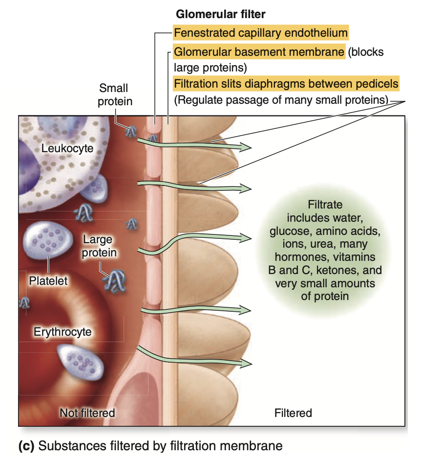
Question 11:濾過障壁
Which of the following components is NOT a part of the filtration barrier in the glomerulus?
a. Fenestrated capillary endothelium
b. Glomerular basement membrane (GBM)
c. Podocytes with slit diaphragms
d. Efferent arteriole
Answer: d. Efferent arteriole
Explanation (日本語):
濾過障壁は、糸球体における血液と濾過液を隔てる構造で、細孔を持つ毛細血管内皮:Fenestrated capillary endothelium(a)、糸球体基底膜:Glomerular basement membrane (GBM)(b)、およびスリットダイアフラムを含む足細胞:Podocytes with slit diaphragms(c)から成ります。選択肢dの「輸出細動脈(Efferent arteriole)」は、血液を糸球体から運び出すもので、濾過に直接関与していません。
Question 12:Podocytes
What is the primary function of podocytes in the glomerulus?
a. Support blood vessel contraction
b. Synthesize erythropoietin
c. Form part of the filtration barrier
d. Secrete renin
Answer: c. Form part of the filtration barrier
Explanation (日本語):
足細胞(Podocytes)は糸球体の濾過障壁の一部を形成し、フィルターとして働きます。他の選択肢について、選択肢aの「血管収縮の支持」はメサンギウム細胞の機能です。選択肢bの「エリスロポエチンの合成」は皮質の間質細胞に関連し、選択肢dの「レニンの分泌」は傍糸球体細胞の役割です。
Question 13:Proximal convoluted tubule
In which part of the nephron does the majority of water, electrolytes, and nutrients reabsorption occur?
a. Proximal convoluted tubule (PCT)
b. Distal convoluted tubule (DCT)
c. Loop of Henle
d. Collecting duct
Answer: a. Proximal convoluted tubule (PCT)
Explanation (日本語):
近位尿細管(Proximal convoluted tubule, PCT)は、水分、電解質、グルコース、アミノ酸などの大部分が再吸収される部位です。他の選択肢について、選択肢bの「遠位尿細管(DCT)」は主にナトリウムと水の調整に関与し、選択肢cの「ヘンレのループ」は尿の濃縮に重要です。選択肢dの「集合管」は、尿の最終調整と濃縮に関与しています。
Question 14:Diabetic glomerulosclerosis
Which condition is characterized by an increased permeability of the glomerular filtration barrier, leading to proteinuria?
a. Diabetic glomerulosclerosis
b. Renal tubular acidosis
c. Hyperaldosteronism
d. Cystic fibrosis
Answer: a. Diabetic glomerulosclerosis
Explanation (日本語):
糖尿病性糸球体硬化症(Diabetic glomerulosclerosis)は、糖尿病の合併症で、糸球体基底膜が肥厚し、濾過障壁の透過性が増加し、蛋白尿が引き起こされます。他の選択肢について、選択肢bの「腎尿細管性アシドーシス」は尿の酸性化異常に関連し、選択肢cの「アルドステロン過剰症」は電解質不均衡に関与します。選択肢dの「嚢胞性線維症」は主に肺や消化器系に影響し、腎障害とは直接関係がありません。
Question 15:Mesangial cells
Which cells in the renal corpuscle have phagocytic properties and help maintain the filtration barrier by removing trapped molecules?
a. Podocytes
b. Mesangial cells
c. Endothelial cells
d. Proximal tubule cells
Answer: b. Mesangial cells
Explanation (日本語):
メサンギウム細胞(Mesangial cells)は、糸球体の構造を支えると同時に、抗原抗体複合体や蛋白質の除去を通じて濾過障壁を維持する役割も果たします。選択肢aの「足細胞(Podocytes)」は濾過障壁の構成要素ですが、食作用は持ちません。選択肢cの「内皮細胞(Endothelial cells)」は血液の濾過に関与しますが、食作用はありません。選択肢dの「近位尿細管細胞(Proximal tubule cells)」は再吸収に主に関与しています。
Question 16:Bowmanカプセル(糸球体小体)
Which structure in the renal corpuscle consists of two layers, with a space in between that collects the filtered fluid?
a. Glomerular capillaries
b. Bowman’s capsule
c. Proximal convoluted tubule
d. Efferent arteriole
Answer: b. Bowman’s capsule
Explanation (日本語):
Bowmanカプセル(糸球体小体)は、内側の臓側層と外側の壁側層から成り、これらの間の空間(カプセル腔)が濾過液を受け取ります。他の選択肢について、選択肢aの「糸球体毛細血管」は濾過の場であり、選択肢cの「近位尿細管」は濾過液の通過経路、選択肢dの「輸出細動脈」は血液の出口であり、これらはカプセル腔とは異なります。

Question 17:Slit diaphragm
What is the primary role of the slit diaphragm formed by podocyte processes in the glomerular filtration barrier?
a. It facilitates the reabsorption of proteins.
b. It restricts the filtration of organic anions.
c. It increases blood pressure within the glomerulus.
d. It supports mesangial cell function.
Answer: b. It restricts the filtration of organic anions.
Explanation (日本語):
スリットダイアフラムは、足細胞の突起によって形成され、有機アニオンの濾過を制限するための負に帯電した構造を持ちます。他の選択肢について、選択肢aの「タンパク質の再吸収」は近位尿細管で行われ、選択肢cの「糸球体内の血圧増加」は輸入・輸出細動脈による調整が関与しています。選択肢dは、メサンギウム細胞の機能には直接関係がありません。
Question 18:Proximal convoluted tubule
In the proximal convoluted tubule, small proteins in the filtrate are reabsorbed through which mechanism?
a. Active transport by mitochondria
b. Simple diffusion across the tubule wall
c. Receptor-mediated endocytosis
d. Filtration through slit diaphragms
Answer: c. Receptor-mediated endocytosis
Explanation (日本語):
近位尿細管では、フィルターを通過した小分子タンパク質は、受容体媒介型エンドサイトーシスによって再吸収されます。他の選択肢について、選択肢aの「ミトコンドリアによる能動輸送」はATPを供給しますが、エンドサイトーシスとは異なります。選択肢bの「単純拡散」は特定の物質輸送には適しておらず、選択肢dの「スリットダイアフラムを通じた濾過」は糸球体での濾過に関与します。
近位尿細管(Proximal Convoluted Tubule, PCT)は、腎臓における再吸収の重要な部位であり、ここでフィルターを通過した小分子タンパク質が再吸収されるメカニズムの一つが**受容体媒介型エンドサイトーシス(Receptor-Mediated Endocytosis)**です。このプロセスについて詳しく解説します。
1. フィルターを通過した小分子タンパク質
- 腎糸球体での濾過によって、血漿中の小分子タンパク質はフィルターを通過し、尿細管の管腔に入り、濾液として移動します。これらのタンパク質は、再吸収されなければ尿中に排出されてしまうため、エネルギーを利用して効率的に再吸収する必要があります。
2. 受容体媒介型エンドサイトーシスのプロセス
- 受容体の認識:近位尿細管細胞の表面には、特定の小分子タンパク質(たとえばアルブミンやホルモンなど)に対する受容体(リセプター)が存在しています。この受容体はタンパク質を特異的に認識し、細胞膜に結合させます。
- エンドサイトーシスの開始:受容体に結合したタンパク質は、細胞膜の一部とともに細胞内に取り込まれ、エンドソームと呼ばれる小胞が形成されます。この過程はエンドサイトーシスと呼ばれ、エネルギーを必要とする能動的なプロセスです。
- エンドソーム内での分解:取り込まれたエンドソームはリソソームと融合し、リソソームの酵素によってタンパク質が分解され、アミノ酸などの小分子に分解されます。
- アミノ酸の再利用:分解されたアミノ酸は、再吸収されて血液中に戻され、体内で再利用されます。
3. 受容体媒介型エンドサイトーシスの意義
- タンパク質の無駄のない再利用:体内の貴重な資源であるタンパク質やその構成成分であるアミノ酸を再吸収することで、栄養素の浪費を防ぎます。
- 腎臓の効率的な濾過と再吸収:腎臓は一日に何十リットルもの血液を濾過しますが、近位尿細管での受容体媒介型エンドサイトーシスによって、大部分のタンパク質が再吸収され、尿中に排出されません。
4. 病態における変化
- 腎疾患や糖尿病などの影響:腎臓疾患や糖尿病では、腎フィルターの機能が損なわれ、再吸収が適切に行われなくなることがあります。これにより、尿中に異常な量のタンパク質が検出される「蛋白尿」などの症状が現れることがあります。
受容体媒介型エンドサイトーシスは、近位尿細管における効率的なタンパク質の再吸収に重要な役割を果たしており、体内のタンパク質やアミノ酸のバランスを維持するために不可欠なプロセスです。
Question 19:mesangial cells
Which of the following is NOT a function of mesangial cells within the renal corpuscle?
a. Physical support of glomerular capillaries
b. Adjusting contraction in response to blood pressure changes
c. Formation of the slit diaphragm in the filtration barrier
d. Phagocytosis of immune complexes
Answer: c. Formation of the slit diaphragm in the filtration barrier
Explanation (日本語):
メサンギウム細胞は、糸球体毛細血管の物理的支持、血圧変化に応じた収縮、免疫複合体の貪食などを行いますが、スリットダイアフラムの形成には関与していません。スリットダイアフラムは足細胞が形成する構造です。他の選択肢はすべてメサンギウム細胞の機能を表しています。
Question 20:糸球体基底膜(GBM)
Which layer of the glomerular filtration barrier is most responsible for blocking large proteins from passing into the filtrate?
a. The endothelial cell layer
b. The glomerular basement membrane (GBM)
c. The parietal layer of Bowman’s capsule
d. The visceral layer
Answer: b. The glomerular basement membrane (GBM)
Explanation (日本語):
糸球体基底膜(GBM)は、主に大きなタンパク質を濾過液から除外する機能を持っています。基底膜はIV型コラーゲンとプロテオグリカンのメッシュワークで構成され、約70 kDa以上の大きさのタンパク質の通過を制限します。他の選択肢について、選択肢aの「内皮細胞層」は血液細胞の通過をブロックし、選択肢cの「Bowmanカプセルの壁側層」は濾過には直接関与しません。選択肢dの「臓側層(足細胞)」は濾過に関与しますが、基底膜ほど大きなタンパク質のブロック機能はありません。
Question 21:PCTのendocytosis
In the proximal convoluted tubule (PCT), small proteins in the filtrate are primarily reabsorbed through which process?
a. Paracellular diffusion
b. Receptor-mediated endocytosis
c. Simple diffusion
d. Osmosis
Answer: b. Receptor-mediated endocytosis
Explanation (日本語):
近位尿細管(PCT)では、フィルターを通過した小分子タンパク質が、受容体媒介型エンドサイトーシスを介して再吸収されます。選択肢aの「傍細胞拡散」は一部の水分や電解質の移動に使用され、選択肢cの「単純拡散」は特定の物質に適していません。選択肢dの「浸透」は水の移動に関与しますが、タンパク質の再吸収には関連しません。
Question 22:ヘンレのループの対向流増幅システム
What role does the countercurrent multiplier system in the loop of Henle play in kidney function?
a. It aids in filtering blood cells from the filtrate
b. It helps concentrate the urine by creating a gradient in osmolarity
c. It increases blood flow through the vasa recta
d. It regulates blood pressure directly
Answer: b. It helps concentrate the urine by creating a gradient in osmolarity
Explanation (日本語):
ヘンレのループの対向流増幅システムは、浸透圧勾配を形成して尿を濃縮する働きを持ちます。これにより、髄質の浸透圧が高くなり、尿から水分を引き出すことが可能になります。他の選択肢について、選択肢aの「血球の濾過」は主に糸球体で行われ、選択肢cの「直血管の血流増加」は髄質の環境維持の一部ですが、対向流増幅とは直接関係がありません。選択肢dの「血圧調整」は主に傍糸球体装置の役割です。
Question 23:遠位尿細管の緻密斑(Macula densa)
What is the function of the macula densa in the distal convoluted tubule?
a. To secrete aldosterone
b. To monitor sodium and chloride levels in the filtrate
c. To regulate erythropoietin production
d. To filter blood
Answer: b. To monitor sodium and chloride levels in the filtrate
Explanation (日本語):
遠位尿細管の緻密斑(Macula densa)は、濾過液中のナトリウムおよび塩化物濃度を監視し、フィードバックに基づいて糸球体濾過率(GFR)の調整を行います。他の選択肢について、選択肢aの「アルドステロンの分泌」は副腎が行い、選択肢cの「エリスロポエチンの生成調整」は腎皮質の間質細胞が関与しています。選択肢dの「血液の濾過」は糸球体の役割であり、緻密斑の機能とは異なります。
Question 24:傍糸球体装置(JGA)
How does the juxtaglomerular apparatus (JGA) regulate blood pressure when it detects low blood pressure?
a. By decreasing renin secretion
b. By releasing aldosterone directly
c. By secreting renin, which eventually increases angiotensin II levels
d. By reducing sodium reabsorption in the distal tubule
Answer: c. By secreting renin, which eventually increases angiotensin II levels
Explanation (日本語):
傍糸球体装置(JGA)は血圧が低下するとレニンを分泌します。レニンはアンジオテンシノーゲンをアンジオテンシンIに変換し、さらにアンジオテンシンIIに変換されることで血圧が上昇します。選択肢aの「レニン分泌の減少」や選択肢dの「遠位尿細管でのナトリウム再吸収の減少」は血圧上昇のメカニズムには該当しません。選択肢bの「アルドステロンの直接分泌」は副腎の役割です。
Question 25:薬物や有機イオンのクリアランス
In which part of the nephron are drugs and organic ions actively secreted to enhance clearance from the blood?
a. Proximal convoluted tubule
b. Distal convoluted tubule
c. Loop of Henle
d. Collecting duct
Answer: a. Proximal convoluted tubule
Explanation (日本語):
薬物や有機イオンのクリアランスは、主に近位尿細管(Proximal convoluted tubule)で行われ、これにより薬物の排泄が促進されます。選択肢bの「遠位尿細管」、選択肢cの「ヘンレのループ」、および選択肢dの「集合管」は、主に異なる再吸収や調整の役割を持っており、薬物や有機イオンの排泄に特化していません。
Question 26:Sickle cell nephropathy
Which condition is associated with sickle cell nephropathy and is caused by low oxygen tension in the renal medulla?
a. Diabetic glomerulosclerosis
b. Hypertensive nephropathy
c. Renal infarcts
d. Nephrotic syndrome
Answer: c. Renal infarcts
Explanation (日本語):
鎌状赤血球腎症(Sickle cell nephropathy)では、低酸素状態により赤血球が鎌状に変形し、腎髄質内での梗塞(Renal infarcts)が引き起こされることがあります。他の選択肢について、選択肢aの「糖尿病性糸球体硬化症」は糖尿病に関連し、選択肢bの「高血圧性腎症」は高血圧に関連しています。選択肢dの「ネフローゼ症候群」は、糸球体障害による蛋白尿が主な特徴であり、鎌状赤血球腎症とは異なります。
Question 27:抗利尿ホルモン(ADH)
What is the primary role of antidiuretic hormone (ADH) in the collecting ducts?
a. To increase blood flow in the vasa recta
b. To increase permeability of the collecting ducts to water
c. To decrease the number of aquaporins in the membrane
d. To stimulate sodium reabsorption in the collecting ducts
Answer: b. To increase permeability of the collecting ducts to water
Explanation (日本語):
抗利尿ホルモン(ADH)は、集合管の水透過性を高め、水がより多く再吸収されるようにします。これにより、体液のバランスが保たれます。選択肢aの「直血管の血流増加」や選択肢dの「ナトリウムの再吸収の刺激」はADHの主な役割ではありません。選択肢cの「アクアポリン数の減少」は逆の作用であり、水透過性を減少させます。
Question 28:介在細胞(Intercalated cells)
Which cells in the collecting ducts are mainly involved in maintaining acid-base balance?
a. Principal cells
b. Macula densa cells
c. Podocytes
d. Intercalated cells
Answer: d. Intercalated cells
Explanation (日本語):
集合管内の介在細胞(Intercalated cells)は酸塩基平衡を維持する役割を持ち、H⁺やHCO₃⁻を分泌します。選択肢aの「主細胞」は主に水と電解質の再吸収に関与し、選択肢bの「緻密斑細胞」と選択肢cの「足細胞」は酸塩基平衡には関与していません。
Question 29:Aquaporins
What structural feature of the collecting duct cells allows them to concentrate urine effectively?
a. A large number of mitochondria
b. Sparse microvilli
c. Aquaporins in the membrane
d. Thick interstitial layer
Answer: c. Aquaporins in the membrane
Explanation (日本語):
集合管細胞にはアクアポリンと呼ばれる水チャネルが存在し、これにより水分が効率的に再吸収され、尿の濃縮が行われます。選択肢aの「多数のミトコンドリア」や選択肢dの「厚い間質層」は集合管における濃縮機能に関係ありません。選択肢bの「希薄な微絨毛」も主な役割には関連しません。
Question 30:Lithotripsy
What is a typical medical application for lithotripsy, related to the urinary system?
a. Treating urinary tract infections
b. Breaking down kidney stones
c. Increasing blood flow in the kidneys
d. Treating bladder cancer
Answer: b. Breaking down kidney stones
Explanation (日本語):
リトトリプシー(Lithotripsy)は、腎臓結石(kidney stones)を破砕するために使用される治療法です。選択肢aの「尿路感染症の治療」や選択肢cの「腎血流の増加」、選択肢dの「膀胱癌の治療」はリトトリプシーの主な用途ではありません。
Question 31:尿管と膀胱の移行上皮
What type of epithelium lines the ureters and bladder, providing an osmotic barrier to protect underlying tissues?
a. Stratified squamous epithelium
b. Simple cuboidal epithelium
c. Transitional (urothelium) epithelium
d. Simple columnar epithelium
Answer: c. Transitional (urothelium) epithelium
Explanation (日本語):
尿管と膀胱は移行上皮(urothelium)に覆われており、これは尿の高浸透圧に対する保護バリアとして機能します。選択肢aの「重層扁平上皮」や選択肢bの「単層立方上皮」、選択肢dの「単層円柱上皮」は尿管や膀胱に見られる主要な上皮構造ではありません。
Question 32:尿運搬経路
What is the correct pathway for urine as it exits the nephron?
a. Collecting duct → Minor calyx → Renal pelvis → Ureter → Bladder
b. Collecting duct → Renal pelvis → Minor calyx → Ureter → Bladder
c. Minor calyx → Collecting duct → Ureter → Renal pelvis → Bladder
d. Collecting duct → Ureter → Minor calyx → Bladder → Renal pelvis
Answer: a. Collecting duct → Minor calyx → Renal pelvis → Ureter → Bladder
Explanation (日本語):
尿は、集合管を通り、腎乳頭から小腎杯(minor calyx)に運ばれます。次に腎盤(renal pelvis)を経由し、尿管(ureter)を通って膀胱(bladder)に至ります。他の選択肢は尿の流路を正しく示していません。
Question 33:尿管の壁構造
What are the layers of the ureter wall from innermost to outermost?
a. Adventitia, muscularis, mucosa
b. Mucosa, muscularis, adventitia
c. Mucosa, adventitia, muscularis
d. Muscularis, mucosa, adventitia
Answer: b. Mucosa, muscularis, adventitia
Explanation (日本語):
尿管の壁は内側から外側に向かって粘膜(mucosa)、筋層(muscularis)、外膜(adventitia)の層構造を持っています。他の選択肢は正しい順序を示していません。
Question 34:Umbrella cells
How do umbrella cells in the bladder adapt when the bladder fills with urine?
a. They decrease the number of aquaporins in the membrane
b. They release H⁺ ions to prevent acidity
c. They fold and unfold their apical membranes to adjust surface area
d. They secrete mucus to protect the bladder wall
Answer: c. They fold and unfold their apical membranes to adjust surface area
Explanation (日本語):
膀胱が満杯になると、傘細胞(Umbrella cells)は頂端膜を折り畳んで面積を増やし、尿に対するバリア機能を保持します。他の選択肢は傘細胞の機能に関連していません。
Question 35:Pyelonephritis
Which condition involves inflammation of the renal pelvis and calyces, often due to bacterial infection?
a. Cystitis
b. Urethritis
c. Pyelonephritis
d. Glomerulonephritis
Answer: c. Pyelonephritis
Explanation (日本語):
腎盂腎炎(Pyelonephritis)は、腎盂や腎杯の炎症を引き起こし、一般的に細菌感染によるものです。選択肢aの「膀胱炎(Cystitis)」は膀胱の炎症、選択肢bの「尿道炎(Urethritis)」は尿道の炎症、選択肢dの「糸球体腎炎(Glomerulonephritis)」は糸球体の炎症です。
教科書の問題(10)
Question 1:arcuate arteries
Blood in the renal arcuate arteries flows next into which vessels?
a. Afferent arterioles
b. Efferent arterioles
c. Glomerular capillaries
d. Interlobar arteries
e. Interlobular arteries
Answer: e. Interlobular arteries
Explanation (日本語):
弓状動脈(arcuate arteries)は、腎臓内で皮質方向に放射状に走る皮質放線動脈(interlobular arteries)に分岐します。選択肢a、b、cは糸球体に直接関わる血管であり、選択肢dの葉間動脈(interlobar arteries)はより広い部位に血液を供給します。
Question 2:podocytes
Which cell type comprises the visceral layer of Bowman capsule?
a. Endothelial cells
b. Juxtaglomerular cells
c. Mesangial cells
d. Podocytes
e. Extraglomerular mesangial (or Lacis) cells
Answer: d. Podocytes
Explanation (日本語):
ボウマン嚢の内臓層は、特異な星状の形をした足細胞(podocytes)で構成されています。選択肢aの内皮細胞は血管壁に位置し、選択肢bの傍糸球体細胞や選択肢cのメサンギウム細胞は、主に血圧や糸球体の構造に関わります。選択肢eのLacis細胞は、糸球体外でのサポート細胞です。
Question 3:厚い上行脚(TAL)
Which type of epithelium lines the thick ascending limb of the loop of Henle?
a. Pseudostratified columnar
b. Simple columnar
c. Simple cuboidal
d. Simple squamous
e. Transitional (urothelium)
Answer: c. Simple cuboidal
Explanation (日本語):
ヘンレのループの厚い上行脚(TAL)は単層立方上皮(simple cuboidal epithelium)で覆われており、ナトリウムや塩素イオンの再吸収に重要です。他の選択肢の上皮構造は、ヘンレのループや集合管の特定部位には当てはまりません。
Question 4:Juxtaglomerular cells
Which cell is a modified smooth muscle cell that secretes renin?
a. Macula densa cells
b. Mesangial cells
c. Podocytes
d. Juxtaglomerular cells
e. Endothelial cells
Answer: d. Juxtaglomerular cells
Explanation (日本語):
傍糸球体細胞(Juxtaglomerular cells)は修飾された平滑筋細胞であり、レニンを分泌して血圧の調整に関与します。選択肢aの緻密斑細胞(Macula densa cells)は、ナトリウム濃度を感知する役割を持ち、選択肢b、c、eはレニン分泌には関わりません。
Question 5:bladder mucosa
Epithelial cell membrane domains containing many stiffened plaques of protein are an important feature in which part of the urinary system?
a. Juxtaglomerular apparatus
b. Bladder mucosa
c. Collecting ducts
d. Renal pyramids
e. Membranous urethra
Answer: b. Bladder mucosa
Explanation (日本語):
膀胱粘膜(bladder mucosa)は、傘細胞がタンパク質の硬化プラークを多く含む膜構造を持ち、尿の高浸透圧環境に対する保護バリアとして機能します。選択肢aの傍糸球体装置や選択肢cの集合管はこの構造を持たず、選択肢dとeも関係ありません。
Question 6:aquaporins
An immunohistochemical technique using antibodies against aquaporins to stain a section of kidney would be expected to stain cells in which structures most intensely?
a. Collecting ducts
b. Lining of the major and minor calyces
c. Proximal convoluted tubules
d. Distal convoluted tubules
e. Glomeruli
Answer: a. Collecting ducts
Explanation (日本語):
アクアポリンは水分子を通過させるチャネルタンパク質であり、集合管(collecting ducts)の細胞に豊富に存在します。集合管は水再吸収の最終段階でADHの影響を受け、水の透過性が高まります。他の選択肢ではアクアポリンの発現が比較的少ないです。
Question 7:prostatic urethra
What type of epithelium lines the prostatic urethra?
a. Simple columnar
b. Pseudostratified columnar
c. Stratified squamous
d. Simple squamous
e. Transitional (urothelium)
Answer: e. Transitional (urothelium)
Explanation (日本語):
前立腺部尿道(prostatic urethra)は移行上皮(transitional epithelium)で覆われており、膀胱や尿道の一部でも見られる上皮です。他の選択肢は前立腺部尿道の上皮には当てはまりません。
Question 8:近位尿細管(PCT)
A 14-year-old patient presents in the nephrology clinic with fatigue, malaise, anorexia, abdominal pain, and fever. She reports a loss of 6 lb in the past 2 months. Serum gamma globulin and the immunoglobulins IgG, IgA, and IgM are all elevated. Her serum creatinine is 1.4 mg/dL (normal 0.6-1.2 mg/dL) and urinalysis of glucose and protein are 2+ on a dipstick test, confirmed by laboratory at 8.0 g/dL and 0.95 g/dL, respectively. A renal biopsy is prepared for light microscopy, and an infiltrate containing lymphocytes, plasma cells, and eosinophils is found among tubules having cells with prominent brush borders. Which one of the following statements correctly pertains to these epithelial cells?
a. Impermeable to water despite presence of ADH
b. The primary site for the reduction of the tubular fluid volume
c. The site of the countercurrent multiplier
d. The site of action of aldosterone
e. Indirectly involved in the release of renin
Answer: b. The primary site for the reduction of the tubular fluid volume
Explanation (日本語):
近位尿細管(PCT)は、尿細管液の体積を大幅に減少させる主な部位であり、糖やタンパク質、その他の栄養素の再吸収が行われます。他の選択肢の機能は、集合管や遠位尿細管で主に見られ、近位尿細管には該当しません。
Question 9:腎臓結石
A 45-year-old man presents with nephrolithiasis or kidney stones. The process of calcium oxalate stone formation as seen in this patient begins with Randall plaques found in the basement membranes of which one of the following structures found only in the renal medulla?
a. Proximal convoluted tubules
b. Distal convoluted tubules
c. Thin loops of Henle
d. Afferent arterioles
e. Collecting ducts
Answer: e. Collecting ducts
Explanation (日本語):
腎臓結石(特にカルシウムシュウ酸塩結石)は、集合管の基底膜に形成されるRandallプラークから始まります。他の選択肢の構造は腎髄質(renal medulla)には限定されません。
Question 10:COL4A5遺伝子の変異
A 15-year-old male presents with hematuria, hearing loss, lens dislocation, and the onset of cataracts. Genetic analysis reveals a mutation in the COL4A5 gene. Transmission EM examination of a renal biopsy confirms that the disorder has affected a component of the renal corpuscles in which damage disrupts normal glomerular filtration. Which one of the following structures would most likely be abnormal in the TEM of this patient’s biopsy?
a. Pedicels
b. Filtration slits
c. Slit diaphragms
d. Glomerular basement membranes
e. Fenestrated endothelium of glomerular capillaries
Answer: d. Glomerular basement membranes
Explanation (日本語):
COL4A5遺伝子の変異は、糸球体基底膜(GBM)の異常を引き起こし、正常な糸球体濾過に影響を及ぼします。他の選択肢であるa〜cは基底膜と連動する構造ですが、基底膜の異常が本症例の主因です。
過去問
Question 1:尿道の長さ
問題文: What is the average length of spongy/penile urethra?
a. 3 cm
b. 5 cm
c. 7 cm
d. 15 cm
Answer: d
Question 2:間在細胞
問題文: These cells of the collecting duct help maintain acid-base balance by secreting hydrogen or bicarbonate.
A. Granular cells
B. Mesangial cells
C. Podocytes
D. Intercalated cells
E. Principal cells
F. Lacis cells
Answer: D. Intercalated cells
解説:
集合管の間在細胞(Intercalated cells)は、酸塩基平衡を保つために水素イオンや重炭酸イオンを分泌します。これにより、体内のpHバランスが調整されます。その他の選択肢が誤っている理由として、顆粒細胞(Granular cells)はレニン分泌に関わり、間質細胞(Mesangial cells)は糸球体の物理的支持を提供します。ポドサイト(Podocytes)はボウマン嚢の内皮細胞であり、主細胞(Principal cells)はナトリウムと水の再吸収を行います。Lacis細胞は傍糸球体装置の構造支持を提供します。
集合管の間在細胞(Intercalated cells)は、腎臓の集合管に存在する細胞の一種で、主に酸塩基平衡(pHバランス)を調整する役割を担っています。体内の酸塩基平衡を保つことは重要で、血液が酸性やアルカリ性に偏りすぎないようにするために、間在細胞は水素イオン(H⁺)や重炭酸イオン(HCO₃⁻)を分泌します。
間在細胞の役割
- 水素イオン(H⁺)の分泌:間在細胞の一部は、血液がアルカリ性に偏りすぎたときに、水素イオンを分泌します。これにより、体内の酸性度が高まり、pHバランスが整えられます。
- 重炭酸イオン(HCO₃⁻)の分泌:一方、血液が酸性に傾いたときには、重炭酸イオンを分泌します。重炭酸イオンは血液中の酸性物質と結びついて中和するため、酸性度が下がり、pHバランスが正常に保たれます。
間在細胞の種類
間在細胞には、以下の2種類があります。
- A型間在細胞(Type A Intercalated cells):主に水素イオンを分泌して、血液をアルカリ性に調整します。このタイプは血液が酸性に傾いたときに活動し、尿に水素イオンを排泄して酸性物質を排出します。
- B型間在細胞(Type B Intercalated cells):主に重炭酸イオンを分泌して、血液がアルカリ性になりすぎるのを防ぎます。このタイプは、血液がアルカリ性に傾いたときに働き、重炭酸イオンを排泄することで酸塩基バランスを保ちます。
酸塩基平衡維持の仕組み
間在細胞が水素イオンや重炭酸イオンを調整することで、体内のpHを一定に保ち、細胞や酵素が正常に働ける環境を作り出しています。
Question 3:末期腎疾患
This is the leading cause of end-stage kidney disease in the United States.
a. Pyelonephritis
b. Diabetic glomerulosclerosis
c. Nephrolithiasis
d. Hypertensive nephrosclerosis
Answer: b. Diabetic glomerulosclerosis, c
解説:
アメリカにおける末期腎疾患(ESKD)の主な原因は糖尿病性糸球体硬化症(Diabetic glomerulosclerosis)です。糖尿病が進行すると、糸球体が損傷され腎機能が低下します。他の選択肢について、腎盂腎炎(Pyelonephritis)は腎臓の感染症、結石症(Nephrolithiasis)は腎結石による病態、高血圧性腎硬化症(Hypertensive nephrosclerosis)は高血圧に伴う腎機能の低下ですが、糖尿病による腎障害の発症率が最も高いです。
Question 4:Macula Densa
問題文: This is the sensory structure of the J apparatus that detects sodium.
a. JG granular cells
b. Podocytes
c. Principal cells
d. Lacis Cells
e. Macula Densa
Answer: e. Macula Densa
解説:
マクラデンサ(Macula Densa)は傍糸球体装置(J apparatus)の中でナトリウム濃度を感知する役割を持ち、腎臓での塩分濃度の調整を行います。他の選択肢が間違っている理由として、顆粒細胞(JG granular cells)はレニンの分泌に関与し、ポドサイト(Podocytes)はボウマン嚢の内膜、主細胞(Principal cells)は集合管でナトリウムと水のバランスを調整します。Lacis細胞は構造支持を提供します。
Question 5:ボウマン嚢の内臓層(visceral layer)
問題文: These cells make up the visceral layer of Bowman Capsule.
a. Simple squamous epithelium
b. Podocytes
c. Principal cells
d. Intercalated cells
e. Granular cells
f. Mesangial cells
g. Lacis cells
Answer: b. Podocytes, a
解説:
ボウマン嚢の内臓層(visceral layer)は、ポドサイト(Podocytes)で構成され、糸球体フィルター機能に重要です。他の選択肢について、単層扁平上皮(Simple squamous epithelium)はボウマン嚢の壁側層に存在し、主細胞(Principal cells)や間在細胞(Intercalated cells)は集合管で機能します。顆粒細胞(Granular cells)はレニン分泌、間質細胞(Mesangial cells)とLacis細胞は構造的支持に関与します。
The outer parietal layer of a glomerular capsule consists of a simple squamous epithelium supported externally by a basal lamina. At the tubular pole, this epithelium changes to the simple cuboidal epithelium that continues and forms the proximal tubule (Figure 19–5).
The visceral layer of a renal corpuscle consists of unusual stellate epithelial cells called podocytes (Figures 19–5c and 19–5d), which together with the capillary endothelial cells com- pose the apparatus for renal filtration. From the cell body of each podocyte several primary processes extend and curve around a length of glomerular capillary. Each primary process gives rise to many parallel, interdigitating secondary processes or pedicels (L. pedicellus, little foot; Figures 19–5c and 19–5d). The pedicels cover much of the capillary surface, in direct con- tact with the basal lamina (Figures 19–5c and 19–6).
Question 6:近位尿細管
問題文: TRUE of the proximal convoluted tubule compared to the distal convoluted tubule.
a. Epithelium is flatter and smaller
b. Less numerous than distal convoluted tubules
c. Appear darker red in color
d. Contains more microvilli
e. Has more mitochondria
Answer: e. Has more mitochondria
解説:
近位尿細管(Proximal Convoluted Tubule, PCT)は、遠位尿細管(Distal Convoluted Tubule, DCT)と比較してミトコンドリアが多く含まれており、エネルギーを多く必要とする再吸収活動を支えています。他の選択肢が間違っている理由として、上皮の大きさはむしろ大きく、またPCTはDCTよりも数が多く、色も濃い赤に見えます。さらに、微絨毛が密に存在するため、表面積が広く、再吸収の効率が高まっています。
近位尿細管(Proximal Convoluted Tubule, PCT)は、腎臓の構造の一部であり、特に重要な再吸収の働きを担っています。PCTは、遠位尿細管(Distal Convoluted Tubule, DCT)と比べて多くのミトコンドリアを含んでおり、大量のエネルギーが必要とされるためです。
近位尿細管(PCT)の特徴
- 再吸収の中心:PCTは、尿が形成される過程で、体に必要な物質(例:水、グルコース、アミノ酸、ナトリウムイオンなど)を再吸収し、血液に戻す働きをします。体が失いたくない栄養やイオンが多く再吸収されるため、重要な役割を担っています。
- エネルギーの消費:PCTでの再吸収は、ナトリウムポンプ(Na⁺/K⁺ポンプ)などを利用して物質を積極的に移動させる「能動輸送」が多く使われます。能動輸送にはATPというエネルギーが必要で、その供給源がミトコンドリアです。
- ミトコンドリアの多さ:ミトコンドリアは細胞の「エネルギー工場」として働き、ATPを生成します。PCTはエネルギー消費量が高いため、ミトコンドリアが他の部位よりも多く存在し、再吸収の活動を支えています。
遠位尿細管(DCT)との比較
- PCT:主に大量の物質を再吸収するため、エネルギーが必要です。そのため、ミトコンドリアが豊富に含まれています。
- DCT:PCTに比べると再吸収の活動が少なく、ミトコンドリアの量も少なめです。DCTは主に体内の電解質や酸塩基バランスの調整を担いますが、再吸収の量はPCTよりも少ないため、エネルギー消費も少ないです。
Question 7:間質細胞(Mesangial cells)
問題文: These cells provide physical support of the glomerulus.
a. Podocytes
b. Mesangial cells
c. Principal cells
d. Intercalated cell type B
e. Intercalated cell type A
Answer: b. Mesangial cells
解説:
間質細胞(Mesangial cells)は糸球体(Glomerulus)内で物理的な支持を提供し、毛細血管の安定を維持します。その他の選択肢について、ポドサイト(Podocytes)はボウマン嚢の内壁を構成し、フィルター機能を担っています。主細胞(Principal cells)は集合管でのナトリウムと水の再吸収を行い、間在細胞(Intercalated cells)は酸塩基平衡の調整に関与しています。
間質細胞(Mesangial cells)は、腎臓の糸球体(Glomerulus)の中にある特殊な細胞で、主に糸球体の構造を物理的に支える役割を担っています。糸球体は、血液を濾過して尿を生成する重要な部分ですが、たくさんの細かい毛細血管が集まっているため、構造が複雑で壊れやすいという特徴があります。間質細胞は、この繊細な構造を保つために欠かせない細胞です。
間質細胞の主な役割
- 物理的な支持:間質細胞は、糸球体の毛細血管の間に位置し、「メサンギウム基質(Mesangial matrix)」と呼ばれる物質を分泌して、糸球体の毛細血管の間を埋めるように支えます。これにより、毛細血管が安定し、正常な血流が保たれるのです。
- 血管の安定維持:糸球体の毛細血管は高い圧力にさらされるため、血管が壊れたり、過剰に膨張したりするリスクがあります。間質細胞はこれを防ぎ、毛細血管の安定を維持することで、血液が適切に濾過される環境を整えています。
- 老廃物の除去(貪食作用):間質細胞には、糸球体内に蓄積された老廃物や異物を取り除く「貪食(phagocytosis)」の機能もあります。これにより、糸球体の環境が清潔に保たれ、濾過機能が正常に働くことができます。
Question 8:Aldosterone
問題文: The rate of sodium absorption in the distal convoluted tubule is regulated by what hormone?
a. Angiotensin II
b. Antidiuretic hormone
c. Aldosterone
d. Vasopressin
e. Renin
Answer: c. Aldosterone
解説:
遠位尿細管でのナトリウム吸収速度は、アルドステロン(Aldosterone)により調整されています。アルドステロンはナトリウム再吸収を増加させ、体液量を調整します。他の選択肢が間違っている理由として、アンジオテンシンII(Angiotensin II)はアルドステロン分泌を刺激するホルモンであり、抗利尿ホルモン(Antidiuretic hormone, ADH)およびバソプレッシン(Vasopressin)は水の再吸収を促進します。レニン(Renin)はアンジオテンシンIIを生成する酵素です。
Question 9:有窓型毛細血管
問題文: What type of capillaries make up the Glomerulus?
a. Continuous
b. Sinusoids
c. Fenestrated
d. None
Answer: c. Fenestrated
解説:
糸球体(Glomerulus)は、有窓型(Fenestrated)の毛細血管で構成されており、ろ過効率が高く、血液からの不要な物質を除去する役割を果たします。他の選択肢について、連続型毛細血管(Continuous)は脳などで見られ、シヌソイド(Sinusoids)は肝臓などに存在します。
Question 10:PCT
問題文: Match each item to a choice.
a. Glucose reabsorption
= PCT
b. Portion of the nephron immediately following loop of Henle
= Collecting ducts or tubules
c. Site of Filtration
= Glomerulus
d. Countercurrent multiplier system
= Loop of Henle
e. Site of action of Antidiuretic Hormone
= DCT
Answer: a
- a. Glucose reabsorption = PCT
- b. Portion of the nephron immediately following loop of Henle = Collecting ducts or tubules
- c. Site of Filtration = Glomerulus
- d. Countercurrent multiplier system = Loop of Henle
- e. Site of action of Antidiuretic Hormone = DCT
解説:
- グルコースの再吸収は近位尿細管(PCT)で行われます。近位尿細管はエネルギーを用いてグルコースやアミノ酸を血流に再吸収します。
- ヘンレのループ(Loop of Henle)の直後に続く部位は集合管(Collecting ducts/tubules)で、尿の最終的な濃縮を行います。
- 糸球体(Glomerulus)は血液のろ過を行う部位です。
- ヘンレのループは対向流増幅システム(Countercurrent multiplier system)を形成し、尿の濃縮に重要な役割を果たします。
- 抗利尿ホルモン(ADH)の作用部位は遠位尿細管(DCT)であり、最終的な水の再吸収を調整します。
Question 11:傍糸球体装置
問題文: Which of the following is not part of the JG apparatus?
a. Lacis cells
b. Macula densa
c. JG granular cells
d. Bowman’s capsule
Answer: d. Bowman’s capsule
解説:
傍糸球体装置(Juxtaglomerular apparatus, JG apparatus)は、ナトリウム濃度や血圧を感知する役割を持つ構造で、主にマクラデンサ(Macula densa)、ラキス細胞(Lacis cells)、および傍糸球体顆粒細胞(JG granular cells)で構成されています。ボウマン嚢(Bowman’s capsule)は糸球体を囲む構造であり、JG装置の一部ではありません。
傍糸球体装置(Juxtaglomerular apparatus, JG apparatus)は、腎臓内でナトリウム濃度や血圧の変化を感知し、それに応じて体の水分・電解質バランスや血圧を調節する働きを持つ重要な構造です。この装置は、腎臓の糸球体の近くに位置し、以下の3つの主要な細胞で構成されています:マクラデンサ(Macula densa)、ラキス細胞(Lacis cells)、および傍糸球体顆粒細胞(JG granular cells)。
1. マクラデンサ(Macula densa)
- マクラデンサは遠位尿細管(Distal Convoluted Tubule, DCT)に存在する特殊な細胞の集まりで、尿の中のナトリウムイオン濃度を感知します。
- ナトリウム濃度の低下が検出されると、マクラデンサは傍糸球体顆粒細胞に信号を送り、レニン(Renin)というホルモンの分泌を促します。
- レニン分泌により、体内でのナトリウムの再吸収や血圧上昇に関与する「レニン-アンジオテンシン-アルドステロン系(RAAS)」が活性化します。
2. 傍糸球体顆粒細胞(JG granular cells)
- 傍糸球体顆粒細胞は、腎臓の輸入細動脈(Afferent arteriole)の壁に存在し、レニンを産生・分泌する細胞です。
- 血圧が低下したり、マクラデンサからの信号を受けたりすると、これらの細胞はレニンを分泌します。
- レニンが分泌されると、血圧上昇やナトリウムの再吸収が促進され、体の水分や電解質バランスが維持されます。
3. ラキス細胞(Lacis cells)
- ラキス細胞(または外部メサンギウム細胞とも呼ばれる)は、主にマクラデンサと傍糸球体顆粒細胞の間に存在し、構造的な支持や細胞間のシグナル伝達を助ける役割を持っています。
- ラキス細胞は、直接的なホルモン分泌機能はありませんが、細胞間の情報伝達や支持を行い、傍糸球体装置の調整を支えています。
Question 12:移行上皮
問題文: The superficial layer of the transitional epithelium may contain large, occasionally binucleated, bulbus cells that are called:
a. Lacis cells
b. Umbrella cells
c. Uroplakins
d. Simple squamous cells
Answer: b. Umbrella cells
解説:
移行上皮(Transitional epithelium)の表層には、大きく、時折二核を持つこともある「アンブレラ細胞(Umbrella cells)」と呼ばれる細胞が存在します。これらの細胞は膀胱や尿路の柔軟性を提供します。他の選択肢について、ラキス細胞(Lacis cells)は傍糸球体装置に関わり、ウロプラキン(Uroplakins)は移行上皮細胞膜の構成タンパク質です。単層扁平上皮(Simple squamous cells)は別の組織で見られます。
Question 13:腎杯
問題文: What is the lining epithelium of the renal calyces?
a. Simple squamous
b. Simple columnar
c. Pseudostratified columnar
d. Simple cuboidal
e. Uroepithelium
f. Non-keratinized stratified squamous
Answer: e. Uroepithelium
解説:
腎杯(Renal calyces)の内面は尿路上皮(Uroepithelium)で覆われています。尿路上皮は移行上皮とも呼ばれ、尿の流入に伴う伸縮性が必要な部位に適応しています。他の選択肢について、単層扁平上皮(Simple squamous)や単層円柱上皮(Simple columnar)は腎杯には見られません。
腎杯(Renal calyces)の内面は、**尿路上皮(Uroepithelium)**で覆われています。尿路上皮は、腎盂、尿管、膀胱など、尿が通過する通路の内側を覆う上皮組織です。この上皮は「移行上皮(transitional epithelium)」とも呼ばれ、尿の流入や貯留に伴って伸縮性を持つ特別な構造をしています。
尿路上皮(移行上皮)の特徴
- 伸縮性:尿路上皮は、尿の量に応じて伸びたり縮んだりする能力を持っています。これにより、尿が腎杯や尿管を通過するときに、内面が傷つかず、スムーズに流れることができます。
- 保護機能:尿路上皮は、尿中の有害な成分や毒素から組織を保護する役割も果たします。尿はpHや成分が変動することがあるため、この保護機能は重要です。
腎杯における尿路上皮の役割
腎杯は腎臓で生成された尿が集まる最初の場所です。腎杯から尿が集められ、腎盂、尿管、膀胱へと順次移動します。このとき、腎杯の内面が尿路上皮で覆われていることで、尿の圧力変動や量の増減に対応できる柔軟な構造となり、尿の排出をスムーズに行うことができます。
Question 14:排尿困難
問題文: This is the pain or difficulty in urination.
a. Dysuria
b. Proteinuria
c. Pyuria
d. Hematuria
Answer: a. Dysuria
解説:
排尿時の痛みや困難は「排尿困難(Dysuria)」と呼ばれます。他の選択肢について、蛋白尿(Proteinuria)は尿中に異常に多量のタンパク質が含まれている状態、膿尿(Pyuria)は尿中に膿が含まれている状態、血尿(Hematuria)は尿中に血液が混ざっている状態を指します。
Question 15:集合管主細胞
問題文: These cells of the collecting ducts are rich in aquaporins.
a. Mesangial cells
b. Granular cells
c. Podocytes
d. Intercalated cells
e. Lacis cells
f. Principal cells
Answer: f. Principal cells, c
解説:
集合管の主細胞(Principal cells)はアクアポリン(Aquaporins)が豊富で、水の再吸収を効率的に行います。他の選択肢について、間質細胞(Mesangial cells)は糸球体の支持に関与し、顆粒細胞(Granular cells)はレニンを分泌します。ポドサイト(Podocytes)はボウマン嚢の内膜、間在細胞(Intercalated cells)は酸塩基平衡の調整に関わり、ラキス細胞(Lacis cells)は構造的支持を提供します。
集合管の主細胞(Principal cells)は、水の再吸収に重要な役割を果たし、**アクアポリン(Aquaporins)**と呼ばれる水チャネルを豊富に含んでいます。アクアポリンは細胞膜に存在し、水分子を通すための専用通路として機能します。これにより、主細胞は水を効率的に再吸収し、体内の水分バランスを調整します。
主細胞とアクアポリンの役割
- アクアポリンの特徴:アクアポリンは、水分子のみを選択的に通過させる特殊なタンパク質です。アクアポリンが主細胞の細胞膜に埋め込まれることで、水の再吸収が可能になります。
- 抗利尿ホルモン(ADH)による調節:アクアポリンの数は、抗利尿ホルモン(ADH, Antidiuretic Hormone)によって調整されます。ADHの分泌が増えると、アクアポリンが主細胞の膜に挿入され、水がより多く再吸収されるようになります。これにより、尿の量が減少し、濃縮されます。
- 水分バランスの調整:アクアポリンを通して水が再吸収されることで、血液中の水分量が調整されます。これにより、体が脱水状態になるのを防ぎ、適切な水分量が維持されます。
アクアポリンによる水の再吸収の仕組み
- 高浸透圧環境:集合管周辺の組織液は浸透圧が高いため、アクアポリンを介して水が細胞内から血液中へと移動しやすくなります。
- 再吸収の効率化:アクアポリンがあることで、水分子は迅速に移動でき、水の再吸収が効率的に行われます。
Question 16:傍糸球体細胞
問題文: These cells produce and store renin.
A. JG cells
B. Principal cells
C. Lacis Cells
D. Intercalated cells
E. Podocytes
Answer: A. JG cells
解説:
傍糸球体細胞(Juxtaglomerular cells, JG cells)は、腎臓においてレニンを生成および貯蔵する役割を持っています。レニンは血圧を調整するために重要なホルモンです。他の選択肢が間違っている理由として、主細胞(Principal cells)は水とナトリウムのバランスを調整し、ラキス細胞(Lacis cells)は構造的支持を提供します。間在細胞(Intercalated cells)は酸塩基平衡を保ち、ポドサイト(Podocytes)はボウマン嚢の内膜に存在してろ過機能を助けます。
Question 17:太い上行脚
問題文: What occurs in the thick ascending limb of Henle?
a. Production of erythropoietin
b. Uptake of water
c. Active uptake of Sodium
d. Passive uptake of sodium
Answer: c. Active uptake of Sodium
解説:
ヘンレのループの太い上行脚(thick ascending limb of Henle)では、ナトリウムの能動的な再吸収が行われ、これにより尿の濃縮が促進されます。その他の選択肢について、エリスロポエチン(erythropoietin)の生成は腎臓の間質細胞で行われ、水の吸収は上行脚ではなく下行脚で起こります。ナトリウムの受動的な取り込みは、能動的な取り込みの反対のプロセスです。
Question 18:糸球体のろ過障壁
問題文: Which of the following is not part of the filtration barrier of the kidney?
a. Mesangial cells
b. Glomerular basement membrane
c. Capillary Endothelial cells
d. Slit diaphragm
Answer: a. Mesangial cells, c
解説:
糸球体のろ過障壁は、毛細血管内皮細胞、糸球体基底膜(Glomerular basement membrane)、およびスリット膜(Slit diaphragm)で構成されています。間質細胞(Mesangial cells)は糸球体の支持を提供するもので、ろ過障壁の一部ではありません。
糸球体のろ過障壁は、腎臓で血液を濾過するための構造で、以下の3つの層で構成されています:毛細血管内皮細胞、糸球体基底膜(Glomerular basement membrane)、および**スリット膜(Slit diaphragm)**です。このろ過障壁は、血液から老廃物や余分な水分を選択的に除去し、尿として排出するための重要な働きを持っています。
糸球体のろ過障壁の構造と役割
- 毛細血管内皮細胞(Capillary endothelial cells)
- 糸球体の毛細血管内皮細胞は、多数の小さな穴(窓、フェネストレーション)を持っています。この穴は、血液を濾過する際に血球が通過しないようにする役割があります。
- 血液中の細胞成分(赤血球や白血球など)は通過できませんが、水分や溶質(ナトリウム、カリウム、グルコースなど)は通過できるため、血液の液体成分を効率的にろ過することができます。
- 糸球体基底膜(Glomerular basement membrane, GBM)
- 糸球体基底膜は、ろ過障壁の中央に位置する膜で、主にコラーゲンやプロテオグリカンから成る強固な構造です。この膜は、血液の中の大きなタンパク質が尿中に漏れないようにブロックする役割を持っています。
- 糸球体基底膜は、サイズ選択性と電荷選択性を持ち、特に負電荷を帯びた物質(例:アルブミンなどのタンパク質)が通過しにくくなっています。
- スリット膜(Slit diaphragm)
- スリット膜は、糸球体の足細胞(ポドサイト)の足突起が絡み合ってできる細かい隙間に存在する膜です。このスリット膜も、血液中の不要な成分を除去するフィルターの一部として働きます。
- スリット膜は、基底膜を通過した溶質や水分をさらに選択的に濾過し、大きなタンパク質が尿中に漏れ出ないようにします。この膜も、ろ過バリアの最終段階として、選択的に物質を通過させます。
Question 19:膀胱後部
問題文: Which part of the urinary bladder is covered by serous peritoneum?
a. Posterior part only
b. Entire organ
c. Anterior part only
d. Lower part only
e. Upper part only
Answer: a. Posterior part only, b
解説:
膀胱の後部のみが漿膜(serous peritoneum)で覆われており、他の部分は被覆されていません。その他の選択肢について、膀胱全体や前部、下部は漿膜で覆われていないため、これらの回答は不正解です。
Question 20:葉間動脈
問題文: This is an artery of the kidney that travels between the renal pyramids via the renal columns of Bertin.
a. Arcuate artery
b. Cortical radial artery
c. Interlobular artery
d. Segmental artery
e. Interlobar artery
Answer: e. Interlobar artery, c
解説:
腎臓の葉間動脈(Interlobar artery)は、腎ピラミッドの間をベルチン柱(Renal columns of Bertin)を通って進みます。その他の選択肢について、弓状動脈(Arcuate artery)は腎皮質と髄質の境界を走行し、皮質放射動脈(Cortical radial artery)や葉間動脈(Interlobular artery)は腎皮質に分布します。
ベルチン柱の役割:
- ベルチン柱は、腎ピラミッドの間に位置する腎皮質の延長で、葉間動脈や葉間静脈が通過するためのスペースを提供します。
- これにより、葉間動脈は腎ピラミッドを避けて腎臓の深部まで到達でき、腎皮質や腎髄質に十分な血液供給を行うことができます。
葉間動脈の分岐:
- 葉間動脈は、腎ピラミッドの基部に到達すると**弓状動脈(Arcuate artery)**へと分岐します。弓状動脈は、腎皮質と腎髄質の境界に沿って横向きに走行し、さらにそこから細かい放線動脈(Interlobular arteries)へと枝分かれしていきます。
- 放線動脈は腎皮質へと枝分かれし、最終的に糸球体(glomerulus)に血液を供給し、尿生成のための濾過プロセスが開始されます。

Quiz
Question
問題文: What is the glomerulus?
a. Afferent arteriole
b. Efferent arteriole
c. Capillary tuft
d. Peritubular capillaries
e. Vasa recta
Answer: c. Capillary tuft
解説:
糸球体(Glomerulus)は「毛細血管の束(Capillary tuft)」として知られ、腎臓内で血液の濾過が行われる場所です。他の選択肢について、輸入細動脈(Afferent arteriole)と輸出細動脈(Efferent arteriole)は糸球体に血液を運び入れたり送り出したりする役割を果たします。周囲の毛細血管(Peritubular capillaries)は尿細管の周りを取り巻き、血液の再吸収を行います。Vasa rectaはヘンレのループ周辺で働く毛細血管の集まりです。
Question
問題文: What is a renal pyramid and its associated cortex referred to?
a. Medulla
b. Lobe
c. Renal columns
d. Nephron
e. Medullary ray
Answer: b. Lobe
解説:
腎臓のピラミッドとそれに関連する皮質は「葉(Lobe)」と呼ばれます。腎葉は腎臓の基本的な構造単位の一つです。その他の選択肢について、髄質(Medulla)は腎ピラミッドを含む部位を指し、腎柱(Renal columns)は腎ピラミッドの間を走ります。ネフロン(Nephron)は腎臓の機能単位、髄放線(Medullary ray)は皮質内の髄質への投影部分です。
Question
問題文: Approximately how many nephrons are there in each kidney?
a. 1,000
b. 10,000
c. 100,000
d. 1,000,000
e. 10,000,000
Answer: d. 1,000,000
解説:
各腎臓には約100万(1,000,000)のネフロンが存在します。ネフロンは尿生成に関わる基本的な単位です。他の選択肢の数は腎臓に含まれるネフロンの数としては少なすぎ、または多すぎます。
Question
問題文: What is the Malpighian corpuscle?
a. Glomerulus
b. Bowman’s capsule
c. Renal corpuscle
d. Loop of Henle
e. Distal convoluted tubule
Answer: c. Renal corpuscle
解説:
マルピーギ小体(Malpighian corpuscle)は腎小体(Renal corpuscle)のことで、糸球体とボウマン嚢から構成されます。腎臓での血液の濾過が行われる場所です。他の選択肢について、糸球体(Glomerulus)とボウマン嚢(Bowman’s capsule)は腎小体の構成要素です。ヘンレのループや遠位尿細管は異なる部位にあります。
Question
問題文: What are the ducts of Bellini?
a. Collecting tubules
b. Distal convoluted tubule
c. Proximal convoluted tubule
d. Loop of Henle
e. Medullary ray
Answer: a. Collecting tubules
解説:
ベリーニ管(Ducts of Bellini)は集合管(Collecting tubules)で、尿の最終的な濃縮が行われる場所です。その他の選択肢について、遠位尿細管(Distal convoluted tubule)と近位尿細管(Proximal convoluted tubule)は異なる部位にあり、ヘンレのループや髄放線も集合管とは異なります。
Question
問題文: What are the foot processes on podocytes?
a. Visceral layer of Bowman’s capsule
b. Parietal layer of Bowman’s capsule
c. Pedicels
d. Juxtaglomerular cells
e. Macula densa
Answer: c. Pedicels
解説:
ポドサイトの「足突起(foot processes)」はペディセル(Pedicels)と呼ばれ、ボウマン嚢の内臓層において糸球体フィルター機能を果たします。その他の選択肢について、ボウマン嚢の壁側層は内臓層とは異なり、傍糸球体細胞(Juxtaglomerular cells)やマクラデンサ(Macula densa)は異なる役割を持っています。
Question
問題文: What vessel is a branch of the interlobular artery?
a. Afferent arteriole
b. Efferent arteriole
c. Capillary tuft
d. Peritubular capillaries
e. Vasa recta
Answer: a. Afferent arteriole
解説:
葉間動脈(Interlobular artery)の分枝である輸入細動脈(Afferent arteriole)は、糸球体に血液を供給する役割を果たします。他の選択肢について、輸出細動脈(Efferent arteriole)は糸球体から血液を排出し、周囲の毛細血管や直血管(Vasa recta)は異なる部位で役割を果たします。
Question
問題文: What type of tissue lines the bladder?
a. Simple squamous epithelium
b. Simple cuboidal epithelium
c. Simple columnar epithelium
d. Stratified squamous epithelium
e. Transitional epithelium
Answer: e. Transitional epithelium
解説:
膀胱の内面は移行上皮(Transitional epithelium)で覆われており、膀胱の拡張に適応しています。他の選択肢について、単層扁平上皮や立方上皮、円柱上皮、重層扁平上皮は膀胱には適しません。
Question
問題文: What is the projection of the medulla into the renal cortex called?
a. Medulla
b. Lobe
c. Renal columns
d. Nephron
e. Medullary ray
Answer: c. Renal columns, e
解説:
髄質が皮質に向かって突出している部分は腎柱(Renal columns)と呼ばれます。他の選択肢について、髄質(Medulla)は腎臓の内部部分で、葉(Lobe)やネフロン(Nephron)、髄放線(Medullary ray)は異なる構造を指します。
Question
問題文: What makes up the Juxtaglomerular apparatus?
a. Pedicels
b. Juxtaglomerular cells
c. Macula densa
d. Both b and c
Answer: d. Both b and c
解説:
傍糸球体装置(Juxtaglomerular apparatus)は、傍糸球体細胞(Juxtaglomerular cells)とマクラデンサ(Macula densa)で構成されています。これらの構造は血圧やナトリウム濃度を感知し、腎臓の機能調整を行います。他の選択肢であるペディセル(Pedicels)はポドサイトの足突起です。
Question
問題文: What vessels arise from the efferent arteriole?
a. Afferent arteriole
b. Efferent arteriole
c. Capillary tuft
d. Peritubular capillaries
e. Vasa recta
Answer: d. Peritubular capillaries, e. Vasa recta
解説:輸出細動脈(Efferent arteriole)からは、周囲の毛細血管(Peritubular capillaries)や直血管(Vasa recta)が分岐し、腎臓での再吸収や血液供給を支えます。他の選択肢について、輸入細動脈(Afferent arteriole)は糸球体に血液を供給し、毛細血管の束(Capillary tuft)は糸球体を構成します。
Question
問題文: Which structure does the proximal convoluted tubule lead to?
a. Glomerulus
b. Bowman’s capsule
c. Renal corpuscle
d. Loop of Henle
e. Distal convoluted tubule
Answer: d. Loop of Henle
解説:
近位尿細管(Proximal Convoluted Tubule)はヘンレのループ(Loop of Henle)につながっています。他の選択肢について、糸球体(Glomerulus)とボウマン嚢(Bowman’s capsule)は腎小体の一部であり、遠位尿細管(Distal convoluted tubule)はヘンレのループの後に続きます。
Question
問題文: What is the correct term for the foot processes on podocytes?
a. Pedis
b. Pedicels
c. Pedalis
d. Pes
e. Pediocyte
Answer: b. Pedicels
解説:
ポドサイト(Podocytes)の足突起は「ペディセル(Pedicels)」と呼ばれ、ボウマン嚢の内膜においてフィルター機能を担っています。他の選択肢は解剖学的に適切な用語ではありません。
Question
問題文: Which of the following is NOT a function of the kidney?
a. Erythropoietin production
b. Vitamin D modification
c. Acid-base balance
d. Aldosterone production
e. Renin production
Answer: d. Aldosterone production
解説:
腎臓はエリスロポエチン(Erythropoietin)生成、ビタミンDの活性化、酸塩基平衡、レニン分泌に関与していますが、アルドステロン(Aldosterone)は副腎皮質で生成されるため、腎臓の機能には含まれません。
Question
問題文: What is the space between the renal pyramids called?
a. Medulla
b. Lobe
c. Renal columns
d. Nephron
e. Medullary ray
Answer: c. Renal columns
解説:
腎ピラミッドの間のスペースは腎柱(Renal columns)と呼ばれます。他の選択肢について、髄質(Medulla)は腎臓の内部、葉(Lobe)はピラミッドとその関連する皮質、髄放線(Medullary ray)は皮質内の構造、ネフロン(Nephron)は腎臓の機能単位です。
Question
問題文: What is the capillary tuft of the nephron?
a. Glomerulus
b. Bowman’s capsule
c. Renal corpuscle
d. Loop of Henle
e. Distal convoluted tubule
Answer: a. Glomerulus
解説:
糸球体(Glomerulus)はネフロンの毛細血管の束であり、腎臓の血液濾過が行われる場所です。他の選択肢について、ボウマン嚢(Bowman’s capsule)は糸球体を囲む構造で、腎小体(Renal corpuscle)は糸球体とボウマン嚢を含む単位です。
Question
問題文: What vessel supplies blood to the glomerulus?
a. Afferent arteriole
b. Efferent arteriole
c. Capillary tuft
d. Peritubular capillaries
e. Vasa recta
Answer: a. Afferent arteriole
解説:
輸入細動脈(Afferent arteriole)は糸球体(Glomerulus)に血液を供給します。他の選択肢について、輸出細動脈(Efferent arteriole)は糸球体から血液を運び出し、周囲の毛細血管や直血管(Vasa recta)は腎臓の別の部位で役割を果たします。
Question
問題文: What type of tissue composes the kidney tubules?
a. Simple squamous epithelium
b. Simple cuboidal epithelium
c. Simple columnar epithelium
d. Stratified squamous epithelium
e. Transitional epithelium
Answer: b. Simple cuboidal epithelium
解説:
腎細管は単層立方上皮(Simple cuboidal epithelium)で構成されており、再吸収や分泌を行うための細胞層を提供します。他の選択肢について、単層扁平上皮(Simple squamous epithelium)は薄い膜状の構造に適し、移行上皮(Transitional epithelium)は膀胱で見られます。
Question
問題文: Where is renin secreted from?
a. Visceral layer of Bowman’s capsule
b. Parietal layer of Bowman’s capsule
c. Pedicels
d. Juxtaglomerular cells
e. Macula densa
Answer: d. Juxtaglomerular cells
解説:
レニンは傍糸球体細胞(Juxtaglomerular cells)から分泌され、血圧調整に重要な役割を果たします。他の選択肢について、ボウマン嚢の内臓層や壁側層、ペディセル(Pedicels)、およびマクラデンサ(Macula densa)はレニン分泌に関与していません。
Question
問題文: What are the “straight vessels”?
a. Afferent arteriole
b. Efferent arteriole
c. Capillary tuft
d. Peritubular capillaries
e. Vasa recta
Answer: e. Vasa recta
解説:
「直血管(Vasa recta)」は腎髄質で直線的に走行し、尿の濃縮における対向流交換機構を支えます。他の選択肢について、輸入細動脈と輸出細動脈、毛細血管の束(Capillary tuft)、および周囲の毛細血管(Peritubular capillaries)は異なる機能を持ちます。
Question
問題文: What is the inner region of the kidney called?
a. Medulla
b. Lobe
c. Renal columns
d. Nephron
e. Medullary ray
Answer: a. Medulla
解説:
腎臓の内部の領域は「髄質(Medulla)」と呼ばれ、腎ピラミッドがこの領域に含まれます。他の選択肢について、葉(Lobe)は腎ピラミッドとその関連する皮質で構成され、腎柱(Renal columns)は腎ピラミッドの間の領域、ネフロン(Nephron)は腎臓の機能単位、髄放線(Medullary ray)は皮質内の髄質への投影部分です。
Question
問題文: What is the double layered cap on the glomerulus?
a. Glomerulus
b. Bowman’s capsule
c. Renal corpuscle
d. Loop of Henle
e. Distal convoluted tubule
Answer: b. Bowman’s capsule
解説:
ボウマン嚢(Bowman’s capsule)は糸球体を包む二重層構造のキャップで、腎小体を形成します。他の選択肢について、糸球体(Glomerulus)は毛細血管の束、腎小体(Renal corpuscle)はボウマン嚢と糸球体を含む構造です。
Question
問題文: Where are podocytes seen?
a. Visceral layer of Bowman’s capsule
b. Parietal layer of Bowman’s capsule
c. Pedicels
d. Juxtaglomerular cells
e. Macula densa
Answer: a. Visceral layer of Bowman’s capsule
解説:
ポドサイト(Podocytes)はボウマン嚢の内臓層に存在し、フィルター機能を果たします。他の選択肢について、壁側層にはポドサイトは存在せず、ペディセル(Pedicels)はポドサイトの足突起、傍糸球体細胞(Juxtaglomerular cells)は血圧調整に関与する細胞、マクラデンサ(Macula densa)はナトリウム濃度を感知します。
Question
問題文: What vessel is formed from an aggregation of the glomerular capillaries?
a. Afferent arteriole
b. Efferent arteriole
c. Capillary tuft
d. Peritubular capillaries
e. Vasa recta
Answer: b. Efferent arteriole
解説:
糸球体の毛細血管が集まって形成されるのが輸出細動脈(Efferent arteriole)です。他の選択肢について、輸入細動脈(Afferent arteriole)は糸球体に血液を供給し、毛細血管の束(Capillary tuft)は糸球体自体、周囲の毛細血管(Peritubular capillaries)や直血管(Vasa recta)は別の役割を果たします。
Question
問題文: What is the functional unit of the kidney?
a. Medulla
b. Lobe
c. Renal columns
d. Nephron
e. Medullary ray
Answer: d. Nephron
解説:
腎臓の機能単位は「ネフロン(Nephron)」と呼ばれ、尿生成の基本単位として働きます。他の選択肢について、髄質(Medulla)は腎臓の内部領域、葉(Lobe)は腎ピラミッドとその関連する皮質、腎柱(Renal columns)は腎ピラミッドの間、髄放線(Medullary ray)は皮質内の髄質への投影部分です。
Question
問題文: Which structure leads to the collecting duct?
a. Glomerulus
b. Bowman’s capsule
c. Renal corpuscle
d. Loop of Henle
e. Distal convoluted tubule
Answer: e. Distal convoluted tubule
解説:
遠位尿細管(Distal convoluted tubule)は集合管(Collecting duct)につながります。他の選択肢について、糸球体(Glomerulus)とボウマン嚢(Bowman’s capsule)は腎小体を構成し、ヘンレのループ(Loop of Henle)は近位尿細管と遠位尿細管の間に位置します。
Question
問題文: What cells are sensitive to sodium concentration?
a. Visceral layer of Bowman’s capsule
b. Parietal layer of Bowman’s capsule
c. Pedicels
d. Juxtaglomerular cells
e. Macula densa
Answer: e. Macula densa
解説:
マクラデンサ(Macula densa)はナトリウム濃度を感知する細胞で、傍糸球体装置の一部です。他の選択肢について、ボウマン嚢の内臓層と壁側層はフィルター機能に関わり、ペディセル(Pedicels)はポドサイトの足突起であり、傍糸球体細胞(Juxtaglomerular cells)はレニン分泌に関与しています。

コメント