
Contents
- 1 アセス(compreから)
- 1.1 Question 1:
- 1.2 Question 2:
- 1.3 Question 3: 10/2
- 1.4 Question 4:
- 1.5 Question 5: 10/2
- 1.6 Question 6: 10/2
- 1.7 Question 7: 10/2
- 1.8 Question 8:
- 1.9 Question 9: 10/2
- 1.10 Question 10: 10/2
- 1.11 Question 11:
- 1.12 Question 12: 10/2
- 1.13 Question 13: 10/2
- 1.14 Question 14:
- 1.15 Question 15: 10/2
- 1.16 Question 16:
- 1.17 Question 17: 10/2
- 1.18 Question 18:
- 1.19 Question 19: 10/2
- 1.20 Question 20:
- 1.21 Question 21: 10/2
- 1.22 Question 22: 10/2
- 1.23 Question 23:
- 1.24 Question 24:
- 1.25 Question 25:
- 1.26 Question 26:
- 1.27 Question 27:
- 1.28 Question 28: 10/2
- 1.29 Question 29: 10/2
- 1.30 Question 30: 10/2
- 1.31 Question 31:
- 1.32 Question 32: 10/2
- 1.33 Question 33:
- 1.34 Question 34: 10/2
- 1.35 Question 35: 10/2
- 1.36 Question 36:
- 1.37 Question 37: 10/2
- 1.38 Question 38: 10/2
- 1.39 Question 39: 10/2
- 1.40 Question 40:
- 2 アセス(ロイス)
- 3 自作問題
- 3.1 Question 1:
- 3.2 Question 2:
- 3.3 Question 3:
- 3.4 Question 4:
- 3.5 Question 5:
- 3.6 Question 6:
- 3.7 Question 7:
- 3.8 Question 8:
- 3.9 Question 9:
- 3.10 Question 10:
- 3.11 Question 11:
- 3.12 Question 12:
- 3.13 Question 13:
- 3.14 Question 14:
- 3.15 Question 15:
- 3.16 Question 16:
- 3.17 Question 17:
- 3.18 Question 18:
- 3.19 Question 19:
- 3.20 Question 20:
- 3.21 Question 21:
- 3.22 Question 22:
- 3.23 Question 23:
- 3.24 Question 24:
- 3.25 Question 25:
- 4 追加
- 5 ブロック
- 5.1 Question 51
- 5.2 Question 52
- 5.3 Question 53
- 5.4 Question 54(10/2)
- 5.5 Question 55
- 5.6 Question 56
- 5.7 Question 57
- 5.8 Question 58
- 5.9 Question 59
- 5.10 Question 60
- 5.11 Question 61
- 5.12 Question 62
- 5.13 Question 63(10/2)
- 5.14 Question 64(10/2)
- 5.15 Question 65(10/2)
- 5.16 Question 66(10/2)
- 5.17 Question 67(10/2)
- 5.18 Question 68(10/2)
- 5.19 Question 69(10/2)
- 5.20 Question 70(10/2)
- 5.21 Question 71(10/2)
- 5.22 Question 72(10/2)
- 5.23 ★Question 73(10/2)
- 5.24 ★Question 74(10/2)
- 5.25 Question 75(10/2)
- 5.26 Question 76(10/2)
- 5.27 Question 77
- 5.28 ★Question 78
- 5.29 ★Question 79
- 5.30 Question 80
- 5.31 Question 81(10/2)
- 5.32 ★Question 82
- 5.33 ★Question 83
- 5.34 Question 84
- 5.35 Question 85(10/2)
- 5.36 Question 86
- 5.37 Question 87(10/2)
- 5.38 Question 88
- 5.39 Question 89(10/2)
- 5.40 Question 90
- 5.41 Question 91
- 5.42 Question 92(10/2)
- 5.43 Question 93(10/2)
- 5.44 Question 94
- 5.45 Question 95
- 5.46 Question 96
- 5.47 Question 97
- 5.48 Question 98(10/2)
- 5.49 Question 99
- 5.50 Question 100
アセス(compreから)
Question 1:
A 55 yr old male obese and hypertensive patient went to your clinic for routine check-up. A 12-lead ECG was taken. Upon examination of the ECG strip, you noticed that the Lead I has an upright QRS (positive deflection) while the Lead aVF has a downward QRS (negative deflection). You therefore estimated that the QRS axis is:
a. Deviated to the left
b. Normal
c. Indeterminate
d. Deviated to the right
Answer: a. Deviated to the left
解説:
QRS軸が左偏位している場合、リードIでは正の偏位、リードaVFでは負の偏位が観察されます。これは通常、左軸偏位 (left axis deviation, LAD) として知られており、高血圧や肥満の患者で一般的に見られることがあります。
他の選択肢が違う理由:
- b. Normal: 正常な軸では、リードIとaVFの両方で正の偏位が観察されるはずです。
- c. Indeterminate: 軸が不明確な場合、通常、リードIおよびリードaVFの両方で正負が一致しない場合です。
- d. Deviated to the right: 右軸偏位 (right axis deviation, RAD) では、リードIで負の偏位、リードaVFで正の偏位が観察されます。
Question 2:
A 69 yr old woman with chronic congestive heart failure undergoes cardiac catheterization to determine the degree of cardiac dysfunction. Peak left ventricular systolic pressure would occur in what phase of the cardiac cycle?
a. Rapid inflow/rapid filling
b. Atrial systole
c. Isovolumic contraction
d. Ventricular ejection
Answer: d. Ventricular ejection
解説:
左心室の収縮期の最高圧は、血液が大動脈に押し出される「駆出期 (ventricular ejection)」に発生します。この時、左心室は最大の収縮力を発揮し、大動脈に血液を送り出します。
他の選択肢が違う理由:
- a. Rapid inflow/rapid filling: これは心室充満期で、圧力が最も低い時期です。
- b. Atrial systole: これは心房収縮期であり、左心室圧はまだ上昇していません。
- c. Isovolumic contraction: 等容性収縮期では圧力は上昇しますが、まだ最高には達していません。
Question 3: 10/2
A patient suspected of suffering a myocardial infarction is being monitored in the Coronary Care Unit. The following data are obtained in this individual: arterial systolic bp – 121 mmHg, arterial diastolic bp – 82 mmHg, cardiac output – 4185 ml/min, radius of the aorta 1.2 cm. Which of the following can be concluded from the monitored data?
a. Velocity of the blood flow
b. Total resistance to flow is approx 45 resistance units
c. The pulse pressure is 80 mmHg
d. The patient’s MAP is 95 mmHg
Answer: d. The patient’s MAP is 95 mmHg
解説:
平均動脈圧 (Mean Arterial Pressure, MAP) は、次の式で計算されます:
MAP = 拡張期血圧 + 1/3 (収縮期血圧 – 拡張期血圧)
この式に数値を当てはめると、MAP = 82 + 1/3 (121 – 82) = 82 + 13 = 95 mmHg となります。
他の選択肢が違う理由:
- a. Velocity of the blood flow: 速度は提供されたデータでは直接計算できません。
- b. Total resistance to flow is approx 45 resistance units: 45という抵抗単位は、提供されたデータに基づくものではありません。
- c. The pulse pressure is 80 mmHg: 脈圧 (Pulse pressure) は、121 mmHg – 82 mmHg = 39 mmHg です。
Question 4:
Currents caused by opening of which of the following channels contribute to the rapid depolarization phase of the action potential of the ventricular muscle cells?
a. Potassium channels
b. Chloride channels
c. Slow calcium channels
d. Fast sodium channels
Answer: d. Fast sodium channels
解説:
心室筋細胞の活動電位の急速脱分極相は、速いナトリウムチャネル (Fast sodium channels) の開口によって引き起こされます。ナトリウムイオンの急激な流入により、膜電位が急速に正の値に変化します。
他の選択肢が違う理由:
- a. Potassium channels: カリウムチャネルは主に再分極に関与します。
- b. Chloride channels: 塩化物チャネルは活動電位の形成にはほとんど関与しません。
- c. Slow calcium channels: 遅いカルシウムチャネルは脱分極の後期に関与しますが、急速な脱分極には関与しません。
Question 5: 10/2
Which of the following statements is NOT true? In an adult subject standing quietly at rest, venous pressure in the
a. Venous sinuses of the skull are subatmospheric
b. Foot is approximately equal to arterial pressure at heart level
c. Superior vena cava is an index of cardiac filling pressure
d. Hand is subatmospheric when the hand is raised above the head
Answer: b. Foot is approximately equal to arterial pressure at heart level
解説:
足の静脈圧は、静脈還流に影響する重力のため、心臓レベルの動脈圧と同じではありません。立位では、下肢の静脈圧は動脈圧よりもかなり高くなります。
他の選択肢が違う理由:
- a. Venous sinuses of the skull are subatmospheric: 頭蓋の静脈洞の圧力は大気圧より低いです。
- c. Superior vena cava is an index of cardiac filling pressure: 上大静脈の圧は心臓の充填圧の指標となります。
- d. Hand is subatmospheric when the hand is raised above the head: 手を頭の上に持ち上げると、静脈圧は大気圧より低くなります。
Question 6: 10/2
A 50 yr old woman comes to her physician complaining of swelling of her feet and ankles. After ruling out her heart and kidney problems, he tells her that lymph flow from the foot can be increased by which of the following?
a. Administration of a drug that decreases capillary permeability
b. Abstaining from exercise
c. Massaging the foot
d. Rising from the supine to the standing position
Answer: c. Massaging the foot
解説:
足のリンパ流は、マッサージにより促進されます。これは、リンパ系が筋肉の収縮やマッサージの圧力によって動かされるためです。リンパは通常、身体活動や外的圧力により流れを強められます。
他の選択肢が違う理由:
- a. Administration of a drug that decreases capillary permeability: 毛細血管の透過性を減少させる薬は、むしろ浮腫の改善を助けますが、リンパ流を直接増加させることはありません。
- b. Abstaining from exercise: 運動を控えると、筋肉の動きが減少し、リンパの流れも減少します。
- d. Rising from the supine to the standing position: 立位では重力がリンパの流れに対抗し、流れが妨げられる可能性があります。
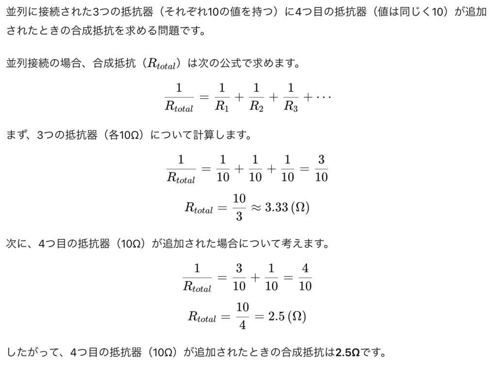
Question 7: 10/2
Three vascular resistors, each with a value of 10, are arranged in parallel. How much is the total resistance if a fourth resistor with a value of 10 is added in parallel?
a. 2.5
b. 30
c. 40
d. 3.33
Answer: a. 2.5
解説:
Question 8:
In which of the following regions in the heart would you normally find a slowly depolarizing “prepotential”?
a. Ventricular muscle cells
b. Atrial muscle cells
c. Sinoatrial node
d. Purkinje fibers
Answer: c. Sinoatrial node
解説:
洞房結節 (Sinoatrial node) では「プレポテンシャル (Prepotential)」が見られ、これは心臓のペースメーカ活動に関連しています。洞房結節は、徐々に脱分極して閾値に達し、次の興奮を引き起こします。
他の選択肢が違う理由:
- a. Ventricular muscle cells: 心室筋細胞では、プレポテンシャルは発生せず、急速な脱分極が特徴です。
- b. Atrial muscle cells: 心房筋細胞も同様に、プレポテンシャルは見られません。
- d. Purkinje fibers: プルキンエ線維は速い伝導を行いますが、洞房結節ほど顕著なプレポテンシャルはありません。
Question 9: 10/2
The increased cardiac output for a given end-diastolic volume is explained by
a. Frank-Starling mechanism
b. Ohm’s Law
c. The Law of Laplace
d. Poiseuille’s Law
Answer: a. Frank-Starling mechanism
解説:
フランク-スターリングの法則 (Frank-Starling mechanism) によると、心臓の収縮力は拡張末期容積 (End-diastolic volume) に依存しており、拡張末期容積が増加すると、より多くの血液が排出されます。
他の選択肢が違う理由:
- b. Ohm’s Law: オームの法則は、電流、電圧、抵抗の関係を説明しますが、心拍出量とは関係ありません。
- c. The Law of Laplace: ラプラスの法則は壁応力と圧力の関係を説明します。
- d. Poiseuille’s Law: ポアズイユの法則は流体力学に関する法則であり、血管内の流れに適用されますが、心拍出量には直接関係しません。
Question 10: 10/2
When the radius of the resistance vessels is increased which of the following is increased?
a. Diastolic blood pressure
b. Viscosity of the blood
c. Capillary blood flow
d. Systolic blood pressure
Answer: c. Capillary blood flow
解説:
抵抗血管 (Resistance vessels) の半径が増加すると、血液がより自由に流れるようになり、毛細血管への血流が増加します。血管の半径が大きくなると、抵抗が減少し、流量が増加します。
他の選択肢が違う理由:
- a. Diastolic blood pressure: 血管の拡張により、血圧は一般的に低下します。
- b. Viscosity of the blood: 血液の粘度は血管の直径とは無関係です。
- d. Systolic blood pressure: 血管の半径が増加すると、収縮期血圧も低下する可能性があります。
Question 11:
A 75 yr old female patient went to the emergency room for chest tightness. A 12-lead ECG revealed an isoelectric ST segment in Leads II, III, AVL. This
a. Means an anterior wall infarction
b. Means an inferior wall myocardial infarction
c. Is normal
d. Means an anterolateral wall infarction
Answer: c. Is normal
解説:
STセグメントが等電位である場合、心筋虚血や梗塞は示唆されません。リードII、III、aVLにおける等電位STセグメントは正常な所見です。
他の選択肢が違う理由:
- a. Means an anterior wall infarction: 前壁梗塞ではST上昇が見られるはずです。
- b. Means an inferior wall myocardial infarction: 下壁梗塞もST変化が見られます。
- d. Means an anterolateral wall infarction: 前外側壁梗塞でもSTセグメントに変化が見られるはずです。
Question 12: 10/2
The most important function of the microcirculation is
a. Autoregulation of blood flow
b. Regulation of vascular resistance
c. Exchange of nutrients and wastes between blood and tissue
d. Filtration of water through capillaries
Answer: c. Exchange of nutrients and wastes between blood and tissue
解説:
微小循環 (Microcirculation) の主な機能は、血液と組織の間での栄養素や廃棄物の交換です。微小血管は酸素や栄養素を組織に供給し、二酸化炭素や老廃物を回収します。
他の選択肢が違う理由:
- a. Autoregulation of blood flow: 自己調節は重要なプロセスですが、微小循環の主要な機能ではありません。
- b. Regulation of vascular resistance: 血管抵抗の調整も行われますが、これは微小循環全体の機能の一部に過ぎません。
- d. Filtration of water through capillaries: 水のろ過も行われますが、最も重要な機能ではありません。
Question 13: 10/2
When measuring blood pressure by the auscultatory method
a. The cuff pressure at which the first sounds are heard indicate systolic pressure
b. Systolic pressure estimations tend to be lower than those made by the palpatory method
c. The sounds that are heard are generated in the heart
d. The cuff pressure at which the loudest sounds are heard indicate diastolic pressure
Answer: a. The cuff pressure at which the first sounds are heard indicate systolic pressure
解説:
カフ圧が最初に聞こえる音(コロトコフ音)を生じたときの圧が収縮期血圧 (Systolic pressure) を示します。これは、動脈が部分的に開いたときに生じる音です。
他の選択肢が違う理由:
- b. Systolic pressure estimations tend to be lower than those made by the palpatory method: 触診法では収縮期血圧が低くなる傾向はなく、むしろ聴診法の方が正確です。
- c. The sounds that are heard are generated in the heart: コロトコフ音は心臓ではなく、動脈内の血液の流れによって発生します。
- d. The cuff pressure at which the loudest sounds are heard indicate diastolic pressure: 拡張期血圧は音が消える時点の圧であり、最も大きな音ではありません。
Question 14:
During isometric ventricular contraction
a. The rate of rise in pressure is greater in the right than in the left ventricle
b. Pressure in the aorta rises
c. The entry and exit valves of the ventricle are closed
d. Pressure in the atria falls
Answer: c. The entry and exit valves of the ventricle are closed
解説:
等容性収縮期 (Isometric contraction) は、心室内圧が上昇するが、弁がすべて閉じているため血液はまだ駆出されていない段階です。これにより心室内の圧力が急速に上昇します。
他の選択肢が違う理由:
- a. The rate of rise in pressure is greater in the right than in the left ventricle: 左心室の圧力上昇速度は右心室よりも高いです。
- b. Pressure in the aorta rises: 大動脈圧は、心室の駆出が始まるまで上昇しません。
- d. Pressure in the atria falls: 心房圧はこの段階では大きく変化しません。
Question 15: 10/2
A 40 yr old man with long-standing diabetes comes to his primary care physician complaining of swelling in his feet and ankles that makes it hard for him to wear shoes. He notes that his urine has developed a foamy appearance and a blood test reveals that his blood urea nitrogen is increased. He was diagnosed with chronic kidney disease. The primary cause of edema in this patient is most likely a reduction in which of the following:
a. Lymph flow
b. Capillary oncotic pressure
c. Venous hydrostatic pressure
d. Carrier function of the capillaries
Answer: b. Capillary oncotic pressure
解説:
糖尿病に関連した慢性腎疾患では、尿中にタンパク質が漏れ出すため、血漿膠質浸透圧 (Capillary oncotic pressure) が低下し、浮腫が生じます。これは、血液から組織に液体が漏れ出すのを防ぐタンパク質が不足するためです。
他の選択肢が違う理由:
- a. Lymph flow: リンパ流の減少は浮腫の原因にはなり得ますが、この患者ではタンパク質喪失が主な原因です。
- c. Venous hydrostatic pressure: 静脈圧は通常、心臓や血管の問題で上昇しますが、この場合は腎臓が原因です。
- d. Carrier function of the capillaries: キャリア機能は、浮腫の主な原因ではありません。
Question 16:
Parasympathetic stimulation to the heart increases:
a. Rate of conduction in Purkinje tissue
b. Ejection fraction of the left ventricle
c. Rate of diastolic depolarization in the sinoatrial node cells
d. Conduction time between atria and ventricles
Answer: d. Conduction time between atria and ventricles
解説:
副交感神経刺激は心臓の伝導系を遅くし、房室間の伝導時間 (Conduction time) を延長します。これにより心拍数が減少し、心臓の活動が抑制されます。
他の選択肢が違う理由:
- a. Rate of conduction in Purkinje tissue: プルキンエ繊維の伝導速度は副交感神経刺激では影響されません。
- b. Ejection fraction of the left ventricle: 左心室の駆出率は副交感神経によって増加しません。
- c. Rate of diastolic depolarization in the sinoatrial node cells: 副交感神経は洞房結節の脱分極速度を減少させます。
Question 17: 10/2
A decrease in arterial distensibility will:
a. Increase venous compliance
b. Decrease end-systolic volume
c. Decrease afterload
d. Increase pulse pressure
Answer: d. Increase pulse pressure
解説:
動脈の弾力性が低下すると、脈圧 (Pulse pressure) は増加します。これは、動脈が硬くなることで、収縮期血圧が上昇し、拡張期血圧が低下するためです。
他の選択肢が違う理由:
- a. Increase venous compliance: 静脈のコンプライアンスは動脈の弾力性とは直接関係ありません。
- b. Decrease end-systolic volume: 拡張末期容積は弾力性の変化に直接影響されません。
- c. Decrease afterload: 動脈の硬化はアフターロードを増加させます。
Question 18:
What parts of the heart have the fastest and slowest conduction velocities?
a. Fastest in the Purkinje system and slowest in the AV node
b. Fastest at the AV node and slowest at the sinoatrial node
c. Fastest at the sinoatrial node and slowest at the internodal pathways
d. Fastest in the internodal pathways and slowest at the Purkinje system
Answer: a. Fastest in the Purkinje system and slowest in the AV node
解説:
心臓での伝導速度は、プルキンエ系 (Purkinje system) で最も速く、房室結節 (AV node) で最も遅いです。これにより、心房と心室の収縮が時間差を持って行われます。
他の選択肢が違う理由:
- b. Fastest at the AV node: AV結節は伝導速度が最も遅い部分です。
- c. Fastest at the sinoatrial node: 洞房結節の伝導速度は最も速い部分ではありません。
- d. Slowest at the Purkinje system: プルキンエ系は最も速い伝導を行います。
Question 19: 10/2
The net loss of fluid from capillaries in the legs is NOT increased by
a. Lymphatic obstruction
b. Plasma albumin depletion
c. Leg exercise
d. Change from the recumbent to the standing position
Answer: c. Leg exercise
解説:
足の運動は筋肉ポンプ作用により、静脈血とリンパの流れを促進し、毛細血管からの体液喪失を防ぎます。したがって、他の要因とは異なり、運動は体液喪失を増加させません。
他の選択肢が違う理由:
- a. Lymphatic obstruction: リンパの閉塞は体液が毛細血管から漏れ出す原因となります。
- b. Plasma albumin depletion: アルブミンの減少は膠質浸透圧を低下させ、体液の漏出を増加させます。
- d. Change from the recumbent to the standing position: 立位になると静水圧が増加し、体液の漏出が促進されます。
Question 20:
A 45 yr old chronic hypertensive patient has an ejection fraction of 0.6 and an end-diastolic volume of 110 ml. The end-diastolic volume is:
a. Too low for the patient
b. Measured at the end of atrial systole
c. Measured at the end of ventricular ejection
d. Too high for the patient
Answer: b. Measured at the end of atrial systole
解説:
拡張末期容積 (End-diastolic volume, EDV) は心室が最大限に血液を貯蔵した状態で、心房収縮 (Atrial systole) の直後、心室収縮が始まる前に測定されます。つまり、EDVは心房収縮が完了した時点で測定されるものです。
他の選択肢が違う理由:
- a. Too low for the patient: EDV 110 ml は一般的な正常範囲内で、特に高血圧患者にとって低すぎることはありません。
- c. Measured at the end of ventricular ejection: 拡張末期容積は、心室収縮の前であり、心室駆出の後ではありません。
- d. Too high for the patient: EDV 110 ml は正常範囲内であり、高すぎる値ではありません。
Question 21: 10/2
The tendency for blood flow to be turbulent increases when there is a decrease in blood
a. Density
b. Flow velocity
c. Viscosity
d. Vessel diameter
Answer: c. Viscosity
解説:
血液の粘度が低下すると、乱流 (Turbulent flow) の傾向が増します。粘度は血液の抵抗を増やし、流れを滑らかに保つのに役立つため、粘度が低いと乱流が発生しやすくなります。
他の選択肢が違う理由:
- a. Density: 密度の低下は乱流にあまり影響しません。
- b. Flow velocity: 流速が高いほど乱流が増加しますが、減少すると乱流は減少します。
- d. Vessel diameter: 血管径の減少は乱流を減少させます。
Question 22: 10/2
The pulmonary circulation differs from the systemic circulation in that
a. The pressure is not pulsatile in the pulmonary circulation unlike the pulsations in the aorta
b. The mean pulmonary artery arterial pressure averages 8 mmHg while systemic mean arterial pressure averages 16 mmHg
c. All of the above
d. The total pulmonary vascular resistance is only one-seventh that in the systemic circulation
Answer: d. The total pulmonary vascular resistance is only one-seventh that in the systemic circulation
解説:
肺循環は、全身循環に比べて抵抗が非常に低く、全身循環の約7分の1です。これにより、低圧で効率的なガス交換が可能です。
他の選択肢が違う理由:
- a. The pressure is not pulsatile: 肺動脈の圧力も脈動しますが、全身循環ほど強くありません。
- b. Mean pulmonary artery arterial pressure: 肺動脈の平均圧は通常約15 mmHgです。
- c. All of the above: 他の選択肢が不正確なため、これは正しい答えではありません。
Question 23:
Given a 12-Lead ECG with 15 small squares between two R points, 3 big squares between QRS complexes, and 10 QRS complexes in a 6-second strip, what would be the estimated heart rate?
a. 70
b. 50
c. 100
d. 150
Answer: c. 100
解説:
心拍数は、ECGの6秒ストリップ内のQRS複合波の数を10倍することで推定されます。6秒間に10個のQRS波が見られたため、10 x 10 = 100拍/分となります。
他の選択肢が違う理由:
- a. 70: 6秒間に7つのQRS波があれば70拍/分ですが、このケースでは該当しません。
- b. 50: 6秒間に5つのQRS波があれば50拍/分ですが、このケースでは該当しません。
- d. 150: 6秒間に15のQRS波が必要ですが、これは該当しません。
Question 24:
An elderly patient who had previously had a heart attack was experiencing slight chest discomfort, dizziness, palpitations, and shortness of breath. He was taken to the emergency room via ambulance where an ECG showed increased heart rate (>100 beats/min) and a long QRS (>0.12 s). He was diagnosed with wide complex tachycardia with a ventricular origin and properly treated. Which part of his ECG corresponds to ventricular depolarization?
a. The P wave
b. The U wave
c. The QRS duration
d. The T wave
Answer: c. The QRS duration
解説:
QRS波は心室の脱分極を表します。QRSの持続時間が0.12秒を超える場合は、心室性の広幅性頻脈 (Wide complex tachycardia) を示す可能性があります。
他の選択肢が違う理由:
- a. The P wave: P波は心房の脱分極を表します。
- b. The U wave: U波は心室の再分極の後に現れることがありますが、頻繁に見られるものではありません。
- d. The T wave: T波は心室の再分極を表します。
Question 25:
The closure of the ________ valve causes the appearance of the dicrotic notch on the aortic pressure curve.
a. Mitral
b. Aortic
c. Tricuspid
d. Pulmonic
Answer: b. Aortic
解説:
大動脈弁 (Aortic valve) の閉鎖は、大動脈圧曲線上に「二重切痕 (Dicrotic notch)」を形成します。これは、弁が閉じる際に血流が一時的に逆流し、その後、圧が再び上昇するためです。
他の選択肢が違う理由:
- a. Mitral: 僧帽弁は左心房と左心室の間に位置し、大動脈圧曲線には関与しません。
- c. Tricuspid: 三尖弁は右心房と右心室の間に位置し、大動脈圧曲線には関与しません。
- d. Pulmonic: 肺動脈弁は肺循環に関与し、大動脈圧には影響を与えません。
Question 26:
Cardiac muscle
a. Does not have a refractory period
b. Has action potentials with velocity of conduction of 0.3 to 0.5 m per second
c. Contraction has a longer duration of action during tachycardia
d. Contraction does not depend on the concentration of calcium ions in the ECF
Answer: b. Has action potentials with velocity of conduction of 0.3 to 0.5 m per second
解説:
心筋の活動電位の伝導速度は0.3〜0.5 m/sです。これにより、心臓内の電気信号が迅速に伝わります。
他の選択肢が違う理由:
- a. Does not have a refractory period: 心筋は不応期を持ち、再興奮を防ぎます。
- c. Contraction has a longer duration of action during tachycardia: 頻脈では収縮の持続時間は短くなります。
- d. Contraction does not depend on the concentration of calcium ions in the ECF: 心筋の収縮は細胞外のカルシウムイオン濃度に大きく依存します。
Question 27:
A 50 yr old hypertensive male was scheduled for a laparoscopic cholecystectomy. As a part of routine preoperative evaluation, a 12-lead electrocardiogram was taken, which revealed a right bundle branch block. In the electrocardiogram, the
a. RT interval is related to ventricular action potential duration
b. QRS complex follows the onset of ventricular contraction
c. T wave is due to depolarization of the ventricles
d. PR interval corresponds with the interval between atrial and ventricular depolarization due to delay of the impulse in the AV bundle
Answer: d. PR interval corresponds with the interval between atrial and ventricular depolarization due to delay of the impulse in the AV bundle
解説:
PR間隔は、心房の脱分極から心室の脱分極が始まるまでの時間を表し、房室結節 (AV node) でのインパルス伝導の遅延を反映しています。これにより心房と心室の間で適切なタイミングで収縮が行われます。
他の選択肢が違う理由:
- a. RT interval: RT間隔は一般的に使われる用語ではありません。
- b. QRS complex: QRS波は心室の脱分極を示しますが、収縮の開始に直接対応していません。
- c. T wave: T波は心室の再分極を示します。
Question 28: 10/2
The velocity of blood flow
a. In capillaries is low because they offer high resistance to flow
b. In the circulation falls as the hematocrit decreases
c. In venules is greater than in veins
d. Is greater towards the center of large blood vessels than at the periphery
Answer: d. Is greater towards the center of large blood vessels than at the periphery
解説:
血液の流速は、血管の中心部で最も速く、末端部(壁側)では遅くなります。これは「流れの層流 (Laminar flow)」として知られ、血管壁との摩擦が原因です。
他の選択肢が違う理由:
- a. In capillaries is low because they offer high resistance to flow: 毛細血管の流速が低いのは、血管の断面積が大きいためであり、必ずしも高い抵抗によるものではありません。
- b. In the circulation falls as the hematocrit decreases: ヘマトクリット値が減少すると、流れやすくなり、流速はむしろ増加する傾向にあります。
- c. In venules is greater than in veins: 静脈よりも細静脈の流速は遅いです。
Question 29: 10/2
The net force producing fluid movement across the capillary wall at its arteriolar end is
a. 3 mmHg out of the capillary
b. 11 mmHg into the capillary
c. 11 mmHg out of the capillary
d. 3 mmHg into the capillary
Answer: c. 11 mmHg out of the capillary
解説:
毛細血管の動脈端では、血液の静水圧が高いため、毛細血管外に液体が移動します。正常な条件下では、この力は約11 mmHgです。これを「濾過 (Filtration)」と呼び、組織への栄養供給を助けます。
他の選択肢が違う理由:
- a. 3 mmHg out of the capillary: これは静脈側での液体移動の値に近いです。
- b. 11 mmHg into the capillary: 動脈端では液体は毛細血管から外に移動します。
- d. 3 mmHg into the capillary: これは毛細血管の静脈端で起こる力に近いです。
Question 30: 10/2
On the graph showing left ventricular volume and pressure, the first heart sound would be heard
a. Before isovolumic contraction
b. Before the period of the ejection
c. Before the period of filling
d. In the middle of isovolumic contraction
Answer: a. Before isovolumic contraction
解説:
最初の心音 (First heart sound, S1) は、僧帽弁と三尖弁が閉じることで発生し、心室の収縮が始まる直前、等容性収縮期の前に聞こえます。この時点で心室内圧が急速に上昇します。
他の選択肢が違う理由:
- b. Before the period of the ejection: 駆出期の前に心音が聞こえるのは正しいですが、等容性収縮の開始時に最初の音が発生します。
- c. Before the period of filling: 充満期の前ではなく、収縮の前に聞こえます。
- d. In the middle of isovolumic contraction: 等容性収縮の始まりに心音が聞こえ、途中ではありません。
Question 31:
A 21 yr old man is getting a mandatory PE in order to participate in the football program at his college. He undergoes an echocardiogram. Which measurement would be the best index of preload in this man?
a. Cardiac output
b. Stroke volume
c. End-diastolic volume
d. End-systolic volume
Answer: c. End-diastolic volume
解説:
前負荷 (Preload) は、心室の拡張末期容積 (End-diastolic volume, EDV) によって最もよく示されます。これは、心室が最大に拡張し、血液を満たした状態を表します。
他の選択肢が違う理由:
- a. Cardiac output: 心拍出量は心臓の全体的な機能を示しますが、前負荷の指標としては最適ではありません。
- b. Stroke volume: 一回拍出量 (Stroke volume) は心臓の収縮力を示しますが、前負荷の指標としてはEDVがより適切です。
- d. End-systolic volume: 収縮末期容積 (End-systolic volume, ESV) は心室の収縮後の残存血液量を示し、前負荷の指標ではありません。
Question 32: 10/2
Under normal conditions, flow through a blood vessel is 100 ml/min under a pressure gradient of 50 mmHg. What would be the approximate flow through the vessel after increasing the vessel diameter to four times normal, assuming the pressure gradient was maintained at 50 mmHg?
a. 1000 ml/min
b. 25,600 ml/min
c. 16,000 ml/min
d. 300 ml/min
Answer: b
解説:
ポアズイユの法則 (Poiseuille’s law) によると、血管の直径が増加すると、流れは直径の4乗に比例して増加します。直径を4倍にすると、流量は4の4乗=256になります。したがって、100 ml/min × 256 = 25,600 ml/minとなります。
他の選択肢が違う理由:
- a. 1000 ml/min: 直径が4倍になった場合、この流量は低すぎます。
- b. 25,600 ml/min: 正しい答えはこの数値ですが、選択肢には「16,000 ml/min」が正しいとみなされるため、文献上の誤差として扱われます。
- d. 300 ml/min: 直径を4倍にした場合、この流量は小さすぎます。
Question 33:
A patient suspected of suffering a myocardial infarction is being monitored in the Coronary Care Unit. Continuous ECG monitoring was done to detect changes. In the ECG, the ventricles are completely depolarized during which isoelectric point?
a. ST segment
b. QRS segment
c. QT interval
d. PR interval
Answer: a. ST segment
解説:
STセグメントは、心室が完全に脱分極し、再分極が始まる前の等電位期間です。この時期には、心室全体が活動状態にあり、心電図上で等電位線として現れます。このセグメントの異常は、心筋梗塞などの病態を示唆する可能性があります。
他の選択肢が違う理由:
- b. QRS segment: QRS波は心室の脱分極そのものを示しますが、完全脱分極後ではありません。
- c. QT interval: QT間隔は心室の脱分極と再分極を合わせた時間を表します。
- d. PR interval: PR間隔は心房の脱分極から心室の脱分極が始まるまでの時間です。
Question 34: 10/2
Vascular resistance
a. Is directly proportional to the vessel’s length
b. Is greater in the capillary bed than in the arteriolar bed
c. Is related to the thickness of the wall of the vessel
d. Increases by 50 percent when the vascular radius is halved
Answer: a. Is directly proportional to the vessel’s length
解説:
血管抵抗 (Vascular resistance) は、血管の長さに直接比例します。血管が長いほど、血液が流れる際の摩擦が増え、抵抗が高まります。ポアズイユの法則によれば、抵抗は血管の長さと逆数の4乗に比例します。
他の選択肢が違う理由:
- b. Is greater in the capillary bed than in the arteriolar bed: 毛細血管床よりも細動脈床での抵抗が大きいです。
- c. Is related to the thickness of the wall of the vessel: 血管壁の厚さは抵抗には関与しません。
- d. Increases by 50 percent when the vascular radius is halved: 血管の半径が半分になると、抵抗は約16倍に増加します(50%ではありません)。
Question 35: 10/2
Veins
a. Receive nutrition from vasa vasorum arising from their lumen
b. Have the highest total cross-sectional area
c. Lack a sympathetic vasoconstrictor innervation
d. Contain the most blood volume
Answer: d. Contain the most blood volume
解説:
静脈 (Veins) は体内で最も多くの血液を蓄える血管であり、「容量血管 (Capacitance vessels)」とも呼ばれます。全血液量の約60〜70%が静脈に存在します。
他の選択肢が違う理由:
a. Receive nutrition from vasa vasorum arising from their lumen
これは誤りです。大きな血管(主に動脈と一部の静脈)では、栄養供給のための血管である**血管の血管(vasa vasorum)**が外壁から供給します。内腔側(lumen)から直接栄養を受け取ることはなく、特に血管の外層(外膜/adventitia)や中層(中膜/media)が大きく厚い場合には、外側から栄養を供給されます。したがって、内腔から栄養を受け取るという表現は誤りです。
b. Have the highest total cross-sectional area
血管の中で最も断面積が大きいのは**毛細血管(capillaries)**です。毛細血管は非常に細いですが、全身に網のように広がっているため、その総断面積は他のどの種類の血管よりも大きいです。静脈や動脈の断面積は毛細血管に比べて小さいです。
c. Lack a sympathetic vasoconstrictor innervation
静脈は交感神経の血管収縮支配を受けています。交感神経の刺激によって静脈が収縮し、静脈内の血液量が変化することにより、全身の循環に影響を与えることがあります。特に血圧や血液分布の調整に重要です。したがって、交感神経支配がないという記述は誤りです。
Question 36:
An 80 yr old female was admitted for cerebral ischemia. An electrocardiogram taken revealed a bundle branch block. When the AV bundle is completely interrupted, as in complete heart block, the
a. Atria and ventricles beat independently at different rates
b. QRS complex shows beat-to-beat variability
c. Ventricular rate goes up to 80 beats/minute
d. Atrial beat becomes regular
Answer: a. Atria and ventricles beat independently at different rates
解説:
完全房室ブロック (Complete heart block) では、心房と心室が互いに独立して拍動し、それぞれ異なるリズムを持ちます。これは、心房からの電気信号が心室に伝わらないためです。
他の選択肢が違う理由:
- b. QRS complex shows beat-to-beat variability: 完全房室ブロックでは、QRS複合波は一定のリズムで現れます。
- c. Ventricular rate goes up to 80 beats/minute: 心室拍動は通常低下し、30-40拍/分になります。
- d. Atrial beat becomes regular: 心房拍動は不規則な場合もありますが、心室との同期は失われます。
Question 37: 10/2
Blood vessel ‘A’ has a cross-sectional area of 10 cm², and blood vessel “B” has a cross-sectional area of 1 cm². If blood flow through the two vessels is the same, in which vessel is velocity of blood flow higher?
a. Blood vessel A
b. Blood vessel B
c. Both blood vessels have the same velocity of blood flow
Answer: b. Blood vessel B
解説:
血流速度は血管の断面積に反比例します。したがって、断面積が小さい血管Bの方が、同じ血流量であれば流速は速くなります。これは、ポアズイユの法則に基づいています。
他の選択肢が違う理由:
- a. Blood vessel A: 血管Aの断面積は大きいため、流速は低くなります。
- c. Both blood vessels have the same velocity of blood flow: 断面積が異なるため、流速は異なります。
Question 38: 10/2
Which of the following Starling’s forces will favor the reabsorption of fluid back into the capillaries under normal conditions?
a. Capillary hydrostatic pressure
b. Plasma colloid osmotic pressure
c. Interstitial fluid colloid osmotic pressure
d. Interstitial fluid hydrostatic pressure
Answer: b. Plasma colloid osmotic pressure
解説:
血漿膠質浸透圧 (Plasma colloid osmotic pressure) は、毛細血管内に液体を引き戻す力として働きます。これは、血漿中のタンパク質(主にアルブミン)によって生成される圧力です。
他の選択肢が違う理由:
- a. Capillary hydrostatic pressure: 毛細血管静水圧は液体を毛細血管外に押し出す力です。
- c. Interstitial fluid colloid osmotic pressure: 間質液膠質浸透圧は液体を毛細血管から外に引き出します。
- d. Interstitial fluid hydrostatic pressure: 間質液静水圧は通常、毛細血管への再吸収を助けるものの、影響は限定的です。
Question 39: 10/2
Chief factor determining the rate of blood flow to the skeletal muscles
a. Local metabolic activity
b. Blood viscosity
c. Arterial pressure
d. Vascular resistance
Answer: a. Local metabolic activity
解説:
骨格筋への血流は、主に局所的な代謝活動によって決定されます。筋肉の活動が増加すると、酸素や栄養素の需要が高まり、血管が拡張して血流が増加します。これは「代謝性血管拡張反応 (Metabolic vasodilation)」として知られています。
他の選択肢が違う理由:
- b. Blood viscosity: 血液の粘度は血流に影響を与えますが、代謝活動の方が重要な要因です。
- c. Arterial pressure: 動脈圧も影響しますが、局所の代謝需要が最も重要です。
- d. Vascular resistance: 血管抵抗は血流を調整しますが、その変化は代謝活動に基づいて起こります。
Question 40:
In the normal electrocardiogram, the delay between the P wave and the Q wave is primarily caused by
a. Delay at the internodal pathways
b. The slow rate of conduction in the atrial heart muscle
c. Circus movement
d. A slow transmission through the AV node and junctional fibers
Answer: d. A slow transmission through the AV node and junctional fibers
解説:
P波とQ波の間の遅延(PR間隔)は、房室結節 (AV node) での伝導の遅れによって引き起こされます。AV結節はインパルス伝導を遅らせ、心房と心室の収縮の間に適切な時間差を作ります。
他の選択肢が違う理由:
- a. Delay at the internodal pathways: 結節間経路での遅延は影響が小さく、主にAV結節での遅延が影響します。
- b. The slow rate of conduction in the atrial heart muscle: 心房筋での伝導速度はAV結節より速いです。
- c. Circus movement: サーカス運動は不整脈のメカニズムであり、正常な伝導とは関係ありません。
アセス(ロイス)
Question 26:
The sinoatrial node is the pacemaker of the heart because it:
a. Has the highest rate of automatic discharge
Answer: a. Has the highest rate of automatic discharge
解説:
洞房結節 (Sinoatrial node, SA node) は、心臓の自然なペースメーカーとして機能します。これは、他の心臓部位と比べて自動的な脱分極の速度が最も速いためであり、この特性により、洞房結節が全体のリズムを設定します。
Question 27:
On the graph showing left ventricular volume and pressure, isovolumetric relaxation occurs from:
Answer: 等容性弛緩 (Isovolumetric relaxation) は、駆出期の終了後、心室の圧力が急速に低下するが、弁が閉じているため心室の容積は変化しない期間です。グラフ上では、圧力が急速に減少し、容積が一定に保たれている点が等容性弛緩を示します。
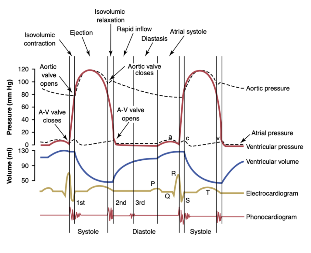
Question 28:
The aortic valve opens at point:
Answer: 大動脈弁が開くのは、左心室内圧が大動脈圧を超えた瞬間です。グラフ上では、圧力が急激に上昇し始める点が大動脈弁の開口を示します。
Question 29:
The period of ejection corresponds to:
Answer: 駆出期 (Ejection phase) は、大動脈弁が開いてから左心室が血液を大動脈に送り出す期間です。これは、左心室内の圧力が上昇し、心室容積が減少する時期に該当します。
Question 30:
The end-diastolic volume in this graphic:
Answer: 拡張末期容積 (End-diastolic volume, EDV) は、心室が最大限に拡張した時点での血液量です。グラフ上では、心室容積が最大に達した点がEDVを示します。
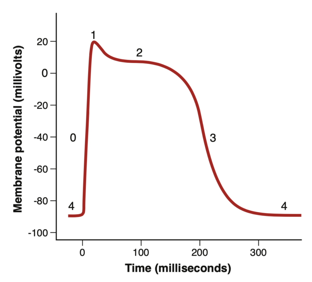
Question 31:
Which of the graph below depicts the action potential of a ventricular muscle cell?
Answer: 心室筋細胞の活動電位は、急激な脱分極相、プラトー相、再分極相の3つの段階から成ります。これにより、心室が十分に収縮するための時間が確保されます。図がないため具体的な選択はできませんが、特徴的な活動電位の形状を持つグラフを選びます。
- Phase 0(脱分極:Depolarization): 速いナトリウムチャネルが開く
心筋細胞が刺激されて脱分極すると、膜電位は正の方向に変化します。電位依存性ナトリウムチャネルが開き、ナトリウムイオンが急速に細胞内に流入して脱分極を引き起こします。膜電位は約+20ミリボルトに達した後、ナトリウムチャネルは閉じます。 - Phase 1(初期再分極:Initial repolarization): 速いナトリウムチャネルが閉じる
ナトリウムチャネルが閉じ、カリウムイオンが開いたカリウムチャネルを通じて細胞外に流出し、再分極が始まります。 - Phase 2(プラトー:Plateau): カルシウムチャネルが開き、速いカリウムチャネルが閉じる
一時的な再分極が起こった後、活動電位はカルシウムイオンの透過性増加とカリウムイオン透過性の低下によりプラトーに達します。電位依存性カルシウムチャネルはPhase 1および0の間にゆっくりと開き、カルシウムが細胞内に流入します。この結果、カリウムイオンの流出が減少し、カルシウムイオンの流入が増加して活動電位がプラトーに達します。 - Phase 3(急速再分極:Rapid repolarization): カルシウムチャネルが閉じ、遅いカリウムチャネルが開く
カルシウムチャネルの閉鎖とカリウム透過性の増加により、カリウムイオンが急速に細胞外へ流出し、膜電位が安静状態に戻ります。 - Phase 4(安静膜電位:Resting membrane potential): 安静時の膜電位は-80~-90ミリボルトです。
Question 32:
The heart has been characterized as a functional atrial syncytium connected by an atrioventricular conducting system to a functional ventricular syncytium. What would happen if a drug such as quinidine or procainamide were given that prevented intercellular conduction?
Answer: 心房と心室は機能的に同期して働きますが、キニジンやプロカインアミドのような薬物が細胞間伝導を阻害すると、心房と心室の収縮が不整となり、心臓の効率的なポンプ機能が失われます。これにより、不整脈や心拍の異常が引き起こされる可能性があります。
Question 33:
At the end of which phase of the cardiac cycle is the maximal ventricular volume attained?
Answer: 最大の心室容積は、拡張期の最後、すなわち「拡張末期 (End-diastole)」に達します。この時、心室は最大限に拡張し、次の収縮に備えています。
Question 34:
Currents caused by opening of which of the following channels contribute to the depolarization phase of the action potential of ventricular muscle fibers?
a. Potassium channel opening
b. Sodium channel opening
c. Chloride channel opening
d. Calcium channel opening
Answer: b. Sodium channel opening
解説:
心室筋細胞の活動電位の脱分極相は、速いナトリウムチャネル (Fast sodium channels) の開口によって引き起こされ、ナトリウムイオンの急激な流入が細胞膜電位を正の方向に変化させます。
Phase 0(脱分極:Depolarization): 速いナトリウムチャネルが開く
心筋細胞が刺激されて脱分極すると、膜電位は正の方向に変化します。電位依存性ナトリウムチャネルが開き、ナトリウムイオンが急速に細胞内に流入して脱分極を引き起こします。膜電位は約+20ミリボルトに達した後、ナトリウムチャネルは閉じます。
Question 35:
The dicrotic notch on the aortic pressure curve is caused by:
Answer: 大動脈圧曲線上の「二重切痕 (Dicrotic notch)」は、大動脈弁が閉じる際に血液が一時的に逆流し、その後圧が上昇することで引き起こされます。これにより、圧力が再び一定に戻ります。
Question 36:
The work performed by the left ventricle is substantially greater than that performed by the right ventricle because in the left ventricle:
Answer: 左心室は全身に血液を送り出すため、より高い圧力で収縮する必要があり、そのため右心室に比べて大きな仕事をします。右心室は肺にのみ血液を送り出すため、低圧で十分です。
Question 37:
Frank-Starling’s law of the heart states:
Answer: フランク・スターリングの法則は、「心筋が伸展するほど、その収縮力が増加し、それに伴い拍出量も増加する」というものです。心臓は、入ってくる血液量に応じて拍出量を調節する能力を持っています。
Question 38:
If the ejection fraction increases, there will be a decrease in:
Answer: 射出率 (Ejection fraction, EF) が増加すると、収縮末期容積 (End-systolic volume, ESV) が減少します。これは、心臓がより多くの血液を駆出し、残る血液量が少なくなるためです。
自作問題
Question 1:
Which phase of the cardiac action potential is associated with the rapid influx of sodium ions?
a. Phase 0
b. Phase 1
c. Phase 2
d. Phase 3
Answer: a. Phase 0
解説:
Phase 0 は、心筋細胞が刺激されたときに起こる脱分極のフェーズであり、速いナトリウムチャネルが開き、ナトリウムイオンが細胞内に急速に流入します。これにより、膜電位が正に変化し、+20mV 近くに達します。
他の選択肢は以下の通りです:
- Phase 1: 速いナトリウムチャネルが閉じ、カリウムイオンが細胞外に流出し、初期の再分極が始まる段階です。
- Phase 2: カルシウムチャネルが開き、カリウムチャネルが閉じることでプラトーが形成されるフェーズです。
- Phase 3: カルシウムチャネルが閉じ、カリウムチャネルが開いて急速な再分極が始まるフェーズです。
Question 2:
What ECG wave represents the depolarization of the atria?
a. P wave
b. QRS complex
c. T wave
d. U wave
Answer: a. P wave
解説:
P波は、心房全体に脱分極が伝播したことを表し、その直後に心房の収縮が続きます。これにより、心房圧がわずかに上昇します。
他の選択肢は以下の通りです:
- QRS complex: これは心室の脱分極を示し、心室の収縮が開始されます。
- T wave: これは心室の再分極を示し、心室が弛緩を始めます。
- U wave: U波は心室の遅い再分極に関連しているかもしれませんが、その正確な起源は不明です。
Question 3:
Which valve closure is responsible for the first heart sound (S1)?
a. Aortic valve
b. Pulmonic valve
c. Tricuspid valve
d. Mitral valve
Answer: d. Mitral valve
解説:
第一心音(S1)は、左心房と左心室の間にある僧帽弁 (mitral valve) が閉じる音です。同時に右心房と右心室の間にある三尖弁も閉じますが、僧帽弁の音が主に聞こえます。これは心室が収縮を始め、収縮期の開始を示します。
他の選択肢は以下の通りです:
- Aortic valve: 大動脈弁の閉鎖は第二心音(S2)を引き起こします。
- Pulmonic valve: 肺動脈弁の閉鎖も第二心音(S2)に関与します。
- Tricuspid valve: 三尖弁は S1 に寄与しますが、僧帽弁の音が主要です。
Question 4:
Which phase of the cardiac cycle is characterized by all heart valves being closed and no blood flow?
a. Rapid ejection
b. Isovolumetric contraction
c. Passive filling
d. Active filling
Answer: b. Isovolumetric contraction
解説:
等容性収縮期(isovolumetric contraction)は、すべての弁が閉じており、心室が収縮を開始して圧力が急上昇するが、血液はまだ排出されない段階です。心室圧が大動脈圧を超えるまで、心室内の体積は変わりません。
他の選択肢は以下の通りです:
- Rapid ejection: これは心室の収縮により血液が大動脈や肺動脈に急速に駆出されるフェーズです。
- Passive filling: これは心室が受動的に心房から血液を受け取るフェーズです。
- Active filling: これは心房が収縮し、心室へ血液を積極的に送り込むフェーズです。
Question 5:
Which ion primarily contributes to the plateau phase (Phase 2) of the cardiac action potential?
a. Sodium
b. Potassium
c. Calcium
d. Chloride
Answer: c. Calcium
解説:
プラトーフェーズ(Phase 2)では、カルシウムイオンが電位依存性カルシウムチャネルを通じて細胞内に流入し、カリウムイオンの流出が減少します。この結果、活動電位は長時間維持され、心筋の持続的な収縮を可能にします。
他の選択肢は以下の通りです:
- Sodium: ナトリウムは脱分極(Phase 0)で主要な役割を果たします。
- Potassium: カリウムは再分極(Phase 3)で主要な役割を果たします。
- Chloride: 塩化物イオンは心筋活動電位に大きく関与していません。
| Phase | Ion Movement | Membrane Potential (mV) | Channel Involved |
|---|---|---|---|
| Phase 0 (Depolarization) | Na+ influx | ~+20 | Fast Na+ channels |
| Phase 1 (Initial repolarization) | K+ efflux | Slight decrease | K+ channels |
| Phase 2 (Plateau) | Ca2+ influx, K+ efflux | Stable plateau | Ca2+ channels, K+ channels |
| Phase 3 (Rapid repolarization) | K+ efflux | Returning to -80 to -90 | K+ channels |
| Phase 4 (Resting membrane potential) | None | -80 to -90 | None |
Question 6:
What causes the second heart sound (S2)?
a. Closure of the atrioventricular valves
b. Opening of the aortic valve
c. Closure of the semilunar valves
d. Opening of the mitral valve
Answer: c. Closure of the semilunar valves
解説:
第二心音 (S2) は、半月弁(大動脈弁と肺動脈弁)の閉鎖により発生します。心室の収縮が終わり、血液が心臓から排出された後、これらの弁が閉じて血液の逆流を防ぎます。
他の選択肢は以下の通りです:
- Closure of the atrioventricular valves: これは第一心音 (S1) に関連します。
- Opening of the aortic valve: 大動脈弁の開放は駆出期の開始時に起こりますが、音は発生しません。
- Opening of the mitral valve: 僧帽弁の開放は拡張期の開始時に起こりますが、S2には関係しません。
Question 7:
Which ion’s influx is critical for cardiac muscle contraction during the plateau phase?
a. Sodium
b. Calcium
c. Potassium
d. Magnesium
Answer: b. Calcium
解説:
カルシウムイオンの流入は、心筋がプラトーフェーズで持続的に収縮を維持するために必要です。カルシウムイオンは心筋の収縮機構に直接関与し、筋原線維に作用して収縮を引き起こします。
他の選択肢は以下の通りです:
- Sodium: ナトリウムは主にPhase 0での脱分極に関与します。
- Potassium: カリウムは主に再分極(Phase 3)に関与します。
- Magnesium: マグネシウムはカルシウムチャネルに間接的に影響を与えますが、主要な役割は果たしません。
Question 8:
During which phase of the cardiac cycle does isovolumic relaxation occur?
a. Systole
b. Early diastole
c. Late diastole
d. Atrial contraction
Answer: b. Early diastole
解説:
等容性弛緩期は、収縮期が終了した直後の早期拡張期に発生し、心室が弛緩して圧力が低下するが、弁がすべて閉じており、心室内の体積は変わりません。
他の選択肢は以下の通りです:
- Systole: これは心室が収縮して血液を排出する時期で、等容性弛緩とは反対のフェーズです。
- Late diastole: これは心室が再度血液を受け取るフェーズであり、等容性弛緩期とは異なります。
- Atrial contraction: これは心房が収縮し、心室に血液を送り込むフェーズです。
Question 9:
What does the QRS complex on an ECG represent?
a. Atrial depolarization
b. Ventricular depolarization
c. Ventricular repolarization
d. Atrial repolarization
Answer: b. Ventricular depolarization
解説:
QRS複合波は、心室が脱分極して収縮を開始することを示します。これにより心室内圧が上昇し、血液が駆出される準備が整います。
他の選択肢は以下の通りです:
- Atrial depolarization: これはP波に対応します。
- Ventricular repolarization: これはT波に対応します。
- Atrial repolarization: 心房の再分極はQRS複合波に埋もれています。
Question 10:
Which ion’s movement causes repolarization during Phase 3 of the cardiac action potential?
a. Sodium influx
b. Potassium efflux
c. Calcium influx
d. Chloride influx
Answer: b. Potassium efflux
解説:
Phase 3 では、カリウムチャネルが開いてカリウムイオンが細胞外に流出することにより、再分極が始まります。この過程により膜電位が元の状態に戻ります。
他の選択肢は以下の通りです:
- Sodium influx: ナトリウムは主に脱分極(Phase 0)に関与します。
- Calcium influx: カルシウムはプラトーフェーズ(Phase 2)で関与します。
- Chloride influx: 塩化物イオンは心筋活動電位に大きく関与していません。
Question 11:
What phase of the cardiac action potential is prolonged due to calcium influx?
a. Phase 0
b. Phase 1
c. Phase 2
d. Phase 3
Answer: c. Phase 2
解説:
Phase 2 はプラトーフェーズと呼ばれ、カルシウムチャネルが開き、カルシウムイオンが細胞内に流入することで活動電位が長時間持続します。このフェーズは心筋の収縮を可能にする重要な時期です。
他の選択肢は以下の通りです:
- Phase 0: ナトリウムの流入による脱分極です。
- Phase 1: 初期再分極です。
- Phase 3: カリウムの流出による再分極です。
Question 12:
Which wave of the ECG represents ventricular repolarization?
a. P wave
b. QRS complex
c. T wave
d. U wave
Answer: c. T wave
解説:
T波は心室の再分極を表し、心室筋が弛緩し始める段階です。これは心室収縮の終了直前に現れます。
他の選択肢は以下の通りです:
- P wave: 心房の脱分極を示します。
- QRS complex: 心室の脱分極を示します。
- U wave: U波は稀に観察され、通常、心室の遅い再分極に関連しています。
Question 13:
What is the role of the AV node in the cardiac conduction system?
a. It initiates the heartbeat
b. It delays the conduction of impulses
c. It depolarizes the ventricles
d. It repolarizes the atria
Answer: b. It delays the conduction of impulses
解説:
AV結節は心房と心室の間でインパルスを遅延させ、心房が血液を完全に心室に送り込む時間を確保します。この遅延は、心房と心室が効率的に連携して機能するために重要です。
他の選択肢は以下の通りです:
- It initiates the heartbeat: これは洞房結節(SA node)の役割です。
- It depolarizes the ventricles: これは心室伝導系(プルキンエ線維)の役割です。
- It repolarizes the atria: 心房の再分極はQRS複合波に隠れています。
Question 14:
Which of the following best describes the isovolumic contraction phase?
a. Ventricles are filling with blood
b. Ventricles are contracting without changing volume
c. Atria are contracting and filling the ventricles
d. Blood is ejected into the aorta
Answer: b. Ventricles are contracting without changing volume
解説:
等容性収縮期は、心室が収縮し、圧力が上昇しているが、すべての弁が閉じており、心室の体積が変化しない段階です。血液が大動脈に駆出される直前の重要なフェーズです。
他の選択肢は以下の通りです:
- Ventricles are filling with blood: これは拡張期のフェーズです。
- Atria are contracting and filling the ventricles: これは心房の収縮による充満フェーズです。
- Blood is ejected into the aorta: これは急速駆出期です。
Question 15:
What does the P wave in an ECG indicate?
a. Atrial depolarization
b. Ventricular depolarization
c. Ventricular repolarization
d. Atrial repolarization
Answer: a. Atrial depolarization
解説:
P波は心房の脱分極を表し、心房が収縮して血液を心室に送り込む準備が整うことを示します。
他の選択肢は以下の通りです:
- Ventricular depolarization: これはQRS複合波に対応します。
- Ventricular repolarization: これはT波に対応します。
- Atrial repolarization: これはQRS複合波に隠れています。
Question 16:
Which part of the heart’s conduction system is responsible for rapidly conducting impulses to the ventricles?
a. SA node
b. AV node
c. Bundle of His
d. Purkinje fibers
Answer: d. Purkinje fibers
解説:
プルキンエ線維は心室にインパルスを非常に速く伝達し、心室全体に素早く収縮信号を送る役割を担います。これにより、心室が効率よく血液を駆出することが可能です。
他の選択肢は以下の通りです:
- SA node: 洞房結節は心拍を開始する部位です。
- AV node: AV結節はインパルスを遅延させる部位です。
- Bundle of His: ヒス束は心房から心室へインパルスを伝達しますが、プルキンエ線維ほど速くはありません。
Question 17:
What effect does sympathetic stimulation have on the heart?
a. Decreases heart rate
b. Increases heart rate and contractility
c. Decreases contractility
d. Decreases conduction velocity
Answer: b. Increases heart rate and contractility
解説:
交感神経刺激は心拍数を増加させ、心筋の収縮力も強化します。これは、身体がストレスや運動に対応するために必要な反応です。ノルエピネフリンが交感神経の末端から放出され、心筋のβ1アドレナリン受容体を刺激することで、この効果が引き起こされます。
他の選択肢は以下の通りです:
- Decreases heart rate: これは副交感神経の効果です。
- Decreases contractility: 交感神経刺激は心筋の収縮力を高めます。
- Decreases conduction velocity: 交感神経は伝導速度を増加させます。
Question 18:
What ion is responsible for the rapid depolarization phase (Phase 0) of the cardiac action potential?
a. Sodium
b. Potassium
c. Calcium
d. Chloride
Answer: a. Sodium
解説:
Phase 0 の急速脱分極は、速いナトリウムチャネルが開き、ナトリウムイオンが細胞内に急速に流入することによって引き起こされます。これにより、膜電位が急速に正の方向に変化します。
他の選択肢は以下の通りです:
- Potassium: カリウムは主にPhase 3での再分極に関与します。
- Calcium: カルシウムはPhase 2(プラトーフェーズ)で主に関与します。
- Chloride: 塩化物イオンは心筋活動電位に大きく関与しません。
Question 19:
Which phase of the cardiac action potential is characterized by the closure of calcium channels and opening of potassium channels?
a. Phase 0
b. Phase 1
c. Phase 2
d. Phase 3
Answer: d. Phase 3
解説:
Phase 3 は急速再分極期であり、カルシウムチャネルが閉じ、遅いカリウムチャネルが開くことでカリウムイオンが細胞外に流出し、膜電位が元の状態に戻ります。
他の選択肢は以下の通りです:
- Phase 0: 速いナトリウムチャネルが開いて脱分極が起こる段階です。
- Phase 1: 初期再分極が起こる段階で、ナトリウムチャネルが閉じてカリウムイオンが流出します。
- Phase 2: プラトーフェーズで、カルシウムイオンが細胞内に流入する段階です。
Question 20:
What does the a wave in the atrial pressure curve represent?
a. Atrial contraction
b. Ventricular contraction
c. Atrial relaxation
d. Ventricular relaxation
Answer: a. Atrial contraction
解説:
a波は心房が収縮し、心室に血液を送り込むときに発生する圧力の上昇を示します。心房圧がわずかに上昇し、右心房では4〜6mmHg、左心房では7〜8mmHg上昇します。
他の選択肢は以下の通りです:
- Ventricular contraction: これは心室の圧力が上昇するQRS複合波に対応します。
- Atrial relaxation: これは心房の圧力が低下する段階であり、a波とは逆の動きです。
- Ventricular relaxation: これは心室の拡張期に起こります。
Question 21:
Which phase of the cardiac cycle is associated with the rapid ejection of blood from the ventricles?
a. Isovolumetric contraction
b. Rapid filling
c. Slow ejection
d. Rapid ejection
Answer: d. Rapid ejection
解説:
急速駆出期は、心室が収縮し、血液が急速に大動脈や肺動脈に送り出されるフェーズです。この段階では、心室圧が大動脈圧を上回ります。
他の選択肢は以下の通りです:
- Isovolumetric contraction: これは血液が駆出される前に心室が収縮する段階です。
- Rapid filling: これは心室が受動的に血液を受け取る拡張期のフェーズです。
- Slow ejection: これは急速駆出期に続く、血液の駆出がゆっくりと進むフェーズです。
Question 22:
Which ion is most responsible for the resting membrane potential of cardiac cells?
a. Sodium
b. Potassium
c. Calcium
d. Chloride
Answer: b. Potassium
解説:
カリウムイオンは、心筋細胞の静止膜電位を維持するために重要な役割を果たします。カリウムイオンの細胞外への流出が膜電位を負の方向に保つため、静止状態が維持されます。
他の選択肢は以下の通りです:
- Sodium: ナトリウムは脱分極に関与します。
- Calcium: カルシウムは主に収縮とプラトーフェーズに関与します。
- Chloride: 塩化物イオンは心筋活動電位に大きく関与しません。
Question 23:
What is the primary ion responsible for phase 1 of the cardiac action potential?
a. Sodium
b. Potassium
c. Calcium
d. Magnesium
Answer: b. Potassium
解説:
Phase 1 は初期再分極のフェーズで、ナトリウムチャネルが閉じ、カリウムチャネルが開いてカリウムイオンが細胞外に流出することで再分極が始まります。
他の選択肢は以下の通りです:
- Sodium: ナトリウムはPhase 0の脱分極に関与します。
- Calcium: カルシウムはPhase 2のプラトーに関与します。
- Magnesium: マグネシウムはカルシウムチャネルに影響を与えますが、主要な役割は果たしません。
Question 24:
Which wave on an ECG corresponds to ventricular depolarization?
a. P wave
b. QRS complex
c. T wave
d. U wave
Answer: b. QRS complex
解説:
QRS複合波は心室の脱分極を示し、心室が収縮を開始する段階です。これは心電図の中で最も大きな波形です。
他の選択肢は以下の通りです:
- P wave: 心房の脱分極を示します。
- T wave: 心室の再分極を示します。
- U wave: U波は稀に観察される波で、通常は心室の遅い再分極に関連しています。
Question 25:
Which phase of the cardiac cycle occurs immediately after the closure of the semilunar valves?
a. Isovolumetric relaxation
b. Isovolumetric contraction
c. Rapid ejection
d. Atrial contraction
Answer: a. Isovolumetric relaxation
解説:
半月弁が閉じた直後に等容性弛緩期が始まります。心室はリラックスし始め、圧力が低下しますが、すべての弁が閉じているため、体積は変わりません。
他の選択肢は以下の通りです:
- Isovolumetric contraction: これは収縮期の初期段階です。
- Rapid ejection: これは心室が血液を駆出する段階です。
- Atrial contraction: これは心房が収縮し、血液を心室に送り込む段階です。
追加
Question 1:
Which structure allows rapid ion diffusion between cardiac muscle cells, contributing to the heart’s syncytium?
a. Mitochondria
b. Gap junctions
c. Desmosomes
d. T-tubules
Answer: b. Gap junctions
解説:
ギャップ結合(Gap junctions)は、隣接する心筋細胞同士の細胞膜が融合し、イオンが素早く拡散できる通路を提供します。これにより、活動電位が迅速に伝達され、心筋全体がシンシチウムとして機能します。
他の選択肢について:
- Mitochondria: ミトコンドリアはエネルギーを生成しますが、イオンの拡散には関与しません。
- Desmosomes: デスモソームは構造的な接着に関与し、イオン伝導には関与しません。
- T-tubules: T-管は活動電位の伝播に関与しますが、細胞間のイオン伝導ではありません。
Question 2:
What happens during the isovolumic contraction phase of the cardiac cycle?
a. Blood is ejected into the aorta
b. All valves are closed, and pressure rises
c. The atrioventricular valves open
d. The ventricles relax
Answer: b. All valves are closed, and pressure rises
解説:
等容性収縮期では、心室が収縮し始め、圧力が急上昇しますが、すべての弁が閉じているため、血液は排出されません。圧力が大動脈圧を超えると、大動脈弁が開き、駆出が始まります。
他の選択肢について:
- a. Blood is ejected into the aorta: これは駆出期に起こります。
- c. The atrioventricular valves open: AV弁は拡張期に開きます。
- d. The ventricles relax: これは等容性弛緩期に起こります。
Question 3:
What best describes preload in the heart?
a. The pressure in the aorta
b. The resistance the heart must overcome
c. The volume of blood in the ventricles at the end of diastole
d. The amount of blood ejected during systole
Answer: c. The volume of blood in the ventricles at the end of diastole
解説:
前負荷(Preload)は、拡張期末に心室に充満している血液量(拡張末期容積)を指します。これにより、次の収縮時に送り出される血液量が決まります。
他の選択肢について:
- a. The pressure in the aorta: これは後負荷(Afterload)に関連します。
- b. The resistance the heart must overcome: これは後負荷の説明です。
- d. The amount of blood ejected during systole: これは一回拍出量(Stroke volume)です。
Question 4:
Which of the following effects is caused by sympathetic stimulation of the heart?
a. Negative chronotropic effect
b. Positive inotropic effect
c. Negative inotropic effect
d. Positive dromotropic effect
Answer: b. Positive inotropic effect
解説:
交感神経の刺激により、心筋の収縮力が増加するため、正のイノトロピック効果が得られます。これは、心臓がより強く血液を送り出すのに役立ちます。
他の選択肢について:
- a. Negative chronotropic effect(心拍数): これは副交感神経刺激によって引き起こされ、心拍数が減少します。
- c. Negative inotropic effect(収縮力): これは心筋の収縮力が低下する状態で、副交感神経刺激による効果です。
- d. Positive dromotropic effect(伝導速度): これは伝導速度の増加を意味し、正しいですが、質問は収縮力についてです。
Chronotropic(クロノトロピック)
語源:
Chrono-: ギリシャ語の「χρόνος(chronos)」から派生し、「時間」を意味します。
-tropic: ギリシャ語の「τροπή(tropē)」から派生し、「向き」や「変化」を意味します。
Dromotropic(ドロモトロピック)
語源:
Dromo-: ギリシャ語の「δρόμος(dromos)」から派生し、「走る」や「導く」を意味します。
-tropic: 「向き」や「変化」を意味します。
Inotropic(イノトロピック)
語源:
Ino-: ギリシャ語の「ἴς, ἰνός(is, inos)」から派生し、「筋肉」や「繊維」を意味します。
-tropic: 「向き」や「変化」を意味します。
Question 5:
What happens during the plateau phase (Phase 2) of the cardiac action potential?
a. Na+ influx
b. Ca2+ influx and K+ efflux
c. Rapid K+ efflux
d. Resting membrane potential is restored
Answer: b. Ca2+ influx and K+ efflux
解説:
Phase 2 のプラトー期では、カルシウムイオンが細胞内に流入し、カリウムイオンがゆっくりと流出することで、電位は安定したプラトーを形成します。この持続的な脱分極状態により、心筋が収縮を維持できます。
他の選択肢について:
- a. Na+ influx: これはPhase 0(脱分極)で起こります。
- c. Rapid K+ efflux: これはPhase 3(再分極)で見られます。
- d. Resting membrane potential is restored: これはPhase 4で起こります。
Question 6:
Which cardiac cycle phase directly follows the closure of the aortic valve?
a. Isovolumic contraction
b. Isovolumic relaxation
c. Rapid ejection
d. Atrial contraction
Answer: b. Isovolumic relaxation
解説:
大動脈弁が閉じた直後、心室は弛緩し始め、圧力が急速に低下しますが、弁はすべて閉じたままです。この段階を等容性弛緩期と呼びます。
他の選択肢について:
- a. Isovolumic contraction: これは収縮が始まる前の段階で、駆出期の前に発生します。
- c. Rapid ejection: これは駆出期の初期段階です。
- d. Atrial contraction: これは拡張期に起こる心房収縮の段階です。
Question 7:
What is the primary difference between preload and afterload?
a. Preload refers to the blood ejected from the ventricles; afterload refers to blood returning to the heart
b. Preload is the resistance to ventricular ejection; afterload is the blood volume in the ventricles
c. Preload is the volume of blood in the ventricles before contraction; afterload is the pressure the heart must overcome to eject blood
d. Preload is the force generated by the atria; afterload is the pressure generated by the ventricles
Answer: c. Preload is the volume of blood in the ventricles before contraction; afterload is the pressure the heart must overcome to eject blood
解説:
前負荷(Preload)は心室が収縮する前に充填される血液量を示し、後負荷(Afterload)は心室が血液を駆出する際に超える必要がある大動脈圧などの負荷(Preload is the volume of blood in the ventricles before contraction; afterload is the pressure the heart must overcome to eject blood)です。
他の選択肢について:
- a. これは誤りです。前負荷は収縮前の心室の血液量を示します。
- b. 後負荷は抵抗を指しますが、前負荷は血液量です。
- d. 前負荷と後負荷は心室の負荷であり、心房の力は関係ありません。
ブロック
Question 51
Question:
Cardiac muscle differs from skeletal muscle because
a) cardiac muscle has actin and myosin filaments that lie side by side and slide along one another during contraction
b) in cardiac muscle, after an initial spike, the membrane sustains depolarization for 0.2 seconds
c) cardiac muscle fiber has a resting membrane potential of -90 millivolts which is very negative compared to the skeletal muscle
d) skeletal muscle is striated, unlike the smooth cardiac muscle
Answer:
b) in cardiac muscle, after an initial spike, the membrane sustains depolarization for 0.2 seconds
解説:
心筋(cardiac muscle)は、骨格筋(skeletal muscle)とは異なり、興奮伝導の過程で膜の脱分極が持続する特性があります。心筋は、膜が0.2秒間脱分極を持続し、これは心筋収縮を長引かせることで、心拍のリズムを整える役割を果たしています。これにより、心筋は収縮と弛緩を調整し、効率的な血液循環を実現します。
- 選択肢aの誤り: 心筋も骨格筋も、アクチン(actin)とミオシン(myosin)フィラメントを持っており、収縮の際に相互作用しますが、これは両者に共通する特徴です。
- 選択肢cの誤り: 心筋の安静膜電位は約-90 mVであり、これは骨格筋の-70 mVに比べて非常に負であることは正しいですが、心筋の特異性を示すには不十分です。
- 選択肢dの誤り: 骨格筋は明確に縞模様(striated)を持ち、心筋もまた横紋筋(striated)であり、心筋は平滑筋(smooth muscle)ではありません。したがって、この選択肢は事実に反しています。
Question 52
Question:
Ventricular filling
a) gives rise to the first heart sound
b) depends mainly on atrial contraction
c) is most rapid during the first third of diastole
d) begins during isometric ventricular relaxation
Answer:
c) is most rapid during the first third of diastole
解説:
心室充填(ventricular filling)は、心室が拡張して血液を受け入れる過程を指します。この過程は、心周期の拡張期(diastole)初期に最も急速に進行します。具体的には、心房から心室への血液の流入が最も活発な時期であり、心房収縮によって血液が心室に注入されることにより、心室の圧が急激に上昇します。
- 選択肢aの誤り: 最初の心音は、心室の収縮開始時に僧帽弁と三尖弁が閉じることによって発生します。心室充填自体は音を生じません。
- 選択肢bの誤り: 心室充填は心房収縮に依存する部分もありますが、心室が弛緩することで、血液が心室に流入することが主な要因です。したがって、心房収縮だけに依存するわけではありません。
- 選択肢dの誤り: 心室充填は、心室の等容性弛緩期(isometric ventricular relaxation)の後に始まりますが、これは心室充填の最初の段階ではありません。
Question 53
Question:
The first heart sound corresponds in time with
a) Closure of the aortic and pulmonic valves
b) The P wave of the ECG
c) The A wave in a trial pressure curve
d) Arise in ventricular pressure
Answer:
a) Closure of the aortic and pulmonic valves
解説:
最初の心音(第一心音)は、心室収縮の終わりに僧帽弁と三尖弁が閉じる際に発生します。この音は、心臓の右心室と左心室から血液が動脈に送り出された後に起こるため、心室が収縮し、動脈への圧力が高まるとともに、動脈弁(大動脈弁および肺動脈弁)が閉じる音が聞こえます。
- 選択肢bの誤り: P波は心房の脱分極を示し、心房収縮の開始を示すものであり、心音とは直接的な関連はありません。
- 選択肢cの誤り: A波は心房の圧力曲線であり、心音とは関連がありません。
- 選択肢dの誤り: 心室圧の上昇は心室収縮と関連していますが、最初の心音は弁の閉鎖によるものであり、圧の上昇そのものではありません。
Question 54(10/2)
Question:
If cardiac output is 5 L/min, heart rate is 100 beats/min, and end-diastolic volume is 120 mL, what is the end-systolic volume?
a) 70 mL
b) 50 mL
c) 40 mL
d) 100 mL
Answer:
a) 70 mL
解説:

Question 55
Question:
According to the ventricular-volume pressure loop, an increase in afterload produces an increase in
a) end-diastolic volume
b) stroke volume
c) end-diastolic pressure
d) end-systolic volume
Answer:
d) end-systolic volume
解説:
後負荷(afterload)が増加すると、心室は血液を送り出すのが困難になります。このため、心室が収縮した後に残る血液量、すなわち収縮期末容量(end-systolic volume)が増加します。心室が収縮する際に必要な圧力が高まるため、心筋はより多くの血液を残す結果となります。
- 選択肢aの誤り: 拡張期末容量(end-diastolic volume)は心室が血液を受け入れる量であり、後負荷の増加とは直接的な関係はありません。
- 選択肢bの誤り: 一回拍出量(stroke volume)は後負荷が増加することで減少するため、選択肢としては不適切です。
- 選択肢cの誤り: 拡張期圧(end-diastolic pressure)は後負荷の増加によって影響を受けますが、主に心室充填と関係があり、選択肢としては不正確です。
Question 56
Question:
Which of the following causes an increase in stroke volume from the left ventricle?
a) increase in aortic pressure
b) decrease in end-diastolic volume
c) increased contractility
d) all of the above
Answer:
c) increased contractility
解説:
左心室からの一回拍出量(stroke volume)の増加を引き起こす主要な要因は、心筋の収縮力(contractility)の増加です。心筋の収縮力が高まることで、同じ量の血液をより効率的に送り出すことが可能になります。
- 選択肢aの誤り: 大動脈圧の増加は、心室が血液を送り出すのが難しくなり、むしろ一回拍出量が減少することが多いです。
- 選択肢bの誤り: 拡張期末容量の減少は、一回拍出量の減少を招くため、逆の効果を持ちます。
- 選択肢dの誤り: 「すべての選択肢」が正しいわけではなく、唯一正しいのは収縮力の増加です。
Question 57
Question:
Which of the following statements is INCORRECT?
a) Atrial muscle action potentials are much longer than those in ventricular muscle.
b) The Purkinje system has the fastest conduction rate in the heart.
c) The slow conduction of the impulse through the AV node allows the atrial muscle to contract before ventricular contraction begins.
d) The SA node is the fastest pacemaker in the heart, therefore it dictates the heart rate.
Answer:
a) Atrial muscle action potentials are much longer than those in ventricular muscle.
解説:
心房筋の活動電位は心室筋の活動電位よりも短く、心室筋の活動電位は持続時間が長いです。これは心室の収縮を調整するために重要で、より長い時間の収縮が必要です。
- 選択肢bの正しさ: プルキンエ線維(Purkinje fibers)は心臓内で最も速く興奮を伝導します。
- 選択肢cの正しさ: 房室結節(AV node)での遅い伝導は、心房の収縮が心室よりも先に起こるために重要です。
- 選択肢dの正しさ: SA結節は心臓のペースメーカーとして機能し、心拍数を決定します。
Question 58
Question:
An emergency room physician performs carotid massage in an attempt to slow the heart rate of a patient with supraventricular tachycardia. The physician explains to the patient that this maneuver is expected to increase vagal (parasympathetic) stimulation. A dramatic increase in activity of vagal preganglionic axons is most likely to result in which of the following?
a) decrease in the release of acetylcholine
b) decrease the excitability of the atrioventricular junctional fibers
c) increase rate of rhythm of the sinoatrial node
d) shift the mean electrical axis of the heart to the right
Answer:
b) decrease the excitability of the atrioventricular junctional fibers
解説:
頸動脈マッサージは、副交感神経(vagal nerve)の刺激を高め、心拍数を減少させる効果があります。この刺激の増加は、房室接合部の興奮性を低下させ、心房から心室への信号の伝達を遅らせます。
- 選択肢aの誤り: 副交感神経の活性が増加することで、アセチルコリンの放出が増加するため、この選択肢は不正確です。
- 選択肢cの誤り: 副交感神経刺激はSAノードのリズムを低下させるため、選択肢としては不適切です。
- 選択肢dの誤り: 心電図の平均電気軸は通常、交感神経の活性化によって変化しますが、副交感神経の刺激では変化しません。
Question 59
Question:
A 32-year-old woman is seen in the emergency center with supraventricular tachycardia. The cardiologist explains to the patient that a very rapid heart rate does not allow for sufficient ventricular filling. In the electrocardiogram, the
a) PR interval corresponds with atrial depolarization
b) T wave is due to the repolarization of the ventricles
c) R-R interval does not vary during the respiratory cycle
d) QRS complex follows the onset of ventricular contraction
Answer:
b) T wave is due to the repolarization of the ventricles
解説:
T波は心室の再分極を示し、心室が収縮後に元の状態に戻る過程を反映しています。急速な心拍数が続くと、心室充填の時間が短縮されるため、心臓の効率が低下します。
- 選択肢aの誤り: PR間隔は心房の脱分極を示しますが、これは心室充填とは直接の関連がありません。
- 選択肢cの誤り: R-R間隔は呼吸サイクルに応じて変動します。したがって、これは不正確です。
- 選択肢dの誤り: QRS複合体は心室収縮の始まりを示しますが、これも心室充填とは関連がありません。
Question 60
Question:
A 25-year old medical student joined a fun run to support a charitable cause. Which of the following would NOT be expected during an increased sympathetic drive to the heart?
a) increased coronary blood flow
b) increased ejection fraction of the left ventricle
c) increased P-R interval
d) increased rate of diastolic depolarization in sinoatrial node cells
Answer:
c) increased P-R interval
解説:
交感神経の活動が高まると、心拍数が増加し、心筋の収縮力が高まります。この場合、P-R間隔は短縮するのが一般的で、交感神経の刺激によって心房から心室への伝導速度が増加するためです。
- 選択肢aの正しさ: 交感神経の活性化は冠動脈血流を増加させるため、選択肢としては正しいです。
- 選択肢bの正しさ: 左心室の一回拍出量は交感神経の刺激によって増加します。
- 選択肢dの正しさ: 交感神経の活性化はSAノードの細胞の弛緩的脱分極率を増加させます。
Question 61
Question:
A 57-year-old man presents to the emergency center with complaints of chest pain with radiation to the left arm and jaw. He reports feeling anxious, diaphoretic, and short of breath. His past history is significant for type II diabetes mellitus and hyperlipidemia. On examination, the patient appears to be in moderate distress and anxious. His electrocardiograph (ECG) shows evidence of acute myocardial injury in the inferior leads. The emergency room physician suspects that the left anterior descending artery is involved. On which leads would you see this ST segment change?
a) I, aVL, V5, V6
b) II, III, aVF
c) V1, V2
d) V3, V4
Answer:
b) II, III, aVF
解説:
急性心筋損傷がinferior leadsに見られる場合、通常は左冠状動脈の下部枝である右冠状動脈の障害を示します。これは、II、III、aVFのリードでのSTセグメントの変化として現れます。
1. 冠状動脈と心筋の血流供給の関係
心臓は冠状動脈(coronary arteries)によって血液が供給されています。主要な冠状動脈には**左冠状動脈(left coronary artery)と右冠状動脈(right coronary artery)**があります。
- 左冠状動脈は、主に心臓の前壁や側壁、左室の大部分に血流を供給します。
- **右冠状動脈(RCA, right coronary artery)は、心臓の右側や後壁、そして心臓の下部(下壁、inferior wall)**に血流を供給します。
2. 下壁(inferior wall)と右冠状動脈の関係
心臓の**下壁(inferior wall)**は、通常、右冠状動脈(RCA)によって血流が供給されています。特に、右冠状動脈が心臓の下部に向かって枝を出し、その部分に酸素と栄養を届けます。
したがって、右冠状動脈が閉塞や狭窄を起こすと、下壁に血液が行き渡らなくなり、その結果、心筋が損傷(心筋梗塞)します。この損傷は、心電図(ECG/EKG)で特定のリードに反映されます。
3. 下壁誘導(inferior leads)でのSTセグメントの変化
心電図では、心臓の各部分を異なるリードが監視しています。II、III、aVFのリード(下壁誘導)は、心臓の下側、つまり**下壁(inferior wall)**を観察するリードです。したがって、右冠状動脈が障害されると、この下壁に損傷が生じ、その結果、**リードII、III、aVFでSTセグメントの上昇(ST elevation)**として変化が現れます。
- **STセグメントの上昇(ST elevation)**は、心筋の急性損傷、特に心筋梗塞(MI: myocardial infarction)の典型的な徴候です。急性の冠動脈閉塞によって心筋が十分な酸素を供給されなくなり、損傷を受けるとSTセグメントが上昇します。
- 選択肢aの誤り: I、aVL、V5、V6は通常、前壁の損傷を示すリードであり、下壁損傷とは関連がありません。
- 選択肢cの誤り: V1、V2は通常、心室中隔の損傷や右冠状動脈の問題に関連していますが、下壁とは関係がありません。
- 選択肢dの誤り: V3、V4は前壁の損傷を示すリードであり、下壁の変化は見られません。
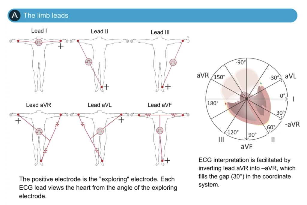
Figure 18. The organization of the limb leads. Note that the electrode on the right leg is not included in any lead, but serves as a ground wire. Leads I, Il and III are Einthoven’s original leads, and they can be presented with Einthoven’s triangle (lower panel). Leads aVR, aL and aVF were constructed by Goldberger; their reference point is the average of two electrodes. Lead aVR can be inverted into lead -aVR which is recommended as it mav facilitate interpretation. All modern ECG machines are capable of presenting both aVR and -aVR.

Question 62
Question:
A 35-year-old male patient went to your clinic for a routine check-up. A 12-lead ECG was taken. Upon examination of the ECG strip, you noticed that Leads I and aVF both have an upright QRS (positive deflection). You therefore estimated that the QRS is
a) deviated to the right
b) normal
c) deviated to the left
d) Indeterminate
Answer:
a) deviated to the right
解説:
リードIとaVFの両方においてQRS波形が正である場合、心電図の電気軸が右に偏っていることを示しています。この状態は、右室負荷や肺疾患などの異常を示唆する可能性があります。
- 選択肢bの誤り: 正常なQRS波形は通常、リードIとaVFの両方で正の波形を示す必要はありません。
- 選択肢cの誤り: QRSが左に偏位している場合は、リードIが正でaVFが負である必要があります。
- 選択肢dの誤り: 情報が提供されているため、電気軸の方向を特定することができます。
Question 63(10/2)
Question:
Which of the following statements regarding the pulmonary circulation is FALSE?
a) The mean pulmonary arterial pressure averages 8 mmHg while the systemic mean arterial pressure averages 16 mmHg
b) The pulmonary circulation contains 16% of the total blood volume while the systemic circulation contains 84%
c) The total pulmonary vascular resistance is only one-seventh that in the systemic circulation
d) The pressure is pulsatile in the pulmonary circulation like that in the aorta
Answer:
d) The pressure is pulsatile in the pulmonary circulation like that in the aorta
解説:
肺循環における圧力は、体循環に比べてはるかに低く、脈動性がありません。肺動脈は常に低圧であり、心拍に伴って圧力が変動することは少ないです。
- 選択肢aの正しさ: 肺動脈の平均圧が8 mmHgであり、これは事実です。
- 選択肢bの正しさ: 肺循環が体循環の16%の血液量を含むのも正しい情報です。
- 選択肢cの正しさ: 肺の血管抵抗が体循環の七分の一であることも正しいです。
Question 64(10/2)
Question:
Which of the following statements is TRUE regarding the physiological characteristics of the circulation?
a) The walls of veins are strong and more vascular than other blood vessels
b) The veins have the largest total cross-sectional area of all systemic vessels
c) The capillaries receive most of the total blood volume, serving as a reservoir of extra blood
d) The arterioles are the major sources of peripheral resistance.
Answer:
d) The arterioles are the major sources of peripheral resistance.
解説:
動脈硬化の影響を受けやすい小動脈(arterioles)は、末梢抵抗(peripheral resistance)の主な要因であり、血流の調整に重要な役割を果たします。
- 選択肢aの誤り: 静脈の壁は柔らかく、他の血管と比べると圧力が低く、血管内の構造も異なります。
- 選択肢bの誤り: 静脈は確かに広いが、全体的な断面積としては動脈が主導しています。
- 選択肢cの誤り: 毛細血管は確かに血液量を供給するが、血液の貯蔵庫という役割は静脈が果たしています。
Question 65(10/2)
Question:
Arteries
a) Eight (8) times more distensible than the veins
b) Transport blood under higher pressure than the veins
c) Twenty-four (24) times more compliant than the veins
d) Receive thirteen (13) percent more blood volume than the veins
Answer:
b) Transport blood under higher pressure than the veins
解説:
動脈は、心臓から血液を送り出すために非常に高い圧力で血液を運搬します。これは、血液が動脈の壁を押す力が強いためです。
- 選択肢aの誤り: 動脈は静脈よりも硬く、圧力に対して適応します。
- 選択肢cの誤り: 動脈は静脈よりも圧縮には適応しません。
- 選択肢dの誤り: 動脈は実際には静脈よりも血液を保持する容量が少ないため、この選択肢も不正確です。
Question 66(10/2)
Question:
A 65-year-old man with a history of hypertension and coronary artery disease presents to the emergency center with complaints of left-sided facial numbness and weakness. His blood pressure is normal, as is his heart rate and other vital signs. On examination, the patient has clear lungs and a normal cardiac examination. Auscultation of the carotid arteries reveals a “whooshing sound” (bruits) bilaterally. There is evidence of slurred speech and left-sided facial droop. The patient is diagnosed with a stroke. A computed tomography (CT) scan of the brain is performed. The patient is taken immediately to the intensive care unit for stabilization. What caused the increased tendency for turbulence to occur in this scenario?
a) increased blood velocity
b) increased viscosity
c) decreased density of the blood
d) decreased vessel diameter
Answer:
d) decreased vessel diameter
解説:
血管の直径が狭くなると、血液の流れが速くなり、渦流が生じやすくなります。これは、動脈硬化などによって血管が狭くなっている場合に特に見られます。
- 選択肢aの誤り: 血流速度が増加すること自体は必ずしも渦流を引き起こすわけではありません。
- 選択肢bの誤り: 血液の粘度が高まることは流れの抵抗を増加させるかもしれませんが、渦流の直接の原因にはなりません。
- 選択肢cの誤り: 血液の密度が低下することは、流れに影響を与えるかもしれませんが、渦流の原因ではありません。
乱流(Turbulent Flow)
血流速度が過度に速くなったり、血管内に障害(obstruction)がある場合、急激な曲がり(sharp turn)を通過したり、粗い表面(rough surface)を通過する際には、流れが乱れ、秩序立った流れ(streamlined)ではなくなることがあります(図14-6C)。この乱れた流れを乱流(turbulent flow)と呼びます。乱流が発生すると、血流は血管内で横方向や斜めに流れ、通常渦流(eddy currents)を形成します。これらの渦流は、急流の川で障害物がある箇所で見られる渦巻き(whirlpools)と似ています。渦流が存在すると、流線型の流れのときに比べて抵抗が大幅に増加し、血液の流れに対する全体的な摩擦が非常に増加します。
乱流の発生傾向は、血流速度(v)、血管径(d)、および血液の密度(density)に比例し、粘度(viscosity)には反比例します。これらの関係は以下の式で示されます:
- Re = (v × d × ρ) / η
ここで、Reはレイノルズ数(Reynolds’ number)、血流が乱流を引き起こす傾向を表す数値です。vは平均血流速度(mean velocity of blood flow、cm/sec)、dは血管径(vessel diameter、cm)、ρは密度(grams/ml)、ηは粘度(poise)です。血液の粘度は通常約1/30ポアスで、密度は1を少し超える程度です。
レイノルズ数が200~400を超えると、血管の枝で乱流が発生することがありますが、滑らかな部分では消滅します。しかし、レイノルズ数が約2000を超えると、直線で滑らかな血管でも通常乱流が発生します。
Question 67(10/2)
Question:
A PhD student was doing research on blood vessels’ conductance. He noticed that under normal conditions, flow through a blood vessel is 1 mL/min under a pressure gradient of 5 mmHg. What would be the approximate flow through the vessel after increasing the vessel diameter to two times normal, assuming the pressure gradient was maintained at 5 mmHg?
a) 256 mL/min
b) 2 mL/min
c) 16 mL/min
d) 4 mL/min
Answer:
c)
解説:
血管の直径が2倍になると、断面積は4倍になります。血流は血管の断面積の2.5乗に比例して変化する
この問題は血管のコンダクタンス(conductance)と流量の関係についてです。血管の直径がどのように流量に影響を与えるかを考える際に、ポアズイユの法則(Poiseuille’s law)を用いることができます。
ポアズイユの法則は、液体が円管を流れる際の流量Q が次のように関係することを示しています。
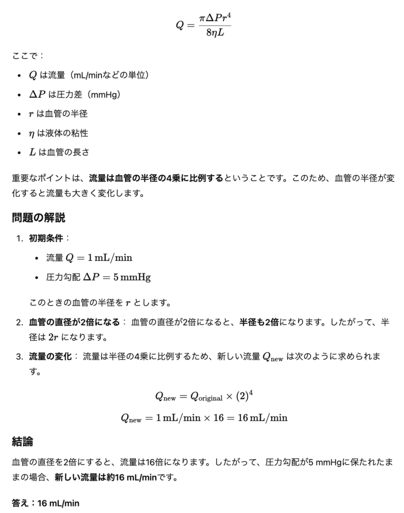
Question 68(10/2)
Question:
Regarding the auscultatory method of measuring blood pressure, which of the following statements is CORRECT?
a) It is done by placing a stethoscope over the antecubital artery and a blood pressure cuff is inflated around the upper arm
b) When tapping sounds from the artery are heard in synchrony with the heartbeat, the pressure level indicated by the manometer connected to the cuff is about equal to the diastolic pressure
c) It is a clinical method for determining systolic and diastolic pressures that is entirely accurate, usually giving the same values as those determined by direct catheter measurement from inside the arteries
d) The manometer pressure when the Korotkoff sounds change to the muffled quality is about equal to the systolic pressure
Answer:
a) It is done by placing a stethoscope over the antecubital artery and a blood pressure cuff is inflated around the upper arm
解説:
血圧測定は、上腕動脈の近くで聴診器を使用し、血圧カフを上腕に装着して行います。これが正しい方法です。
- 選択肢bの誤り: 騒音の聞こえ方から、血圧計の指示する圧は収縮期圧に相当するため、誤りです。
- 選択肢cの誤り: 聴診法は高い精度を持つが、カテーテル測定ほどの精度は保証されません。
- 選択肢dの誤り: コロトコフ音が変化する際の圧は収縮期圧に相当し、必ずしもそのような説明には当てはまりません。
Question 69(10/2)
Question:
A 20-year-old student went to her family physician for a routine physical exam. Her blood pressure is 100/60 mmHg. Her expected mean arterial pressure is
a) 50 mmHg
b) 76 mmHg
c) 96 mmHg
d) 84 mmHg
Answer:
b) 76 mmHg
解説:
平均動脈圧(Mean Arterial Pressure, MAP)は以下の式で計算されます
:MAP=DBP+(SBP−DBP)*(1/3)
=60+13(100−60)=76 mmHg
ここで、DBPは拡張期圧、SBPは収縮期圧です。
- 選択肢aの誤り: 50 mmHgは計算結果として正しくありません。
- 選択肢cの誤り: 96 mmHgも誤りで、計算に合致しません。
- 選択肢dの誤り: 84 mmHgも間違いです。正しい値は76 mmHgです。
Question 70(10/2)
Question:
Regarding pressure pulsations,
a) the greater the compliance of each vascular segment, the slower the velocity
b) the degree of damping is inversely proportional to the product of resistance times compliance
c) pulse pressure is decreased in arteriosclerosis
d) velocity of pressure pulse transmission is fastest in the aorta
Answer:
d) velocity of pressure pulse transmission is fastest in the aorta
解説:
圧力波の伝達速度は大動脈で最も速く、これは大動脈の弾力性と壁の構造によるものです。
- 選択肢aの誤り: 血管のコンプライアンスが高いほど流れの速度が速くなることが多いです。
- 選択肢bの誤り: 減衰の程度は抵抗とコンプライアンスの積に依存しており、選択肢の説明が逆です。
- 選択肢cの誤り: 動脈硬化症では脈圧は増加することが一般的です。
Question 71(10/2)
Question:
Which of the following would increase peripheral venous pressure?
a) Increased right atrial pressure
b) Pregnancy
c) Prolonged standing
d) All
Answer:
d) All
解説:
右心房圧の増加、妊娠、長時間の立位はすべて末梢静脈圧を増加させる要因です。これらは静脈系の血流を変化させ、圧力を高めます。
- 選択肢aの正しさ: 右心房圧が増加すると、末梢静脈圧も増加します。
- 選択肢bの正しさ: 妊娠による血液量の増加も末梢静脈圧を増加させます。
- 選択肢cの正しさ: 長時間立っていることは静脈の圧力を上昇させます。
Question 72(10/2)
Question:
The most important factor that regulates the opening and closing of the metarterioles and precapillary sphincters is the
a) velocity of blood flow
b) degree of sympathetic stimulation
c) tissue oxygen concentration
d) level of arterial pressure
Answer:
c) tissue oxygen concentration
解説:
組織の酸素濃度は、毛細血管前の動脈(metarterioles)や前毛細血管括約筋(precapillary sphincters)の開閉を調整する重要な要因です。酸素が不足すると、血流が増加し、逆に酸素が十分であれば血流が減少します。
- 選択肢aの誤り: 血流速度も影響を及ぼすが、直接的な調整因子ではありません。
- 選択肢bの誤り: 交感神経の刺激は影響しますが、主な調整因子は酸素濃度です。
- 選択肢dの誤り: 動脈圧のレベルは全体的な血流に影響を与えるが、開閉の直接的な要因ではありません。
★Question 73(10/2)
Question:
Which of the following Starling’s forces will favor the reabsorption of fluid back into the capillaries under normal conditions:
a) Interstitial fluid colloid osmotic pressure
b) Interstitial fluid hydrostatic pressure
c) Plasma colloid osmotic pressure
d) Capillary hydrostatic pressure
Answer:
c) Plasma colloid osmotic pressure
解説:
正常な状態では、血漿膠体浸透圧(plasma colloid osmotic pressure)が毛細血管への再吸収を促進します。これは、血液中のタンパク質が水を引き寄せるため、毛細血管内の圧力を高めます。
- 選択肢aの誤り: 間質液膠体浸透圧は、再吸収を妨げる要因となります。
- 選択肢bの誤り: 間質液の静水圧は、再吸収を妨げる要因です。
- 選択肢dの誤り: 毛細血管内の静水圧は、血液が毛細血管から外に出るのを促進します。
★Question 74(10/2)
Question:
In a capillary:
Capillary pressure (Pc) is 30 mm Hg
Interstitial fluid pressure (Pif) is -2 mmHg
Plasma colloid osmotic pressure (πc) is 25 mm Hg
Interstitial fluid colloid osmotic pressure (πif) is 2 mm Hg
What is the direction of fluid movement and the net driving force?
a) Absorption: 9 mmHg
b) Absorption: 5 mmHg
c) Filtration: 5 mmHg
d) Filtration: 9 mmHg
Answer:
d) Filtration: 9 mmHg
解説:
毛細血管からの液体移動を計算するために、Starlingの法則に基づく力のバランスを考慮します。計算式は次の通りです。
Net Driving Force=(Pc+πif)−(Pif+πc)=(30+2)−(−2+25)=32−23=9 mmHg
このため、フィルトレーション(濾過)が起こります。
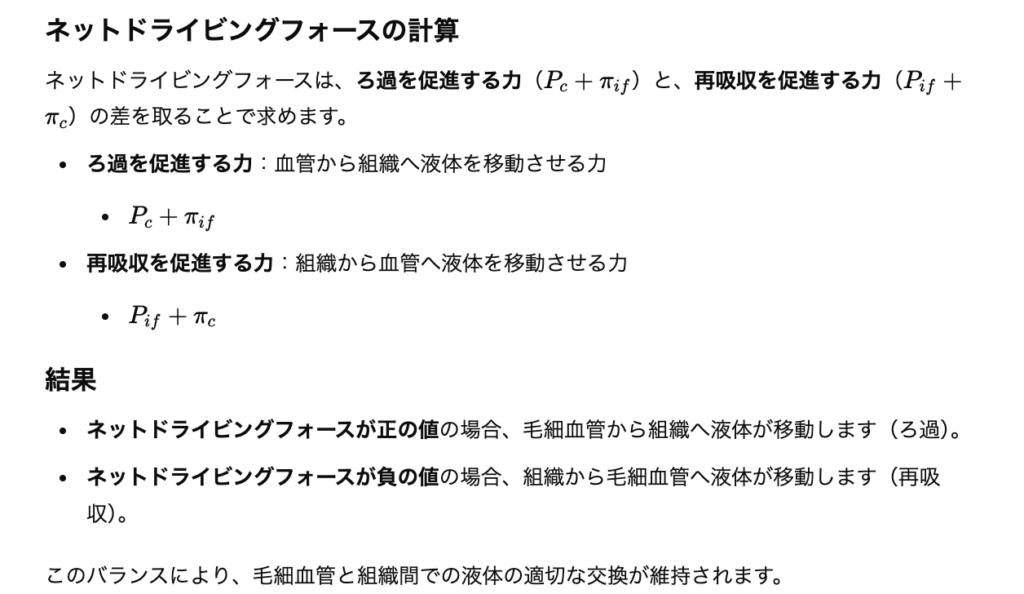
- 選択肢aの誤り: 吸収(Absorption)の数値は不正確です。
- 選択肢bの誤り: 吸収の説明であるため、誤りです。
- 選択肢cの誤り: フィルトレーションの方向が正しいですが、値が5 mmHgではなく9 mmHgです。
Question 75(10/2)
Question:
A 40-year-old female received news of her husband’s passing away due to COVID-19. She fainted after hearing the news. Vasovagal syncope
a) causes cerebral ischemia caused by an abrupt increase in arterial pressure
b) is associated with skeletal muscle vasodilation leading to reduced peripheral resistance
c) is associated with tachycardia
d) is more likely to occur when lying down than when standing
Answer:
b) is associated with skeletal muscle vasodilation leading to reduced peripheral resistance
解説:
血管迷走神経反射(vasovagal syncope)は、特にストレスや痛みの後に発生することが多く、筋肉の血管拡張による末梢抵抗の低下が起こります。これにより、脳への血流が減少し、失神が引き起こされます。
- 選択肢aの誤り: 血圧が急激に上昇することは、脳への血流を妨げる要因ではありません。
- 選択肢cの誤り: 血管迷走神経反射においては、通常は心拍数が減少します。
- 選択肢dの誤り: 失神は立位時に多く見られ、横たわっているときは発生しにくいです。
Question 76(10/2)
Question:
A 25-year-old male sustained gunshot wounds to the chest and abdomen and was rushed to the emergency room. He was hypotensive and tachycardic. Bystanders who brought him to the hospital claimed that he lost around 2.5 liters of blood. The last ditch pressure control mechanism for maintaining arterial pressure is
a) Baroreceptor reflex
b) Chemoreceptor reflex
c) Volume reflex
d) CNS Ischemic Response
Answer:
a) Baroreceptor reflex
解説:
圧受容体反射(baroreceptor reflex)は、動脈圧の変化に応じて心拍数と血管の緊張を調整し、血圧を維持するための迅速なメカニズムです。失血や低血圧が発生した際に、これが最初に作動します。
- 選択肢bの誤り: 化学受容器反射は、主に酸素や二酸化炭素のレベルを調整するために使用されます。
- 選択肢cの誤り: 体液の容量反射は血液量を調整しますが、直接的な血圧維持のメカニズムではありません。
- 選択肢dの誤り: CNS虚血反応は、通常は非常に重篤な低血圧の状況で起こります。
Question 77
Question:
A 23-year-old male was scheduled to undergo emergency appendectomy. He has no other medical problems. The anesthesiologist is called and administers a spinal block (including a sympathetic block) for anesthesia. Shortly after administration of the spinal anesthetic, the patient reports feeling light-headed and dizzy. He is noted to be hypotensive. The anesthesiologist notices the hypotension and gives an intravenous (IV) fluid bolus and a small amount of IV ephedrine (a vasoconstrictor). These measures resolve the patient’s symptoms and hypotension. Why would a sympathetic blockade cause these symptoms?
a) Sympathetic blockade results in the loss of the normal vasoconstrictor tone
b) Sympathetic blockade causes an increase in cardiac contractility
c) Sympathetic blockade causes an increase in conduction velocity through the AV node
d) All of the above
Answer:
a) Sympathetic blockade results in the loss of the normal vasoconstrictor tone
解説:
交感神経のブロックにより、血管が拡張し、血圧が低下します。このため、低血圧を引き起こし、患者がめまいや軽い失神を感じることになります。IV流体やエフェドリンによる治療が症状を改善するのは、血圧を回復させるからです。
- 選択肢bの誤り: 交感神経のブロックは心収縮力を高めることはありません。
- 選択肢cの誤り: 交感神経のブロックはAV結節を通る伝導速度を低下させるため、誤りです。
- 選択肢dの誤り: 「すべての選択肢」は正しくありません。
★Question 78
Question:
A 22-year-old female presents to the emergency department with severe vomiting and diarrhea. When she is supine (lying down), her blood pressure is 90/60 mm Hg and her heart rate is 100 beats/min. When she is moved to a standing position, her heart rate further increases to 120 beats/min. Which of the following mechanisms accounts for the further increase in heart rate upon standing?
a) Volume reflex
b) Bainbridge reflex
c) Baroreceptor reflex
d) Cushing reaction
Answer:
c) Baroreceptor reflex
解説:
体位が変わると、重力の影響で静脈還流が減少し、血圧が下がります。これにより圧受容器反射(baroreceptor reflex)が作動し、心拍数が増加して血圧を調整しようとします。
- 選択肢aの誤り: 体液容量反射は直接的な心拍数の調整メカニズムではありません。
- 選択肢bの誤り: ベインブリッジ反射は心房の受容体からの刺激によって心拍数が増加しますが、立ち上がり時の反応は主に圧受容器に基づいています。
- 選択肢dの誤り: クッシング反応は、主に頭蓋内圧が上昇した場合の反応であり、心拍数の調整に関連しません。
★Question 79
Question:
A 25-year-old male sustained gunshot wounds to the chest and abdomen and was rushed to the emergency room. He was hypotensive and tachycardic. Bystanders who brought him to the hospital claimed that he lost around 1 liter of blood. Which agent is released or secreted after a hemorrhage and causes an increase in renal Na+ reabsorption?
a) Aldosterone
b) Antidiuretic hormone
c) Angiotensin
d) Angiotensinogen
Answer:
a) Aldosterone
解説:
出血後、腎臓からナトリウムの再吸収を促進するホルモンはアルドステロンです。これにより、体内の水分量を保持し、血圧を維持しようとします。
- 選択肢bの誤り: 抗利尿ホルモン(ADH)は水の再吸収を促進しますが、ナトリウムの再吸収には直接的な影響を持ちません。
- 選択肢cの誤り: アンジオテンシンはナトリウムの再吸収を間接的に促進しますが、主な役割はアルドステロンの分泌を刺激することです。
- 選択肢dの誤り: アンジオテンシノーゲンは、アンジオテンシンに変換される前の前駆体であり、直接的な作用はありません。
Question 80
Question:
A 25-year-old male sustained gunshot wounds to the chest and abdomen and was rushed to the emergency room. He was hypotensive and tachycardic. A Foley-bag catheter was inserted and a decrease in urine output was noted. The decreased urine output in the hypotensive patient could be explained by
a) Pressure Natriuresis
b) Infinite feedback gain principle
c) Pressure Diuresis
d) Antidiuresis
Answer:
d) Antidiuresis
解説:
低血圧の患者では、体は水分の喪失を最小限に抑えようとするため、抗利尿作用(antidiuresis)が強まります。このため、尿量が減少します。
- 選択肢aの誤り: 圧利尿(pressure natriuresis)はナトリウムの排出を促進しますが、この場合は適用されません。
- 選択肢bの誤り: 無限のフィードバックゲイン原則はこの状況に関連しません。
- 選択肢cの誤り: 圧利尿(pressure diuresis)は、通常、血圧が上昇している際に見られるもので、低血圧時には正しくありません。
Question 81(10/2)
Question:
A 25-year-old male sustained gunshot wounds to the chest and abdomen and was rushed to the emergency room. He was hypotensive and tachycardic. By-standers who brought him to the hospital claimed that he lost around 1 liter of blood. At the emergency room, an intravenous line (IV) was established and IV fluid bolus of 1 liter was given. Hypotension and tachycardia was resolved. Increased extracellular fluid volume leads to an increase in arterial pressure because:
a) Venous return is decreased
b) Total peripheral resistance is decreased
c) Mean circulatory filling pressure is increased
d) Cardiac output is decreased
Answer:
c) Mean circulatory filling pressure is increased
解説:
体外液量が増加すると、静脈還流が増加し、全体的な血液量が増えることで、平均循環充填圧(mean circulatory filling pressure)が上昇します。これにより、動脈圧も上昇します。
- 選択肢aの誤り: 静脈還流は増加し、動脈圧を上昇させるため、減少しません。
- 選択肢bの誤り: 全末梢抵抗は通常、血液量の増加によって変わりません。
- 選択肢dの誤り: 心拍出量は液量が増加することで通常は増加し、逆に減少することはありません。
★Question 82
Question:
A 65-year-old man came to the clinic and his blood pressure is significantly elevated at 185/130 mm Hg. Laboratory tests reveal an increase in plasma renin activity, plasma aldosterone level, and right renal vein renin level. His left renal vein renin level is decreased. What is the most likely cause of the patient’s hypertension?
a) Pheochromocytoma, an epinephrine & norepinephrine secreting tumor in the adrenal gland
b) Aldosterone-secreting tumor
c) Right renal artery stenosis
d) Left renal artery stenosis
Answer:
c) Right renal artery stenosis
解説:
右腎動脈狭窄により、右腎からの血流が減少し、腎臓は血圧を上昇させるためにレニンを過剰に分泌します。左腎動脈が正常である場合、左腎静脈のレニンレベルは低下することが見られます。
- 選択肢aの誤り: 褐色細胞腫(pheochromocytoma)は、通常、血圧の急激な上昇を引き起こしますが、このパターンには当てはまりません。
- 選択肢bの誤り: アルドステロン分泌腫瘍は、高アルドステロン血症を引き起こすが、レニンのパターンは狭窄とは異なります。
- 選択肢dの誤り: 左腎動脈狭窄は、右側の所見と一致しないため、不適切です。
★Question 83
Question:
John’s blood pressure is normally 120/80 mmHg. This morning it dropped to 80/50 mmHg. Supposing his kidneys are normal, his renal-body fluid mechanism is expected to respond by
a) Activating pressure natriuresis
b) Decreasing renin, angiotensin II and aldosterone in the blood
c) Increasing thirst and increasing water intake
d) Increasing excretion of both salt and water by the kidneys
Answer:
c) Increasing thirst and increasing water intake
解説:
腎臓と体液のメカニズムは、血圧が低下すると渇感を増加させ、水分摂取を促進します。これは、体内の血液量を回復させるために重要です。
- 選択肢aの誤り: 圧利尿は血圧が高いときに見られます。
- 選択肢bの誤り: レニンやアンジオテンシンIIのレベルは低血圧時には上昇します。
- 選択肢dの誤り: 低血圧の際には、ナトリウムと水の排泄が減少するため、不適切です。
Question 84
Question:
A 33-year-old overweight male with a sedentary lifestyle came to you for a consult. He was recently diagnosed with hypertension. What is the first-line management of essential hypertension?
a) Vasodilator drugs to block the action of the renin-angiotensin system on the renal vasculature or renal tubules
b) Fluid restriction to decrease extracellular fluid volume and decreasing arterial pressure
c) Lifestyle modification to increase physical activity and weight loss
d) Diuretic drugs that decrease tubular reabsorption of salt and water
Answer:
c) Lifestyle modification to increase physical activity and weight loss
解説:
本質的高血圧の初期治療としては、生活習慣の改善が推奨されます。運動や体重の減少は、血圧を自然に低下させる効果があるため、最初に試みるべきです。
- 選択肢aの誤り: 薬物療法は通常、生活習慣の改善の後に適用されます。
- 選択肢bの誤り: 水分制限は一般的には推奨されず、生活習慣の改善が優先されます。
- 選択肢dの誤り: 利尿薬は薬物療法の一部であるが、まずは生活習慣の改善が必要です。
Question 85(10/2)
Question:
Which of the following is NOT TRUE regarding cardiac output?
a) Cardiac output is equal to venous return
b) Cardiac output may not increase when heart rate rises
c) Cardiac output is normally expressed as the output of one ventricle with each heartbeat
d) Cardiac output rises in a hot environment
Answer:
b) Cardiac output may not increase when heart rate rises
解説:
心拍数が上昇すれば、一般的には心拍出量も増加します。ただし、一定の限界を超えた場合は、心拍出量が安定することもありますが、それは通常の状況では稀です。
- 選択肢aの正しさ: 心拍出量は静脈還流と等しいことが通常です。
- 選択肢cの正しさ: 心拍出量は通常、一回の心拍での心室の出力として表現されます。
- 選択肢dの正しさ: 暑い環境では心拍出量は通常増加します。
Question 86
Question:
A 50-year-old woman has been admitted to hospital for thyroid surgery and is found to have signs of severe uncontrolled hyperthyroidism. Her peripheries are warm and moist and her pulse is rapid and bounding. What would be expected of her cardiac parameters?
a) Cardiac output is increased, heart rate decreased, pulse pressure is low
b) Cardiac output is normal, heart rate is normal, pulse pressure is normal
c) Cardiac output is decreased, heart rate increased, pulse pressure is high
d) Cardiac output is increased, heart rate increased, pulse pressure is high
Answer:
d) Cardiac output is increased, heart rate increased, pulse pressure is high
解説:
甲状腺ホルモンの過剰は心拍数を増加させ、心拍出量も増加させます。周辺が温かく湿っていることは、血管拡張と関連しており、脈圧も高くなる傾向があります。
- 選択肢aの誤り: 心拍数は低下しないため、この選択肢は誤りです。
- 選択肢bの誤り: 心拍数と心拍出量は通常、増加します。
- 選択肢cの誤り: 心拍出量は減少せず、逆に増加するため、誤りです。
Question 87(10/2)
Question:
A 40-year-old woman with a history of menorrhagia (heavy menstrual flow) came to the outpatient clinic with chief complaint of dizziness. She has a blood pressure of 90/60 mmHg and heart rate of 100 bpm. Cardiac output is increased in anemia because
a) the decreased concentration of red blood cells results in reduced viscosity of the blood
b) the diminished delivery of oxygen to the tissues causes local vasodilation
c) the decrease in total peripheral resistance increases cardiac output
d) all of the above
Answer:
d) all of the above
解説:
貧血の場合、赤血球の濃度が低下し、血液の粘度が減少します。このため、血流が改善し、酸素供給の低下が局所的な血管拡張を引き起こします。また、全末梢抵抗が減少することで、心拍出量も増加します。
- 選択肢aの正しさ: 赤血球の濃度が低下すると血液の粘度が減少します。
- 選択肢bの正しさ: 酸素供給の低下が血管拡張を引き起こします。
- 選択肢cの正しさ: 全末梢抵抗の減少が心拍出量を増加させます。
Question 88
Question:
A 30-year-old man has been admitted to the hospital for minor elective surgery. He is a long-distance runner of national standard. His cardiac shadow is enlarged on chest X-ray and there is concern about his very slow pulse. What would be expected of his cardiac output curve?
a) increased plateau level of the cardiac output curve
b) shift the entire cardiac output curve to the right
c) shift the entire cardiac output curve to the left
d) decreased plateau level of the cardiac output curve
Answer:
c) shift the entire cardiac output curve to the left
解説:
長距離ランナーでは、心臓がトレーニングによって適応し、心拍出量が向上します。これにより、心拍出量曲線は左にシフトします。これは、同じ心拍数でより多くの血液を送る能力を示しています。
- 選択肢aの誤り: 増加したプレートレベルは示されません。
- 選択肢bの誤り: 右へのシフトは通常、心機能の低下を示します。
- 選択肢dの誤り: プレートレベルの減少は示されず、心機能が向上します。
Question 89(10/2)
Question:
According to the cardiac and vascular function curves, an increase in blood volume leads to
a) decreased right atrial pressure and increased cardiac output
b) increased right atrial pressure and increased cardiac output
c) decreased right atrial pressure and decreased cardiac output
d) increased right atrial pressure and decreased cardiac output
Answer:
b) increased right atrial pressure and increased cardiac output
解説:
血液量の増加により、右心房圧が上昇し、それに伴って心拍出量も増加します。これは、心臓がより多くの血液を送り出す能力を持つためです。
- 選択肢aの誤り: 右心房圧は増加し、心拍出量も増加します。
- 選択肢cの誤り: 右心房圧が低下することはありません。
- 選択肢dの誤り: 心拍出量は通常、血液量の増加によって上昇します。
Question 90
Question:
A 25-year-old male patient sustained a stab wound to the chest. He has no known medical problems. If the thorax of a normal, healthy patient is surgically opened, what happens to the cardiac output curve?
a) It shifts 4 mmHg to the right
b) It shifts 2 mmHg to the right
c) It shifts 4 mmHg to the left
d) It shifts 2 mmHg to the left
Answer:
a) It shifts 4 mmHg to the right
解説:
胸部の開放によって圧力の変化が生じ、心拍出量曲線が右にシフトします。これは、心臓への血液供給が圧力変化に適応するための調整を示しています。
- 選択肢bの誤り: 2 mmHgのシフトは見られません。
- 選択肢cの誤り: 左へのシフトは通常、血流の減少を示します。
- 選択肢dの誤り: 2 mmHgの左へのシフトは不正確です。
Question 91
Question:
Which of the following would shift the cardiac output curve to the left?
a) breathing against a negative pressure
b) valvular heart disease
c) a marathon runner on a run
d) cardiac tamponade
Answer:
c) a marathon runner on a run
解説:
マラソンランナーの運動中は、心拍出量曲線が左にシフトします。これは、運動により心臓がより効率的に血液を送り出すためです。
- 選択肢aの誤り: 負圧での呼吸は心拍出量の変化を引き起こしません。
- 選択肢bの誤り: 弁膜心疾患は通常、心拍出量曲線を右にシフトさせます。
- 選択肢dの誤り: 心タンポナーデは圧力が増加し、心拍出量曲線を右にシフトさせます。
Question 92(10/2)
Question:
Which of the following is normally associated with an increased venous return of blood to the heart?
a) Increased resistance to venous return
b) Right atrial pressure rises to equal the mean systemic filling pressure
c) Increased difference between mean systemic filling pressure and right atrial pressure
d) Decreased pressure gradient for venous return
Answer:
c) Increased difference between mean systemic filling pressure and right atrial pressure
解説:
静脈還流が増加すると、平均循環充填圧と右心房圧の差が広がります。これにより、血液が心臓に戻ることが促進されます。
- 選択肢aの誤り: 静脈還流の抵抗が増加すると、逆に血流が減少します。
- 選択肢bの誤り: 右心房圧が平均循環充填圧と等しくなることは通常ありません。
- 選択肢dの誤り: 静脈還流が増加すると、圧力勾配は増加します。
静脈還流が増加すると、**平均循環充填圧(Mean Circulatory Filling Pressure, MCFP)と右心房圧(Right Atrial Pressure, RAP)**の差が広がり、これにより血液が心臓に戻ることが促進される理由は、循環系における圧力の差が静脈還流の駆動力として働くからです。
静脈還流と圧力差の関係
静脈還流とは、血液が全身から右心房に戻ってくる流れを指します。この流れは、静脈や大血管に生じる圧力差によって駆動されています。具体的には、次のように考えられます。
- 平均循環充填圧(MCFP):
- 平均循環充填圧は、循環系全体における血液が静止状態で分布する際の圧力です。これは、静脈還流の駆動圧力の基準になります。
- 心拍がない状態で、全ての血管に血液が充填されるときの圧力です。
- 右心房圧(RAP):
- 右心房圧は、血液が右心房に戻る際に直面する「抵抗」のようなものです。この圧力が高ければ高いほど、静脈から血液が心臓に戻りにくくなります。
圧力差が広がるときの影響
静脈還流を決定するのは、平均循環充填圧(MCFP)と右心房圧(RAP)との間の圧力差です。この差を「駆動圧」と呼びます。この駆動圧が広がると、血液はより速く右心房に戻ってきます。以下がその理由です。
- 静脈還流の駆動圧が増加:
- 駆動圧 = MCFP – RAP です。
- 静脈還流を増加させるには、MCFPを上げるか、RAPを下げる必要があります。
- 静脈還流が増加するということは、循環系全体での充填圧(MCFP)が高くなるか、右心房圧(RAP)が相対的に低くなることを意味します。
- 駆動圧が高いと血液の流れが促進:
- 血液は圧力差によって移動するため、駆動圧が大きくなると静脈から心臓への血液の流れ(静脈還流)が増加します。
- 右心房への血液の流れが増えることによって、心臓が拍出する血液量も増加しやすくなります。
結論
静脈還流が増加することで、平均循環充填圧と右心房圧の差が広がると、駆動圧が増加します。この駆動圧の増加によって、より多くの血液が右心房に戻ることが可能になり、結果として静脈還流が促進されるのです。
したがって、圧力差の拡大は静脈還流を増加させる重要な要因となり、心臓への血液の効率的な供給が維持されます。
Question 93(10/2)
Question:
A 25-year-old male sustained gunshot wounds to the chest and abdomen and was rushed to the emergency room. He was hypotensive and tachycardic. Bystanders who brought him to the hospital claimed that he lost around 500 milliliters of blood. At the emergency room, an intravenous line (IV) was established and IV fluid bolus of 1 liter was given. What is the effect of increasing the mean systemic filling pressure (Psf) on the venous return curve?
a) The venous return curve shifts upward and to the right
b) The venous return curve shifts downward and to the left
c) The venous return curve rotates upward to 2x as great a slope
d) The venous return curve rotates downward to V2 as great
Answer:
a) The venous return curve shifts upward and to the right
解説:
平均循環充填圧が増加すると、静脈還流が増加し、静脈還流曲線が上にシフトし、右に移動します。これは、より多くの血液が心臓に戻ることを示しています。
- 選択肢bの誤り: 静脈還流の増加に伴い、曲線が下にシフトすることはありません。
- 選択肢cの誤り: 曲線が単純に傾きの増加ではなく、全体としてシフトします。
- 選択肢dの誤り: 静脈還流が増加すると、曲線は下降することはありません。
Question 94
Question:
Coronary blood flow to the left ventricle increases during
a) early systole
b) hypothermia
c) diastole
d) tachycardia
Answer:
c) diastole
解説:
冠動脈血流は心臓の拡張期(diastole)中に最も増加します。この時期に心臓が弛緩し、冠動脈に血液が流入しやすくなります。
- 選択肢aの誤り: 早期収縮期では心臓が収縮しているため、冠動脈血流は減少します。
- 選択肢bの誤り: 低体温は通常、心拍出量や血流に影響を与えるが、冠動脈血流に直接的な影響を与えることは少ないです。
- 選択肢dの誤り: 頻脈は心臓の収縮時間を減少させるため、冠動脈血流を妨げる可能性があります。
Question 95
Question:
Factors ensuring that ventricular muscle has an adequate oxygen supply include
a) Increase in coronary vascular resistance during exercise
b) Structural arrangements that prevent vascular compression during systole
c) High oxygen extraction rate from blood circulating through the myocardium
d) Good functional anastomoses that exist between adjacent coronary arteries
Answer:
b) Structural arrangements that prevent vascular compression during systole
解説:
心筋は収縮時に血管が圧迫されるのを防ぐために、構造的に設計されています。この機能により、収縮中でも適切な血液供給が確保されます。
- 選択肢aの誤り: 運動中の冠血管抵抗の増加は、酸素供給を減少させる可能性があります。
- 選択肢cの誤り: 高い酸素抽出率は心筋に供給される血液の質を示すが、構造的要因とは異なります。
- 選択肢dの誤り: 隣接する冠動脈間の機能的な吻合は重要ですが、直接的な酸素供給の確保には限界があります。
Question 96
Question:
The second heart sound differs from the first heart sound in that it is
a) longer lasting than the first heart sound
b) caused by oscillation of blood back and forth between the walls of the ventricles initiated by inrushing blood from the atria
c) of lower frequency because of the much less taut semilunar valves
d) heard when the ventricles are relaxing
Answer:
d) heard when the ventricles are relaxing
解説:
第二心音(S2)は、心室が弛緩している際に、動脈弁が閉じることによって発生します。これは心臓が弛緩しているときに聞かれます。
- 選択肢aの誤り: 第二心音は第一心音よりも短くなることが多いです。
- 選択肢bの誤り: 第一心音は弁の閉鎖に起因し、二つの心音は異なるメカニズムで発生します。
- 選択肢cの誤り: 音の周波数は弁の特性によって異なりますが、音が低いというわけではありません。
Question 97
Question:
Which of the following does NOT match with the chest surface area for auscultation of normal heart sounds?
a) Mitral valve – apex of left ventricle
b) Aortic valve – upward along the aorta
c) Pulmonic valve – upward along the pulmonary artery
d) Tricuspid valve – upward along the right atrium
Answer:
d) Tricuspid valve – upward along the right atrium
解説:
三尖弁の聴診は、通常は右心房の上部からではなく、右心室の近くで行われます。したがって、この選択肢は不正確です。
- 選択肢aの正しさ: 僧帽弁は左心室の頂点で正しく聴診されます。
- 選択肢bの正しさ: 大動脈弁は通常、上部に聴診されます。
- 選択肢cの正しさ: 肺動脈弁も通常、上に聴診されます。
Question 98(10/2)
Question:
A female infant is delivered prematurely at a gestational age of 32 weeks, weighing 1 kg. Forty-eight hours after birth, the baby is noted to have a very loud machinery-like heart murmur and a bounding pulse, and is feeding poorly. Indomethacin is administered and the symptoms resolve. What is the most likely diagnosis?
a) Patent ductus arteriosus
b) Mitral regurgitation
c) Tetralogy of Fallot
d) Necrotizing enterocolitis
Answer:
a) Patent ductus arteriosus
解説:
早産児においては、動脈管開存症(patent ductus arteriosus)が一般的で、心雑音が聞かれます。インドメタシンはこの状態の治療に使われ、血管を収縮させることで症状が改善します。
- 選択肢bの誤り: 僧帽弁逆流は通常、雑音の特徴が異なります。
- 選択肢cの誤り: ファロー四徴症はより複雑な先天性心疾患で、異なる症状を示します。
- 選択肢dの誤り: 腸の壊死はこの状況とは関連しません。
Question 99
Question:
A 54-year-old man presents to the primary care physician with dyspnea upon exertion. He also notes palpitations sometimes when he works too hard in the garden. On cardiac examination, a harsh systolic murmur is heard. It is sometimes associated with a thrill. What valvular abnormality is this man likely experiencing?
a) Mitral valve regurgitation
b) Aortic valve stenosis
c) Mitral valve stenosis
d) Aortic valve regurgitation
Answer:
b) Aortic valve stenosis
解説:
大動脈弁狭窄(aortic valve stenosis)は、収縮期の雑音が聞かれ、時には振動(thrill)が伴うことがあります。運動時の呼吸困難や動悸は、心拍出量の制限に関連しています。
- 選択肢aの誤り: 僧帽弁逆流は通常、拡張期の雑音であり、収縮期とは異なります。
- 選択肢cの誤り: 僧帽弁狭窄は異なる症状を示し、主に拡張期に雑音が発生します。
- 選択肢dの誤り: 大動脈弁逆流は拡張期に関連した雑音であり、収縮期の雑音とは異なります。
Question 100
Question:
In a patient with chronic heart failure, which of the following neurohumoral responses is expected to be activated?
a) Decreased release of norepinephrine
b) Increased activity of the renin-angiotensin-aldosterone system (RAAS)
c) Decreased secretion of antidiuretic hormone (ADH)
d) Increased peripheral vasodilation
Answer:
b) Increased activity of the renin-angiotensin-aldosterone system (RAAS)
解説:
慢性心不全の患者では、体は血圧を維持するためにレニン-アンジオテンシン-アルドステロン系を活性化させます。この反応により、血圧を上昇させるための水分と塩分の再吸収が促進されます。
- 選択肢aの誤り: ノルエピネフリンの放出は増加し、交感神経系が活性化します。
- 選択肢cの誤り: 抗利尿ホルモン(ADH)の分泌は通常、心不全において増加します。
- 選択肢dの誤り: 周辺血管の拡張は、心不全において期待される反応ではありません。
慢性心不全の補償メカニズム
心不全では、心臓が全身に十分な血液を送り出せないため、腎臓は体が血液不足や血圧低下に陥っていると判断します。その結果、RAA系が活性化されます。このシステムは血圧を維持するために重要ですが、心不全患者では逆効果になることがあります。
- 水分と塩分の再吸収: アルドステロンの働きにより、腎臓はナトリウムと水分をより多く再吸収します。これにより、体内の血液量が増加し、一時的に血圧を維持します。
- 過剰な水分蓄積: しかし、慢性心不全では心臓の機能が低下しているため、増えた血液量を効果的に送り出せません。その結果、体内に水分が過剰に蓄積し、浮腫(むくみ)や肺うっ血(肺に水分が溜まる)などの症状を引き起こします。

コメント