
Contents
- 1 アセス
- 1.1 Question 1:
- 1.2 Question 2:
- 1.3 Question 3:
- 1.4 Question 4:
- 1.5 Question 5:
- 1.6 Question 6:
- 1.7 Question 7:
- 1.8 Question 8:
- 1.9 Question 9:
- 1.10 Question 10:
- 1.11 Question 11:
- 1.12 Question 12:
- 1.13 Question 13:
- 1.14 Question 14:
- 1.15 Question 15:
- 1.16 Question 16:
- 1.17 Question 17:
- 1.18 Question 18:
- 1.19 Question 19:
- 1.20 Question 20:
- 1.21 Question 21:
- 1.22 Question 22:
- 1.23 Question 23:
- 1.24 Question 24:
- 1.25 Question 25:
- 1.26 Question 26:
- 1.27 Question 27:
- 1.28 Question 28:
- 1.29 Question 29:
- 1.30 Question 30:
- 1.31 Question 31:
- 1.32 Question 32:
- 1.33 Question 33:
- 1.34 Question 34:
- 1.35 Question 35:
- 1.36 Question 36:
- 1.37 Question 37:
- 1.38 Question 38:
- 1.39 Question 39:
- 1.40 Question 40:
- 1.41 Question 41:
- 1.42 Question 42:
- 1.43 Question 43:
- 1.44 Question 44:
- 1.45 Question 45:
- 1.46 Question 46:
- 1.47 Question 47:
- 1.48 Question 48:
- 1.49 Question 49:
- 1.50 Question 50:
- 2 ブロック
- 2.1 Question 1 of 200
- 2.2 Question 2 of 200
- 2.3 Question 3 of 200
- 2.4 Question 4 of 200
- 2.5 Question 5 of 200
- 2.6 Question 6 of 200
- 2.7 Question 7 of 200
- 2.8 Question 8 of 200
- 2.9 Question 10 of 200
- 2.10 Question 11 of 200
- 2.11 Question 12 of 200
- 2.12 Question 13 of 200
- 2.13 Question 14 of 200
- 2.14 Question 15 of 200
- 2.15 Question 16 of 200
- 2.16 Question 17 of 200
- 2.17 Question 18 of 200
- 2.18 Question 19 of 200
- 2.19 Question 20 of 200
- 2.20 Question 21 of 200
- 2.21 Question 22 of 200
- 2.22 Question 23 of 200
- 2.23 Question 24 of 200
- 2.24 Question 26 of 200
- 2.25 Question 27 of 200
- 2.26 Question 29 of 200
- 2.27 Question 31 of 200
- 2.28 Question 32 of 200
- 2.29 Question 35 of 200
- 2.30 Question 39 of 200
- 2.31 Question 42 of 200
- 2.32 Question 43 of 200
- 2.33 Question 44 of 200
- 2.34 Question 45 of 200
- 2.35 Question 47 of 200
- 2.36 Question 48 of 200
- 2.37 Question 50 of 200
アセス
Question 1:
Ventral primary rami that gives rise to the intercostal nerves arise from which vertebral levels?
- T1-T11
- C3-T12
- C5-T12
- T1-T12
Correct Answer: T1-T11
Explanation:
T1-T11の腹側一次枝(ventral primary rami)は肋間神経(intercostal nerves)を形成します。これらの神経は肋骨に沿って走行し、胸壁と関連する筋肉、皮膚に感覚と運動を供給します。選択肢のT1-T12は誤りで、T12は腹側枝として肋下神経(subcostal nerve)を形成するため、肋間神経には含まれません。他の選択肢であるC3-T12やC5-T12は肋間神経に関係がなく、脊髄の異なる部位に関連する神経分布を示します。
Question 2:
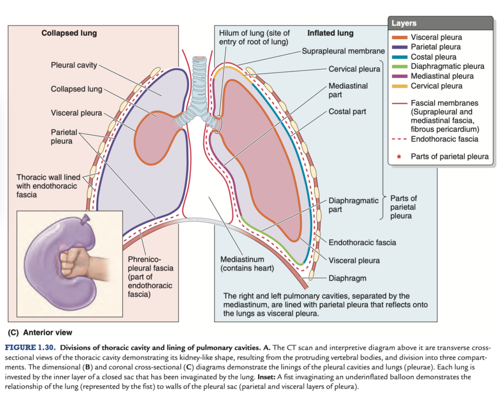
Which pair of parietal pleura is NOT attached or adjacent to the endothoracic fascia?
- Mediastinal and cervical parts
- Costal and cervical parts
- Mediastinal and diaphragmatic parts
- Costal and diaphragmatic parts
Correct Answer: Mediastinal and cervical parts
Explanation:
胸膜(pleura)は、胸腔内の臓器を覆う膜であり、壁側胸膜(parietal pleura)は胸壁と隣接しています。特に、壁側胸膜の胸骨部分(costal part)は胸壁の内側にある胸壁筋膜(endothoracic fascia)に密接しています。しかし、縦隔部分(mediastinal part)と頸部部分(cervical part)は胸壁筋膜と直接接しておらず、他の構造に隣接しています。したがって、この組み合わせは胸壁筋膜に接していません。他の選択肢は全て胸壁筋膜と関わっています。
Question 3:
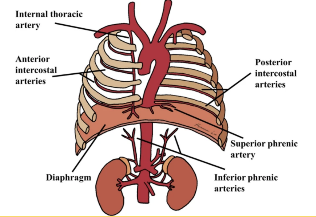
Which of the following arteries terminate at the 6th intercostal space and become the superior epigastric artery on the anterior abdominal wall?
- Anterior intercostal artery
- Lateral thoracic artery
- Posterior intercostal artery
- Internal thoracic artery
Correct Answer: Internal thoracic artery
Explanation:
内胸動脈(internal thoracic artery)は、第6肋間スペースで上腹壁動脈(superior epigastric artery)と筋横隔動脈(musculophrenic artery)に分岐します。前肋間動脈(anterior intercostal artery)や後肋間動脈(posterior intercostal artery)は、主に胸壁と肋間筋を供給します。外側胸動脈(lateral thoracic artery)は胸筋や乳房などに供給され、6肋間スペースに関与しません。
Question 4:
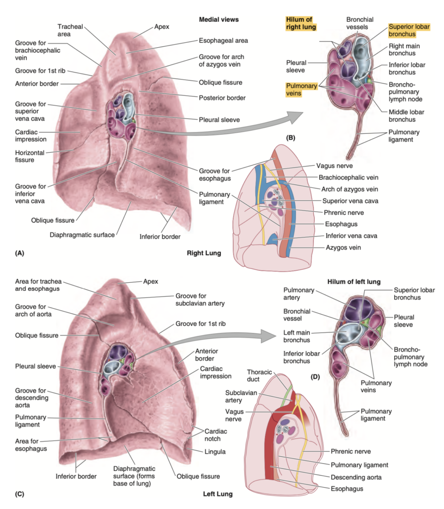
Which of the following is True about the right lung?
- Contains the cardiac notch
- Does not contain an oblique fissure
- Divided by a single fissure into a superior and inferior lobe
- The horizontal fissure divides the superior and middle lobe
Correct Answer: The horizontal fissure divides the superior and middle lobe
Explanation:
右肺は水平裂(horizontal fissure)によって上葉と中葉に分けられます。心切痕(cardiac notch)は左肺の特徴です。また、右肺には斜裂(oblique fissure)も存在し、これによって中葉と下葉が分けられます。したがって「単一の裂で上葉と下葉が分けられる」という記述も誤りです。
Question 5:
Which of the following is TRUE about the lower margin of the visceral pleura?
- Passes 12th rib paravertebral
- Passes 12th rib mid-axillary line (MAL)
- Passes 6th rib mid-clavicular line (MCL)
- Passes 8th rib MCL
- Passes 10th rib paravertebral
- Passes 8th rib MAL
Correct Answer: Passes 8th rib MCL
Explanation:
臓側胸膜(visceral pleura)の下縁は、鎖骨中線(mid-clavicular line, MCL)で第6肋骨、腋窩中線(mid-axillary line, MAL)で第8肋骨、脊椎傍線(paravertebral line)で第10肋骨まで達します。したがって「第8肋骨MCL」は誤りで、「第8肋骨MAL」が正しいです。他の選択肢は、適切な線に対して誤った肋骨番号を提示しています。
Question 6:
The lungs are immediately surrounded by which structure?
- Parietal pleura
- Pleural cavity
- Pericardium
- Visceral pleura
- Clavi-pectoral fascia
Correct Answer: Visceral pleura
Explanation:
肺は臓側胸膜(visceral pleura)に直接覆われています。臓側胸膜は肺の表面に密着しており、肺を保護しています。壁側胸膜(parietal pleura)は胸壁に接しており、その間に胸膜腔(pleural cavity)があります。心膜(pericardium)や鎖骨胸筋膜(clavipectoral fascia)は肺とは関係ありません。
Question 7:
Which of the following is TRUE about the costal line of pleural reflection?
- Passes 12th rib mid-axillary line (MAL)
- Passes 8th rib paravertebral line
- Passes 12th rib paravertebral line
- Passes 10th rib mid-clavicular line (MCL)
- Passes 10th rib MAL
- Passes 8th rib at MCL
Correct Answer: Passes 8th rib at MCL
Explanation:
肋骨の胸膜反射線(costal line of pleural reflection)は、臓側胸膜が壁側胸膜に反転するポイントを示しています。鎖骨中線(MCL)では、第8肋骨の高さで胸膜反射が起こります。その他の選択肢は、誤った肋骨レベルまたは誤った線での反射位置を示しています。
鎖骨中線(mid-clavicular line, MCL):
- 肋骨胸膜反射線は、第 8肋骨 まで達します。
- 肺の下縁も第6肋骨付近に位置しており、胸膜はこれよりさらに下方に延びています。
腋窩中線(mid-axillary line, MAL):
- 第 10肋骨 の高さまで達します。
- この場所では、横隔膜と接している部分が広がり、胸腔と腹腔の間に位置します。
脊椎傍線(paravertebral line):
- 第 12肋骨 の高さに達し、背中側では胸壁と胸膜が接する最も下のポイントになります。
Question 8:
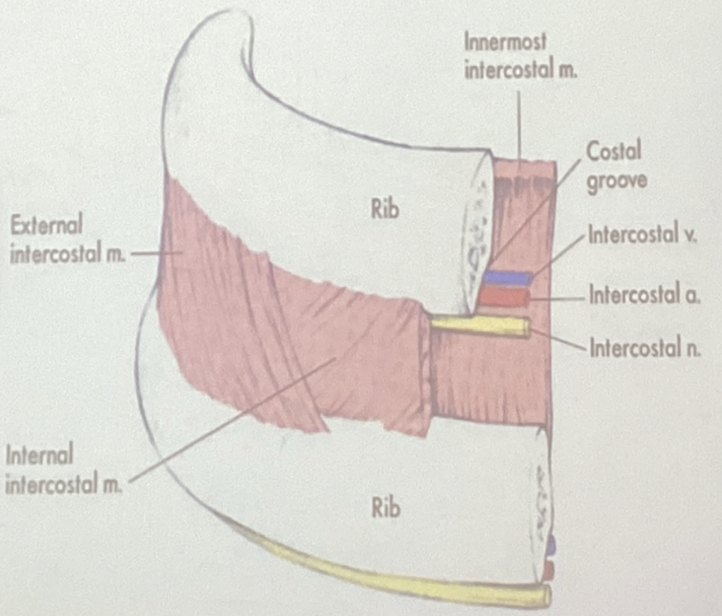
The intercostal vessels and nerves are found in which of the following layers of the thoracic wall?
- Between internal & innermost intercostal muscles
- Between external & internal intercostal membranes
- Between innermost intercostal muscle & endothoracic fascia
- Between external & internal intercostal muscles
Correct Answer: Between internal & innermost intercostal muscles
Explanation:
肋間血管と神経(intercostal vessels and nerves)は、内肋間筋(internal intercostal muscles)と最内肋間筋(innermost intercostal muscles)の間に存在します。外肋間膜(external intercostal membrane)や胸壁筋膜(endothoracic fascia)はこの神経血管束に関与しません。したがって、正しい選択肢は「内肋間筋と最内肋間筋の間」です。
Question 9:
Where is the angle of Louis located?
- Between the manubrium and the sternal body
- Between the sternoclavicular joints
- Beside the articular facet of a typical rib
- A potential space below the inferior margins of the lung where pleural fluid will accumulate
Correct Answer: Between the manubrium and the sternal body
Explanation:
ルイの角(angle of Louis)は、胸骨柄(manubrium)と胸骨体(sternal body)の境界にある解剖学的ランドマークです。胸骨角(sternal angle)とも呼ばれ、第2肋間を識別するために使用されます。他の選択肢は、胸骨角とは異なる解剖学的構造を示しています。
ルイの角(angle of Louis)は、第 4胸椎(T4) と 第5胸椎(T5) の間に位置しています。これは胸骨柄(manubrium)と胸骨体(body of sternum)の接合部にあり、解剖学的ランドマークとして重要です。
Question 10:
A potential space below the inferior margins of the lung where pleural fluid will accumulate is called?
- Costodiaphragmatic recess
- Pericardial sac
- Pulmonary ligament
- Costomediastinal recess
Correct Answer: Costodiaphragmatic recess
Explanation:
肋骨横隔陥凹(costodiaphragmatic recess)は、肺の下縁と横隔膜との間に位置する潜在的なスペースです。胸水(pleural fluid)はこの部位に蓄積することがあります。心膜腔(pericardial sac)は心臓を覆う膜であり、肺とは関係がありません。肺靭帯(pulmonary ligament)は肺の支持構造であり、肋骨縦隔陥凹(costomediastinal recess)は縦隔の近くに位置する別のスペースです。
Question 11:
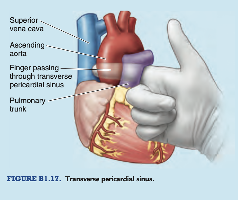
The surgeon entered the pericardial sac and was able to insert his finger through the transverse pericardial sinus. Which paired great vessels will you be able to palpate and ligate anterior to his finger?
- Ascending aorta and pulmonary artery
- Superior vena cava and pulmonary artery
- Pulmonary artery and pulmonary vein
- Superior vena cava and ascending aorta
Correct Answer: Ascending aorta and pulmonary artery
Explanation:
横走心膜洞(transverse pericardial sinus)は上行大動脈(ascending aorta)と肺動脈(pulmonary artery)の間に存在します。この構造を通して、外科医はこれらの大血管を触診し、結紮することができます。上大静脈(superior vena cava)や肺静脈(pulmonary veins)はこの位置には存在しないため、他の選択肢は誤りです。
Surgical Significance of Transverse Pericardial Sinus
The transverse pericardial sinus is especially important to cardiac surgeons. After the pericardial sac is opened anteriorly, a finger can be passed through
the transverse pericardial sinus posterior to the ascending aorta and pulmonary trunk (Fig. B1.17). By passing a surgical clamp or a ligature around these large vessels, inserting the tubes of a coronary bypass machine, and then tightening the ligature, surgeons can stop or divert the circulation of blood in these arteries while performing cardiac surgery, such as coronary artery bypass grafting.
Question 12:
Which structure constricts the esophagus to prevent reflux of gastric contents?
- Sternopericardial ligament
- Central tendon
- Left crus of diaphragm
- Right crus of diaphragm
Correct Answer: Right crus of diaphragm
Explanation:
食道裂孔(esophageal hiatus)は横隔膜の右脚(right crus of diaphragm)が形成し、胃から食道への逆流を防ぐ役割を果たします。T10椎骨のレベルにあります。左脚(left crus)はこの機能には直接関与しません。中央腱(central tendon)や胸骨心膜靭帯(sternopericardial ligament)は、この食道の収縮とは関係がありません。
Question 13:
The diaphragm receives blood supply directly from the aorta through which artery?
- Musculophrenic artery
- Superior phrenic artery
- Inferior phrenic artery
- Pericardiophrenic artery
Correct Answer: Inferior phrenic artery
Explanation:
下横隔動脈(inferior phrenic artery)は大動脈から直接供給され、横隔膜に血液を供給します。上横隔動脈(superior phrenic artery)も横隔膜に血液を供給しますが、必ずしも大動脈から直接ではありません。筋横隔動脈(musculophrenic artery)や心膜横隔動脈(pericardiophrenic artery)は、主に胸壁や心膜に血液を供給します。
下横隔動脈(inferior phrenic artery) は直接、大動脈(aorta)から分岐して横隔膜に血液を供給する主要な動脈です。この動脈は横隔膜の下側に血液を供給し、横隔膜の主要な血液供給源の一つです。
- Musculophrenic artery(筋横隔動脈) は内胸動脈(internal thoracic artery)から分岐し、横隔膜の一部にも供給しますが、大動脈から直接供給されるわけではありません。
- Superior phrenic artery(上横隔動脈) も大動脈から分岐しますが、これは横隔膜の上側に供給されます。
- Pericardiophrenic artery(心膜横隔動脈) は内胸動脈から分岐し、主に心膜と横隔膜の一部に血液を供給しますが、大動脈から直接の供給ではありません。
Question 14:
Which of the following is TRUE about pulmonary circulation?
- Oxygenated blood leaves the lungs via the pulmonary veins
- Oxygenated blood enters the lungs via the pulmonary arteries
- Deoxygenated blood leaves the lungs via the pulmonary veins
- Oxygenated blood enters the lungs via the pulmonary veins
Correct Answer: Oxygenated blood leaves the lungs via the pulmonary veins
Explanation:
肺循環では、肺で酸素を取り込んだ血液が肺静脈(pulmonary veins)を通って心臓に戻ります。肺動脈(pulmonary arteries)は酸素を含まない血液を肺に運ぶ役割を果たしています。他の選択肢は、血液の酸素化の流れについて誤った説明をしています。
Question 15:
Which rib is considered a vertebrochondral rib?
- 9th rib
- 11th rib
- 12th rib
- 1st rib
Correct Answer: 9th rib
Explanation:
椎軟骨肋(vertebrochondral ribs)は第8〜10肋骨を指します。これらは直接胸骨に結合せず、他の肋軟骨に結合するため「仮性肋骨」とも呼ばれます。11番目および12番目の肋骨は「浮遊肋骨」(floating ribs)と呼ばれ、前方で接続されていません。1番目の肋骨は典型的な真肋骨(true rib)で、直接胸骨に接続されています。
Question 16:
Which of the following is TRUE about the caval opening of the diaphragm?
- Passage of azygous vein
- At the level of T10
- Passage for inferior vena cava
- Passage for esophagus
- Passage of right phrenic nerve
Correct Answer: Passage for inferior vena cava
Explanation:
大静脈孔(caval opening)は横隔膜のT8レベルに位置し、下大静脈(inferior vena cava)が通ります。また、右横隔神経(right phrenic nerve)もここを通ります。食道裂孔(esophageal hiatus)はT10レベルにあり、奇静脈(azygous vein)は大静脈孔を通らないため、他の選択肢は誤りです。
Question 17:
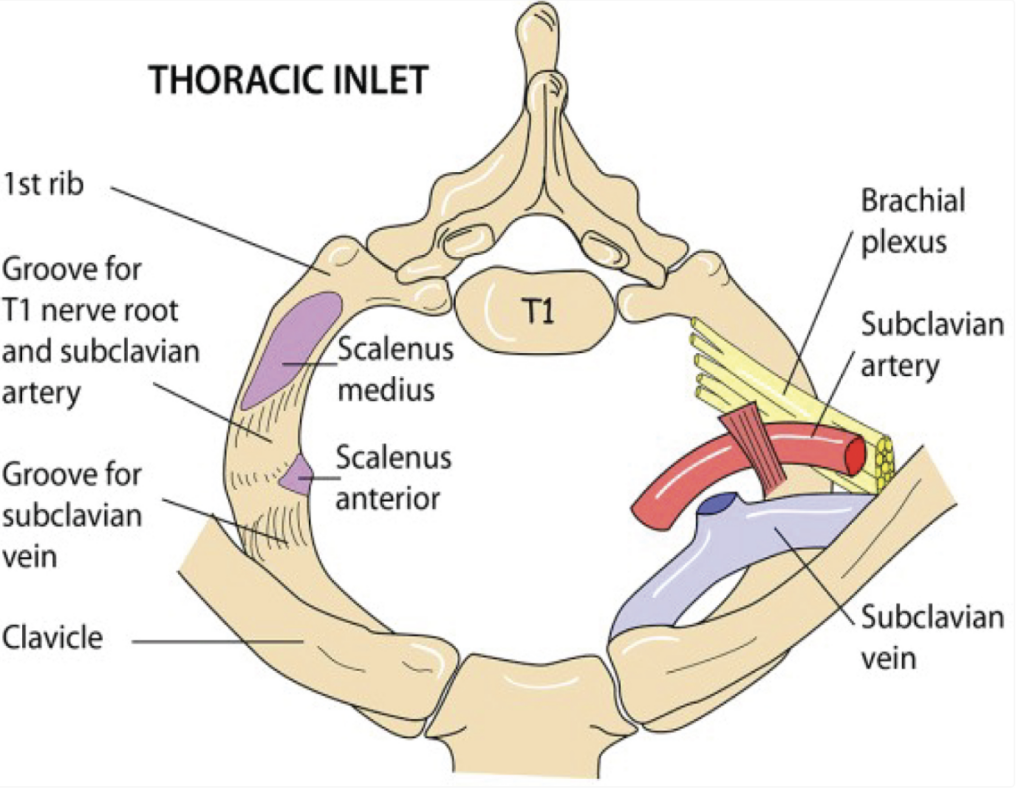
What structure bounds the anatomical thoracic inlet laterally?
- First pair of ribs
- Superior border of manubrium
- T1 vertebra
- Medial border of clavicle
Correct Answer: First pair of ribs
Explanation:
胸郭入口(thoracic inlet)の外側境界は第1肋骨です。胸骨上縁(superior border of manubrium)は前方、T1椎骨は後方の境界を形成します。鎖骨内側縁(medial border of clavicle)は胸郭入口の一部ではありません。
Superior border of manubrium:
間違いの理由: 胸骨柄(manubrium)の上縁は、胸郭入口(thoracic inlet)の前方の境界を形成しますが、胸郭入口の側方の境界ではありません。
T1 vertebra:
間違いの理由: T1椎骨は胸郭入口の後方の境界を形成しますが、側方の境界には関与していません。
Medial border of clavicle:
間違いの理由: 鎖骨の内側縁は胸郭入口の一部ではありません。側方の境界は第1肋骨によって形成されます。
Question 18:
TRUE of pulmonary plexus?
- Sympathetic innervation from T1-T8
- Parasympathetic innervation from cranial nerve XI
- Parasympathetic innervation from cranial nerve X
- Parasympathetic innervation from T1-T8
Correct Answer: Parasympathetic innervation from cranial nerve X
Explanation:
肺神経叢(pulmonary plexus)は、副交感神経系の支配を迷走神経(cranial nerve X)から受けています。交感神経はT1-T5レベルから供給されるため、T1-T8は正しくありません。他の選択肢も副交感神経の供給源について誤りがあります。
Question 19:
What thoracic wall muscle is attached from the 2nd to the 6th costal cartilages?
- Serratus posterior inferior
- Subcostalis
- Levatores costarum
- Transverse thoracis
Correct Answer: Transverse thoracis
Explanation:
胸横筋(transversus thoracis)は、第2〜6肋軟骨に付着する胸壁の筋肉です。この筋肉は胸郭の内側にあり、呼気時に胸郭を引き下げる役割を果たします。他の選択肢は異なる筋肉で、胸郭の他の部分に付着しています。
Question 20:
A 57-year-old homeless person is rushed to the ER for shortness of breath after being stabbed in the right chest. The ER physician plans to do thoracentesis(胸腔穿刺) at the 2nd ICS right parasternal line. What layers will the needle pierce?
- Skin, subcutaneous tissue, pectoralis major muscle, external intercostal aponeurosis, internal intercostal muscle, pleura
- Skin, subcutaneous tissue, external intercostal muscle, internal intercostal muscle, pleura
- Skin, subcutaneous tissue, pectoralis major muscle, internal intercostal muscle, parietal pleura, visceral pleura
- Skin, subcutaneous tissue, pectoralis major muscle, external intercostal aponeurosis, parietal pleura
Correct Answer: Skin, subcutaneous tissue, pectoralis major muscle, external intercostal aponeurosis, internal intercostal muscle, pleura
Explanation:
胸腔穿刺(thoracentesis)の際、胸壁の層を貫通する順番は、皮膚、皮下組織、大胸筋(pectoralis major muscle)、外肋間筋膜(external intercostal aponeurosis)、内肋間筋(internal intercostal muscle)、そして胸膜(pleura)です。したがって、これに合致するのは最初の選択肢です。他の選択肢は、いくつかの層が欠けているか、順序が間違っています。
Question 21:
The pleural reflection outlines the extent of the pulmonary cavities EXCEPT in which of the following?
- Sternum
- Vertebrae
- ribs
- Diaphragm
Correct Answer: ribs
Explanation:
胸膜反射(pleural reflection)は、胸膜が胸腔内で折り返し、肺の周囲を覆う位置を示します。胸膜反射線は、肺の広がりとその限界を示す重要な解剖学的ランドマークです。胸膜反射は、胸骨(sternum)、椎骨(vertebrae)、および横隔膜(diaphragm) に沿って定義されますが、肋骨そのものには特定の反射線が存在しません。
他の選択肢が正しい理由:
- Sternum(胸骨): 胸膜反射線は、前方で胸骨に沿って走ります。
- Vertebrae(椎骨): 背部では椎骨に沿って胸膜が反射しています。
- Diaphragm(横隔膜): 下方では、横隔膜の上に沿って胸膜が広がります。
したがって、「肋骨(ribs)」は胸膜反射線には直接関与していません。
Question 22:
Which of the following statements is correct regarding the thoracic skeleton?
- Includes 11 pairs of ribs
- Ribs and costal cartilages form the largest part
- Protects the thoracic viscera only
- Has 11 intervertebral discs
Correct Answer: Ribs and costal cartilages form the largest part
Explanation:
胸郭骨格(thoracic skeleton)の主要部分は肋骨(ribs)と肋軟骨(costal cartilages)から構成され、これが胸郭の大部分を形成します。肋骨は12対あり、11対ではありません。また、胸郭は胸腔内臓器のみならず、他の重要な臓器も保護しています。胸椎には12の椎間板があり、11枚ではありません。
Question 23:
The 2nd pair of cartilages articulate through which part of the sternum?
- Body of sternum
- Sternum and xiphoid process
- Manubrium only
- Manubrium and body of sternum
Correct Answer: Manubrium and body of sternum
Explanation:
第2肋軟骨は、胸骨の上部のマニュブリウム(manubrium)と胸骨体(body of sternum)との間で関節を形成します。したがって、「Manubrium and body of sternum」が正しい選択肢です。他の選択肢は適切な関節部位を示していません。
Question 24:
The vagus nerve and its branches gives rise to the general visceral efferent that innervates the smooth muscle found in the tracheobronchial tree.
- True
- False
Correct Answer: True
Explanation:
迷走神経(vagus nerve)とその枝は、気管支・気管の平滑筋に対する一般内臓運動神経(general visceral efferent)を供給します。これにより、呼吸系の筋肉を支配しています。従って、「True」が正しい選択肢です。
Question 25:
The fibrous thickening of the fascia that separates the parietal pleura from the thoracic wall and located at the part of the lungs that protrude out or RI(肋間の略) is called?
- Endothoracic fascia
- Cervical pleura
- Supracostal pleura
- Sibson’s fascia
- Supraclavicular fascia
Correct Answer: Sibson’s fascia
Explanation:
Sibson’s fascia(シブソン筋膜)は、胸膜の壁側(parietal pleura)と胸壁との間に存在する繊維状の厚化部分です。他の選択肢はこの特定の部位に関連しません。たとえば、内胸膜(endothoracic fascia)は胸壁全体に広がっており、頸部胸膜(cervical pleura)や肩鎖靭帯(supraclavicular fascia)は別の部位を示します。
胸腔の上部(胸郭出口付近)で、壁側胸膜(parietal pleura)と胸壁を隔てる筋膜の線維性肥厚部分です。この筋膜は、肺が突出する部分を覆い、胸壁と胸膜腔を保護する役割を果たしています。
Question 26:
Which of the following structures is found in both superior and posterior mediastina?
- Thoracic duct
- Phrenic nerve
- Superior vena cava
- Trachea
Correct Answer: Thoracic duct
Explanation:
胸管(thoracic duct)は、上部および後部縦隔(mediastina)の両方に存在します。他の選択肢は、それぞれ特定の縦隔部位に限られています。例えば、横隔神経(phrenic nerve)は主に上部縦隔に位置し、上大静脈(superior vena cava)や気管(trachea)はそれぞれの位置が限定されています。
Question 27:
The caval opening of the diaphragm opens at what level of vertebrae?
- 10th thoracic vertebra
- 8th thoracic vertebra
- 12th thoracic vertebra
- 6th thoracic vertebra
Correct Answer: 8th thoracic vertebra
Explanation:
大静脈孔(caval opening)は、T8レベルに位置します。他の選択肢は大静脈孔の位置とは異なります。例えば、T10は食道裂孔の位置であり、T12は大動脈裂孔の位置です。
Question 28:
What specific branch of a typical intercostal nerve innervates the skin of the lateral thoracic area?
- Anterior division of lateral cutaneous
- Medial division of anterior cutaneous
- Lateral division of anterior cutaneous
- Muscular
Correct Answer: Anterior division of lateral cutaneous
Explanation:
典型的な肋間神経(intercostal nerve)の側方皮膚領域に分布するのは、前方の側方皮膚神経(anterior division of lateral cutaneous)です。他の選択肢はこの皮膚領域を支配しません。
Question 29:
Which of the following structures are found in the superior mediastinum? (multiple choices)
- Arch of aorta
- Azygos vein
- Thoracic duct
- Subclavian artery
- Thymus
- Hemi-azygous vein
Correct Answers: Arch of aorta, Subclavian artery, Thymus
Explanation:
上部縦隔(superior mediastinum)には、大動脈弓(arch of aorta)、鎖骨下動脈(subclavian artery)、胸腺(thymus)が含まれます。奇静脈(azygos vein)や半奇静脈(hemi-azygous vein)は後部縦隔(posterior mediastinum)に存在し、胸管(thoracic duct)は後部縦隔に位置します。
| 区分 | 位置 | 内容物 |
|---|---|---|
| 上縦隔 (Superior Mediastinum) | 横隔面より上(胸骨角とT4-T5椎体の交点) | 胸腺、大血管(腕頭静脈、SVC、大動脈弓とその枝)、迷走神経、横隔神経、交感神経幹、気管、食道 |
| 下縦隔 (Inferior Mediastinum) | 横隔面と横隔膜の間 | 前縦隔、中縦隔、後縦隔で構成 |
| 前縦隔 (Anterior Mediastinum) | 胸骨と心膜の間 | 胸腺の一部、脂肪組織、リンパ節 |
| 中縦隔 (Middle Mediastinum) | 心臓およびその周辺 | 心臓、心膜、主要な血管(上大静脈、下大静脈、肺動脈、肺静脈) |
| 後縦隔 (Posterior Mediastinum) | 胸椎の前方 | 胸大動脈、胸管およびリンパ管、後縦隔リンパ節、奇静脈および半奇静脈、食道と食道神経叢 |
Question 30:
Which of the following structures enters the aortic hiatus?
- Thoracic duct
- Vagus nerves
- Accessory azygos vein
- Inferior vena cava
Correct Answer: Thoracic duct
Explanation:
大動脈裂孔(aortic hiatus)には胸管(thoracic duct)が通過します。下大静脈(inferior vena cava)は横隔膜の別の部分を通ります。迷走神経(vagus nerves)や付属奇静脈(accessory azygos vein)は別の部位に関連しています。
| 開口部 | 位置 | 通過する構造 |
|---|---|---|
| 大静脈孔 (Caval Opening) | T8-T9椎骨間の椎間板のレベル、横隔膜の腱中心に位置 | 下大静脈(IVC)、右横隔神経の末梢枝、少数のリンパ管(肝臓から中部横隔膜および縦隔リンパ節に向かう) |
| 食道裂孔 (Esophageal Hiatus) | T10椎骨のレベル、横隔膜の右脚(右クルラ)内 | 食道、前方および後方迷走神経幹、左胃動脈の食道枝、少数のリンパ管 |
| 大動脈裂孔 (Aortic Hiatus) | T12椎骨の下縁のレベル、横隔膜の後部に位置 | 大動脈、胸管、時には奇静脈および半奇静脈 |
Question 31:
The inferior thoracic aperture anterior laterally is bounded by which of the following structures?
- Xiphisternal joint
- 11th and 12th pairs of ribs
- 12th thoracic vertebra
- Costal cartilages of ribs 7 to 10
Correct Answer: Costal cartilages of ribs 7 to 10
Explanation:
下部胸郭開口部(inferior thoracic aperture)の前外側境界は第7〜10肋軟骨(costal cartilages)によって形成されています。他の選択肢はこの境界には関与しません。たとえば、第11および12肋骨は後方境界を形成します。
Question 32:
The ventral primary rami of the subcostal nerve arises from?
- T11
- T12
- T1
- T10
Correct Answer: T12
Explanation:
下肋間神経(subcostal nerve)はT12神経根から発生します。T11は第11肋間神経に対応し、T1やT10は異なる神経に関連しています。
Question 33:
Which of the following statements is TRUE?
- True ribs attach indirectly to the sternum
- Ribs 8-10 are considered as false ribs
- Floating ribs attach indirectly to the sternum by connecting their own costal cartilages to the costal cartilages of the rib immediately above it
- Ribs 1-10 are considered true ribs
Correct Answer: Ribs 8-10 are considered as false ribs
Explanation:
第8〜10肋骨は「仮性肋骨」(false ribs)と呼ばれ、胸骨に間接的に接続されています。浮遊肋骨(floating ribs)である第11および12肋骨は胸骨には接続されていません。また、真肋骨(true ribs)は第1〜7肋骨を指します。
True ribs attach indirectly to the sternum: 真肋は直接胸骨に接続するため、誤りです。
Floating ribs attach indirectly to the sternum by connecting their own costal cartilages to the costal cartilages of the rib immediately above it: 浮遊肋は胸骨に接続せず、肋軟骨も持たないため誤りです。
Ribs 1-10 are considered true ribs: 真肋は第1〜7肋骨までであり、8〜10は仮肋なので、誤りです。
Question 34:
The female mammary gland extends transversely from the lateral border of the sternum to the mid axillary line and vertically coming from what ribs?
- 3rd to 7th
- 4th to 8th
- 1st to 5th
- 2nd to 6th
Correct Answer: 2nd to 6th
Explanation:
女性の乳腺は胸骨の外側縁から腋窩中線(mid axillary line)まで、縦には第2〜6肋骨にわたって広がっています。これにより、乳腺の位置と広がりが示されています。他の選択肢は異なる肋骨範囲を示しています。
Question 35:
The scalene attaches to the scalene tubercle which is found?
- Between the grooves of the subclavian artery and vein on the 1st rib
- On the head of the first rib
- Between the grooves of the subclavian artery and vein on 2nd rib
- Along the shaft of the clavicle
Correct Answer: Between the grooves of the subclavian artery and vein on the 1st rib
Explanation:
斜角筋(scalene)は、第1肋骨の鎖骨動脈(subclavian artery)と静脈(vein)の溝の間にある斜角筋結節(scalene tubercle)に付着します。第2肋骨や鎖骨のシャフトではなく、第1肋骨のこの特定の部位が正しいです。
Question 36:
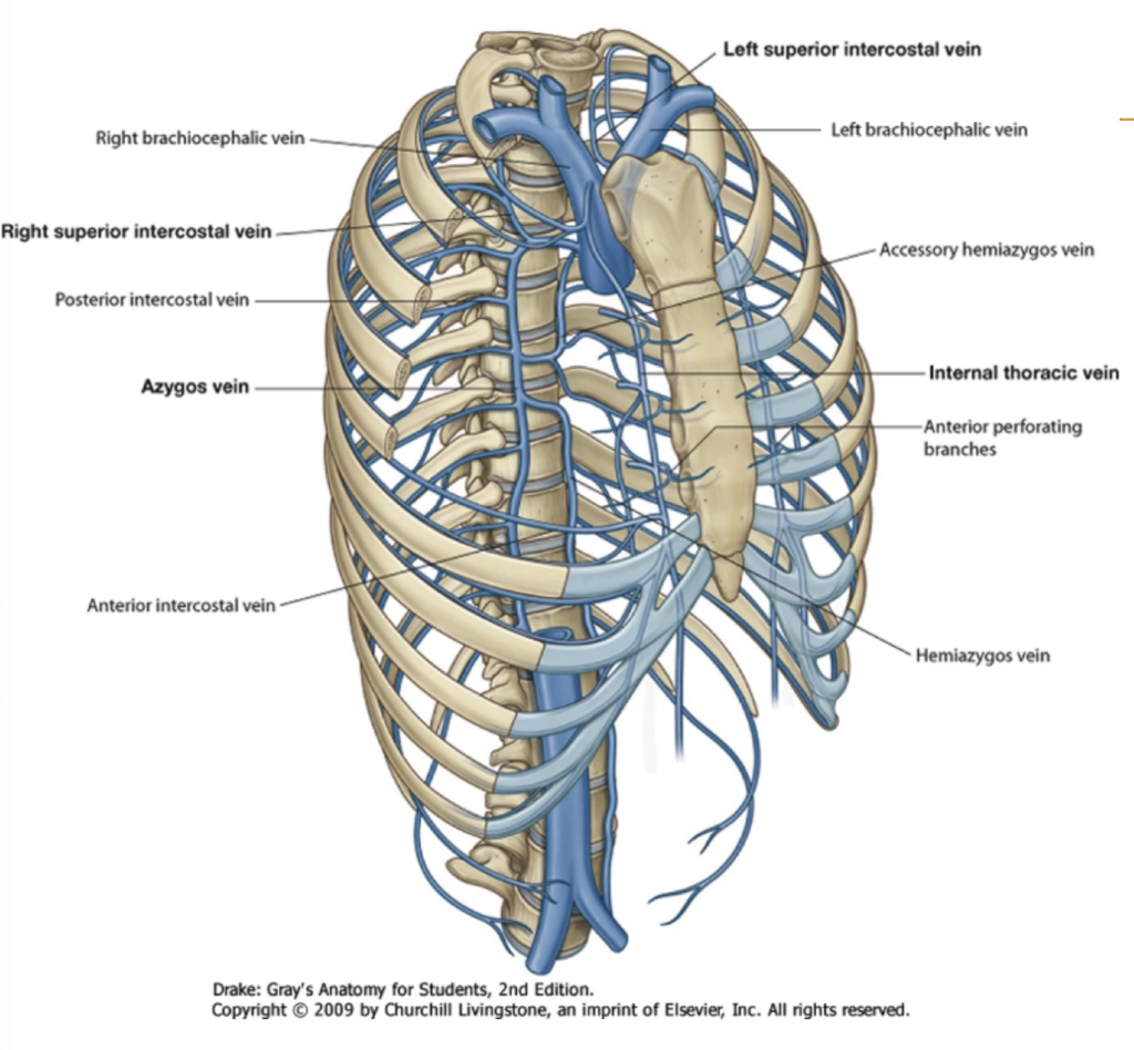
The final tributary(支流) of the right superior intercostal vein before draining to the superior vena cava is?
- Left brachiocephalic vein
- Accessory hemi-azygous vein
- Azygos vein
- Hemiazygos vein
Correct Answer: Azygos vein
Explanation:
右側の上部肋間静脈(right superior intercostal vein)は、上大静脈(superior vena cava)に注ぐ前に、奇静脈(azygos vein)に合流します。他の選択肢は異なる静脈系に関連しています。
Question 37:
Primary site of gas exchange?
- Terminal bronchioles
- Respiratory bronchioles
- Alveoli
- Alveolar duct
Correct Answer: Alveoli
Explanation:
ガス交換の主要な部位は肺胞(alveoli)です。末梢気管支(terminal bronchioles)や呼吸気管支(respiratory bronchioles)は、ガス交換を行うための通路を提供しますが、実際のガス交換は肺胞で行われます。 alveolar ductも通過点ですが、主要なガス交換部位ではありません。
Question 38:
Which of the following structures indicate the inferior limit of the central part of the thoracic cavity?
- Xiphoid process
- Xiphisternal joint
- Subcostal margin
- Infrasternal angle
Correct Answer: Xiphisternal joint
Explanation:
中央部の胸腔(thoracic cavity)の下縁は、胸骨剣状突起(xiphoid process)からの延長にある胸骨剣状関節(xiphisternal joint)によって示されます。胸骨剣状突起(xiphoid process)は胸腔の最下部を示すが、胸骨剣状関節がより具体的な指標です。肋骨下縁(subcostal margin)や肋骨下角(infrasternal angle)は、胸腔の他の部分に関連しています。
Question 39:
Which of the following is TRUE about the aortic hiatus of the diaphragm (select multiple)?
- Passage of thoracic duct
- Passage of inferior vena cava
- Passage of the aorta at the level of T12
- Level of T10
- Level of T8
- Passage of azygos vein
- Passage of esophagus
Correct Answers: Passage of thoracic duct, Passage of the aorta at the level of T12, Passage of esophagus
Explanation:
大動脈裂孔(aortic hiatus)は、T12レベルに位置し、大動脈(aorta)や食道(esophagus)が通過します。胸管(thoracic duct)もここを通ります。T10やT8のレベルは異なる裂孔に関連し、下大静脈(inferior vena cava)や奇静脈(azygos vein)は別の位置を通過します。
Question 40:
How many pairs of costal facets does the sternum have?
- 7
- 12
- 10
Correct Answer: None of the above
Explanation:
胸骨(sternum)には肋骨面(costal facets)がありません。肋骨面は通常、胸椎(thoracic vertebrae)に存在します。したがって、選択肢の中には正しい回答がありません。
Question 41:
The pulmonary artery receives blood from the
- Left ventricle
- Right ventricle
- Right atrium
- Left atrium
Correct Answer: Right ventricle
Explanation:
肺動脈(pulmonary artery)は右心室(right ventricle)から血液を受け取ります。右心房(right atrium)からは右心室に血液が送られます。左心室(left ventricle)や左心房(left atrium)は大動脈系に関連しています。
Question 42:
The line of reflection where the mediastinal pleura ends anteriorly and sharply becomes continuous with the costal pleura
- Vertebral line
- Mediastinal line
- Costal line
- Sternal line
Correct Answer: Costal line
Explanation:
中縦隔胸膜(mediastinal pleura)が前方で終わり、肋膜(costal pleura)と連続する部分は「肋骨線(costal line)」です。他の選択肢はこの線の具体的な位置を示していません。
Question 43:
The intercostobrachial nerve comes from what other main nerve?
- 1st thoracic spinal
- 2nd thoracic spinal
- 2nd intercostal
- 1st and 2nd intercostal
Answer: 2nd intercostal
解説:
肋間上腕神経 (Intercostobrachial nerve) は、主に第2肋間神経 (2nd intercostal nerve) から分岐します。この神経は胸壁の感覚を担い、腕の内側に感覚を伝える役割を持っています。乳房切除術などの手術時に、この神経が損傷されることがあります。
他の選択肢が間違っている理由:
a. 第1胸神経 (1st thoracic spinal nerve) とは直接的な関連がありません。
b. 第2胸神経 (2nd thoracic spinal nerve) という用語は厳密には使用されず、正確には第2肋間神経です。
d. 第1肋間神経は関与していません。
Question 44:
An endothoracic fascia between the 1st rib and the 7th cervical vertebra that prevents sudden intrathoracic pressure change from the neck is called
- Sternopericardial ligament
- Phrenicopleural fascia
- Pulmonary ligament
- Suprapleural fascia
Correct Answer: Suprapleural fascia
Explanation:
首部からの急激な胸腔内圧変化を防ぐのは「上胸膜筋膜(suprapleural fascia)」です。その他の選択肢は異なる機能や位置に関連しています。
Question 45:
Which of the following ribs is considered as vertebrocostal?
- 10th
- 8th
- 11th
- 7th
Correct Answer: 7th
Explanation:
「椎肋骨(vertebrocostal)」肋骨とは、胸椎(thoracic vertebrae)と肋骨(costal)に接続している肋骨を指します。第7肋骨がこれに該当します。第8、10、11肋骨は他の分類に該当します。
解説: Vertebrocostal ribs(椎肋骨)は、脊椎(椎骨)から出発し、前方では肋軟骨を介して胸骨に直接接続する肋骨を指します。これに該当するのは、第1〜7肋骨です。第7肋骨はこの範囲に含まれるため、vertebrocostal rib として分類されます。
他の選択肢について:
- 第8肋骨と第10肋骨は false ribs(仮肋)と呼ばれ、胸骨に直接接続せず、他の肋骨の軟骨を介して間接的に胸骨とつながっています。
- 第11肋骨は floating rib(浮遊肋)で、胸骨には全く接続しません。
Question 46:
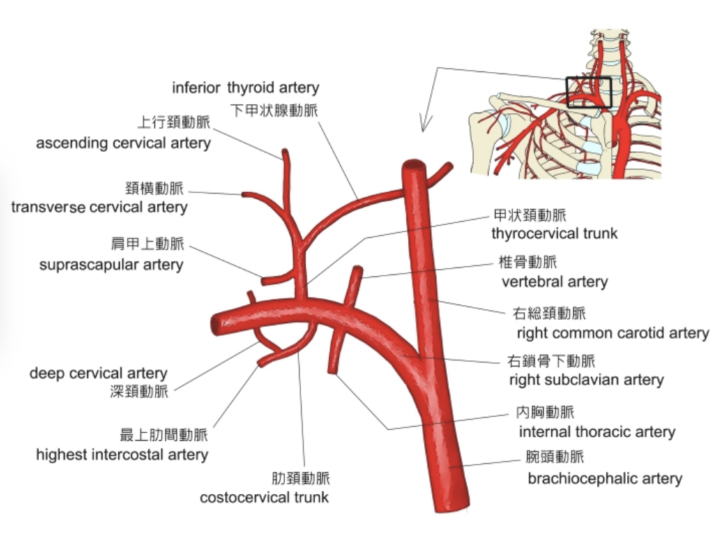
The supreme intercostal artery comes from what specific branch of the subclavian artery?
- Costocervical trunk
- Thyrocervical trunk
- Internal thoracic
- Lateral thoracic
Correct Answer: Costocervical trunk
Explanation:
最上肋間動脈(supreme intercostal artery)は、鎖骨下動脈(subclavian artery)の「頸肋動脈(costocervical trunk)」から分岐します。他の選択肢は異なる動脈です。
Question 47:
Which of these IS not part of the conducting zone?
- Trachea
- Larynx
- Respiratory bronchioles
- Bronchi
Correct Answer: Respiratory bronchioles
Explanation:
呼吸ゾーン(respiratory zone)に属するのは呼吸気管支(respiratory bronchioles)です。導入ゾーン(conducting zone)は気管(trachea)、喉頭(larynx)、気管支(bronchi)を含みます。したがって、呼吸気管支は導入ゾーンの一部ではありません。
Question 48:
What do you call a concave area at the superior border of the manubrium?
- Sternal angle of Louis
- Synchondrosis of the 1st rib
- Jugular notch
- Clavicular notch
Correct Answer: Jugular notch
Explanation:
胸骨上部の凹んだ領域は「頚静脈切痕(jugular notch)」と呼ばれます。その他の選択肢は異なる解剖学的構造に関連しています。
Question 49:
Where does the right superior intercostal vein drain directly?
- Superior vena cava
- Azygos
- Right subclavian
- Right brachiocephalic
Correct Answer: Azygos
Explanation:
右側上部肋間静脈(right superior intercostal vein)は、直接奇静脈(azygos vein)に排出されます。その他の選択肢は異なる静脈系に関連しています。
Question 50:
Which of the following structures can be found in the posterior mediastinum?
- Azygos vein
- Left common carotid artery
- Arch of aorta
- Superior vena cava
- Thoracic duct
- Esophagus
- Vagus nerve
- Thymus
Correct Answers: Azygos vein, Thoracic duct, Esophagus, Vagus nerve
Explanation:
後部縦隔(posterior mediastinum)には、奇静脈(azygos vein)、胸管(thoracic duct)、食道(esophagus)、迷走神経(vagus nerve)が含まれます。他の構造物は上部縦隔(superior mediastinum)や心臓の近くに位置しています。
ブロック
Question 1 of 200
The structure that lies at the level of the 2nd costal cartilage between T4 and T5 is?
- a) Body of sternum
- b) Xiphoid process
- c) Angle of Louis
- d) Xiphisternal joint
Correct Answer: c) Angle of Louis
Explanation:
「ルイ角(Angle of Louis)」は、第2肋骨軟骨(2nd costal cartilage)のレベルで、T4とT5の間に位置しています。他の選択肢は異なる部位に位置します。胸骨体(body of sternum)はより下部にあり、剣状突起(xiphoid process)や胸骨剣状関節(xiphisternal joint)は異なるレベルに位置しています。
Question 2 of 200
A rib that has a rounded smooth superior border and a sharp thin inferior border is?
- a) BOTH TYPICAL AND ATYPICAL RIBS
- b) TYPICAL RIBS
- c) ATYPICAL RIBS
- d) NONE
Correct Answer: c) ATYPICAL RIBS
Explanation:
「典型的な肋骨(typical ribs)」は、上部が比較的平坦で、下部は角度が緩やかです。対して、「非典型的な肋骨(atypical ribs)」、例えば第1肋骨(1st rib)や第2肋骨(2nd rib)は、上部が滑らかで丸みを帯び、下部が鋭く薄いです。
Question 3 of 200
Ribs with the following characteristics: a head, neck, tubercle, angle and shaft, are considered?
- a) TYPICAL RIBS
- b) ATYPICAL RIBS
Correct Answer: a) TYPICAL RIBS
Explanation:
「典型的な肋骨(typical ribs)」は、頭部(head)、頸部(neck)、突起(tubercle)、角度(angle)、およびシャフト(shaft)を持っています。これに対し、「非典型的な肋骨(atypical ribs)」はこれらの特徴を持たないか、異なる形状をしています。
Question 4 of 200
The condition where 3 or more ribs are fractured in 2 points is?
- a) Pneumothorax
- b) Scoliosis
- c) Pleural effusion
- d) Flail chest
Correct Answer: d) Flail chest
Explanation:
「フレイルチェスト(flail chest)」は、3本以上の肋骨が2カ所で骨折し、胸郭が不安定になる状態を指します。他の選択肢はこの症状を特徴としていません。
Question 5 of 200
The ANTERIOR INTERCOSTAL A: INTERNAL THORACIC ARTERY supplies the?
- a) Posterior 3rd-11th intercostal space
- b) Anterior 7th-10th intercostal space
- c) Anterior 1st-6th intercostal space
- d) Posterior 1st-2nd intercostal space
Correct Answer: c) Anterior 1st-6th intercostal space
Explanation:
「前肋間動脈(anterior intercostal artery)」は、内部胸部動脈(internal thoracic artery)から供給され、1〜6肋間スペース(intercostal space)に分布します。7〜10肋間スペースは、筋膜神経動脈(musculophrenic artery)から供給されます。
Question 6 of 200
The ANTERIOR INTERCOSTAL A: MUSCULOPHRENIC ARTERY supplies the?
- a) Anterior 7th-10th intercostal space
- b) Posterior 3rd-11th intercostal space
- c) Anterior 1st-6th intercostal space
- d) Posterior 1st-2nd intercostal space
Correct Answer: a) Anterior 7th-10th intercostal space
Explanation:
「筋膜神経動脈(musculophrenic artery)」は、前方の7〜10肋間スペースに血液を供給します。その他の肋間スペースへの供給は、他の動脈によって行われます。
Question 7 of 200
The POSTERIOR INTERCOSTAL A: HIGHEST INTERCOSTAL ARTERY supplies the?
- a) Posterior 1st-2nd intercostal space
- b) Posterior 3rd-11th intercostal space
- c) Anterior 1st-6th intercostal space
- d) Anterior 7th-10th intercostal space
Correct Answer: a) Posterior 1st-2nd intercostal space
Explanation:
「最上肋間動脈(highest intercostal artery)」は、1〜2肋間スペースの後方に供給します。3〜11肋間スペースの後方への供給は、他の動脈によって行われます。
Question 8 of 200
What do you call a rough, elevated area found on the upper surface of the 2nd atypical rib?
- a) Tuberosity for Serratus Anterior Muscle
- b) Scalene Medius Muscle Tubercle
- c) Costal Groove
- d) Crest of the Head
Correct Answer: b) Scalene Medius Muscle Tubercle
Explanation:
「第2非典型肋骨(2nd atypical rib)」の上面にある粗い隆起部は、「中斜角筋突起(scalene medius muscle tubercle)」です。その他の選択肢は異なる部位や構造に関連しています。
解説: 第2非典型肋骨(2nd atypical rib)の上面にある粗い隆起部は、「中斜角筋突起(Scalene Medius Muscle Tubercle)」です。中斜角筋がこの部分に付着しているため、隆起が形成されています。
他の選択肢について:
a) Serratus Anterior Muscle の結節は、第1肋骨に存在します。
c) Costal Groove(肋骨溝)は、肋骨の内側にあり、血管や神経が通る溝です。
d) Crest of the Head(肋骨頭の稜)は、肋骨の頭部にある部分で、関節面を持つ部位です。
Correct Answer: a) T3-T4
Explanation:
「胸骨上部関節(manubriosternal joint)」は、T3-T4椎間板(intervertebral disc)のレベルに位置しています。胸骨柄と胸骨体の接合部で、通常「ルイの角(Angle of Louis)」として触知可能で視認されます。T4-T5は誤りです。
Question 10 of 200
The left superior intercostal vein drains into which of the following veins?
- a) Azygous
- b) Internal Thoracic
- c) Superior Vena Cava
- d) Brachiocephalic
Correct Answer: d) Brachiocephalic
Explanation:
「左側上部肋間静脈(left superior intercostal vein)」は、「腕頭静脈(brachiocephalic vein)」に排出されます。他の静脈はこの静脈に直接排出しません。
Question 11 of 200
What do you call an empty double layer of fold found at the inferior root of the lung?
- a) Pulmonary Ligament
- b) Suprapleural Membrane
- c) Costal Pleura
- d) Hilar Pleura
Correct Answer: a) Pulmonary Ligament
Explanation:
「肺靭帯(pulmonary ligament)」は、肺の下部で見られる二重層の折り畳み部分です。その他の選択肢は異なる構造や機能を持っています。
Question 12 of 200
Which of the following pleural reflection lines is described as rounder and gradual that becomes continuous with the mediastinal pleura posteriorly?
- a) Vertebral
- b) Mediastinal
- c) Costal
- d) Sternal
Correct Answer: a) Vertebral
Explanation:
「椎骨線(vertebral line)」は、背部でより丸く、緩やかに曲がりながら縦隔胸膜(mediastinal pleura)と連続します。他の選択肢は異なる反射線に関連しています。
| 反射線の名前 | 特徴 | 位置 |
|---|---|---|
| 椎骨反射線(Vertebral line) | より丸く、緩やかにカーブし、後方で縦隔胸膜と連続する。 | 脊柱近くの縦隔胸膜と肋骨胸膜の境界。 |
| 縦隔反射線(Mediastinal line) | 前方で縦隔胸膜が肋骨胸膜と鋭く連続する。 | 前面の胸骨近く、縦隔胸膜と肋骨胸膜が交わる場所。 |
| 肋骨反射線(Costal line) | 肋骨に沿って走り、横隔膜胸膜と連続する。 | 肋骨沿い、横隔膜と胸膜の境界部分。 |
| 胸骨反射線(Sternal line) | 前面で胸骨沿いに縦隔胸膜が肋骨胸膜と鋭く連続し、左右の胸膜腔が接近している場所。 | 胸骨の前面、縦隔胸膜が肋骨胸膜に変わる部分。 |
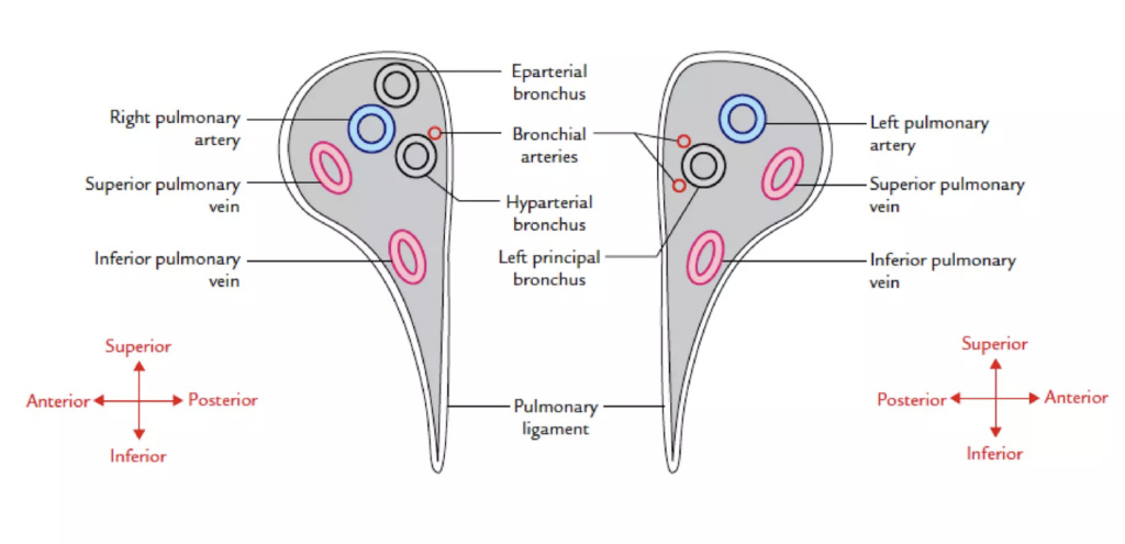
Question 13 of 200
Which of the following structures is located anterior most at the root of the lung?
- a) Inferior Pulmonary Vein
- b) Bronchus
- c) Superior Pulmonary Vein
- d) Pulmonary Artery
Correct Answer: c) Superior Pulmonary Vein
Explanation:
「肺の根部(root of the lung)」では、「上肺静脈(superior pulmonary vein)」が最も前面に位置しています。その他の構造物はより後方または中央に位置します。
Question 14 of 200
Which of the following statements is considered correct regarding the broncho-pulmonary segment?
- a) Drained by Intersegmental veins
- b) The Segmental Bronchi and Arteries are found in each segment
- c) Usually 20 – 22 in Number
- d) Surgically Resectable
Correct Answer: b) The Segmental Bronchi and Arteries are found in each segment
Explanation:
「気管支肺葉区(broncho-pulmonary segment)」は、各セグメントに気管支(segmental bronchi)と動脈(segmental arteries)が存在します。選択肢aは不正確で、気管支肺葉区は、通常20〜22セグメントではなく、通常は10セグメントであるため、選択肢cも不正確です。選択肢dは正しいですが、bの説明がより具体的です。
気管支肺葉区(broncho-pulmonary segment) は、肺の機能的および解剖学的単位であり、各セグメントには独自の**区域気管支(segmental bronchi)と動脈(arteries)**が含まれています。これにより、特定のセグメントを独立して手術で切除できる特徴を持っています。
他の選択肢が間違っている理由:
- a) Drained by Intersegmental veins(区域間静脈で排出される):
- この選択肢は正確ではありません。気管支肺葉区は**区域内静脈(segmental veins)**によって排出され、区域間静脈は隣接するセグメントを通過しないため、主に外側に向かって排出されます。
- c) Usually 20 – 22 in Number(通常20〜22個のセグメントがある):
- 気管支肺葉区の数は通常左右合わせて18〜20個です。右肺は10個、左肺は8〜10個であり、20〜22個というのは多すぎます。
- d) Surgically Resectable(外科的に切除可能):
- この選択肢も正しいですが、bの選択肢の方がより具体的で正確です。気管支肺葉区が独自の気管支と動脈を持つため、外科的に切除が可能です。
Question 15 of 200
What broncho-pulmonary segment is found in the middle lobe of the right lung?
- a) Anterior
- b) Posterior
- c) Medial
- d) Superior
Answer: c. Medial
解説:
右肺の中葉 (middle lobe) は、2つの気管支肺区 (bronchopulmonary segments) に分けられます。それは外側 (Lateral segment) と 内側 (Medial segment) です。このため、選択肢の中で中葉に存在する気管支肺区は「Medial segment」となります。
他の選択肢が間違っている理由:
a. 前方 (Anterior) は上葉に位置する気管支肺区です。
b. 後方 (Posterior) も上葉または下葉に位置します。
d. 上方 (Superior) は下葉に存在する気管支肺区で、中葉には存在しません。
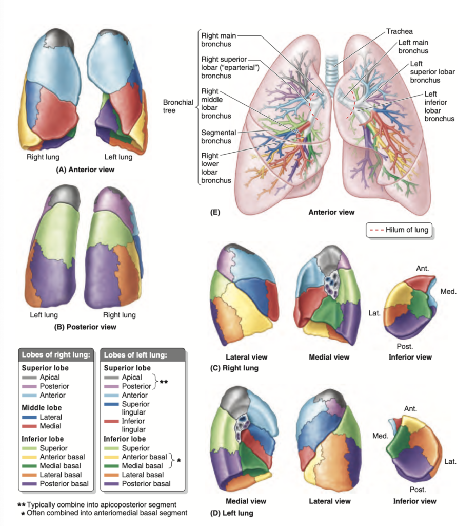
Question 16 of 200
What broncho-pulmonary segment of the left lung helps form the cardiac notch?
- a) Inferior Lingular
- b) Anterior Segmental
- c) Posterior Basal
- d) Anteromedial Basal
Correct Answer: a) Inferior Lingular
Explanation:
「左肺の心臓切痕(cardiac notch)」を形成するのは、「下部リニュラ(inferior lingular)」気管支肺葉区です。その他の選択肢は心臓切痕には関与しません。
Question 17 of 200
What broncho-pulmonary segment of the left lung is considered the largest seen from the lateral view?
- a) Apico-Posterior
- b) Anteromedial Basal
- c) Superior Lingual
- d) Anterior Segmental
Correct Answer: a) Apico-Posterior
Explanation:
「左肺の側面から見た最大の気管支肺葉区(broncho-pulmonary segment)」は、「上部背面(Apico-Posterior)」セグメントです。これは左肺の上部に位置し、大きく見えるためです。
**Apico-Posterior Segment(上後区)**は左肺の上葉に位置し、側面から見たときに最も大きく見える気管支肺葉区(broncho-pulmonary segment)です。このセグメントは上部に広がり、側面からの視野で特に目立ちます。
他の選択肢が間違っている理由:
- b) Anteromedial Basal(前内側基底区): 左肺の下葉に属し、側面から見るとあまり大きく見えません。
- c) Superior Lingual(上舌区): 左肺の上葉の一部ですが、Apico-Posteriorに比べて小さく、側面からの視野では目立ちません。
- d) Anterior Segmental(前区): これも左肺の上葉に位置しますが、Apico-Posteriorほど大きくありません。
Question 18 of 200
Which lobe of the lung receives the first branch of the pulmonary artery before it enters the hilum?
- a) Left Inferior
- b) Right Inferior
- c) Left Superior
- d) Right Middle
Correct Answer: d) Right Middle
Explanation:
「右肺(right lung)」の「中葉(middle lobe)」は、肺門(hilum)に入る前に肺動脈の最初の枝を受け取ります。他の選択肢は異なる位置または葉に関連しています。
右肺の中葉(Right Middle Lobe)は、肺門(hilum)に入る前に、肺動脈の最初の枝を受け取ります。肺動脈は、酸素を取り入れるために肺に血液を供給する主な血管であり、右中葉がその最初の供給先となります。
「右肺の中葉(Right Middle Lobe)は、肺門(hilum)に入る」という表現は、肺動脈の分枝が肺門を通って右中葉に血液を供給するという意味です。具体的に説明すると、次のような流れです:
- **肺門(Hilum)**とは:
- 肺門は、肺の中央にある領域で、ここを通って肺動脈、肺静脈、気管支、リンパ管、神経が肺に出入りします。いわば、肺に物資や血液を供給する「玄関口」のような場所です。
- **肺動脈(Pulmonary Artery)**とは:
- 心臓から送られてくる酸素が少ない血液を肺に運び、ガス交換(酸素の取り込みと二酸化炭素の排出)を行うための血管です。
- 肺門を通る肺動脈の分枝:
- 右肺において、肺動脈の枝がまず右中葉に供給され、その後に他の肺葉(右上葉、右下葉)へ血液が送られます。つまり、右中葉が最初に肺動脈の供給を受けるということです。
左肺(left lung)では、**上葉(Left Superior Lobe)**が肺動脈の最初の分枝を受け取ります。
具体的には、肺動脈は肺門(hilum)に入る前に、左肺の上葉へ最初に血液を供給する枝を分けます。このため、右肺と左肺で最初に供給される葉が異なります。右肺では中葉、左肺では上葉が最初に供給されるという違いがあります。
Question 19 of 200
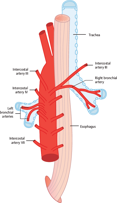
Where do the two left bronchial arteries come from?
- a) Arch of the Aorta
- b) Thoracic Aorta
- c) Posterior Intercostal Artery
- d) Left Superior Bronchial Artery
Correct Answer: b) Thoracic Aorta
Explanation:
「左側の2本の気管支動脈(left bronchial arteries)」は、「胸部大動脈(thoracic aorta)」から分枝します。選択肢aは大動脈弓(arch of the aorta)であり、選択肢cとdは正しい出発点ではありません。
- a) Arch of the Aorta は大動脈弓ですが、ここから直接気管支動脈は分枝しません。
- c) Posterior Intercostal Artery は肋間動脈であり、肺や気管支に直接的な血液供給はしていません。
- d) Left Superior Bronchial Artery は存在しますが、胸部大動脈から分岐しています。
**右側の気管支動脈(right bronchial artery)**は、左側と異なり、通常1本しか存在しません。右側の気管支動脈は、通常、以下のいずれかの血管から分枝します:
- 第三後肋間動脈(third posterior intercostal artery)
- **奇静脈(azygos vein)**に隣接する部分から直接分枝することもあります。
右側の気管支動脈は、左側と同様に気管支や肺の結合組織に血液を供給しますが、その起源は一貫しておらず、胸部大動脈から直接分枝することはまれです。
Question 20 of 200
Where does the right bronchial vein drain?
- a) Accessory Hemiazygous
- b) Azygous
- c) Hemiazygous
- d) Left Superior Intercostal
Correct Answer: b) Azygous
Explanation:
「右気管支静脈(right bronchial vein)」は、「奇静脈(azygous vein)」に排出されます。他の選択肢は異なる静脈系に関連しています。
他の選択肢について:
- a) Accessory Hemiazygous(副半奇静脈):左側の胸腔の一部の静脈を排出しますが、右気管支静脈には関与しません。
- c) Hemiazygous(半奇静脈):主に左側の胸壁からの静脈血を排出します。
- d) Left Superior Intercostal(左上肋間静脈):左側の肋間静脈からの静脈血を排出しますが、右側の気管支静脈には関係しません。
Question 21 of 200
What is the nerve innervation of the peripheral part of the diaphragmatic pleura?
- a) Intercostal
- b) Phrenic
- c) Vagus
- d) Pulmonary Plexus
Correct Answer: a) Intercostal
Explanation:
「横隔膜胸膜の周辺部(peripheral part of the diaphragmatic pleura)」の神経支配は「肋間神経(intercostal nerves)」です。他の選択肢は異なる部位に神経支配を行います。
b) Phrenic Nerve(横隔神経):横隔膜の中央部分の感覚や運動を支配しますが、横隔膜胸膜の周辺部には関与しません。
c) Vagus Nerve(迷走神経):主に内臓の感覚や運動に関与し、胸膜には直接関与しません。
d) Pulmonary Plexus(肺神経叢):主に気管支や肺に関与し、胸膜には直接関与しません。
横隔膜自体の神経支配は**横隔神経(Phrenic Nerve)**です。
横隔神経について:
- 脊髄のC3, C4, C5レベルから起こります(特にC4が主な寄与)。
- 横隔神経は運動神経として横隔膜を支配し、横隔膜の収縮と呼吸運動に関与します。また、横隔膜の感覚神経としても機能し、特に中央部分の感覚を伝えます。
Question 22 of 200
What thoracic wall muscle arises from the spinous process of C6 to T12 vertebrae?
- a) Serratus Posterior Inferior
- b) Serratus Posterior Superior
- c) Transverse Thoracis
- d) Levatores Costarum
Correct Answer: a) Serratus Posterior Inferior
Explanation:
「後下部鋸筋(Serratus Posterior Inferior)」は、C6からT12の棘突起(spinous processes)から起こります。他の選択肢は異なる起始部や機能を持っています。
- Serratus Posterior Inferior(後鋸筋下部) は、C6からT12の棘突起(spinous processes)から起こります。この筋肉は、下部肋骨に付着し、呼気を助ける役割を果たします。
- Serratus Posterior Superior(後鋸筋上部) は、上位の胸椎から起こり、吸気を助けます。
- Transverse Thoracis(胸横筋) は、胸骨の内面から起こり、肋間筋の一部として呼吸に関与します。
- Levatores Costarum(肋骨挙筋) は、胸椎の横突起から起こり、肋骨を持ち上げて吸気を助けます。
Question 23 of 200
What do you call a fibrous extension of the endothoracic fascia that attaches to the first rib and C7 vertebra?
- a) Costovertebral Pleura
- b) Suprapleural Membrane
- c) Phrenicopleural Fascia
- d) Mediastinal Pleura
Correct Answer: b) Suprapleural Membrane
Explanation:
「第一肋骨とC7椎骨に付着する内胸膜筋膜の繊維性延長(fibrous extension of the endothoracic fascia)」は「超胸膜膜(Suprapleural Membrane)」と呼ばれます。他の選択肢は異なる部位や構造を指しています。
Suprapleural Membrane(超胸膜膜) は、内胸膜筋膜(endothoracic fascia)の繊維性延長部分であり、第一肋骨とC7椎骨に付着しています。この膜は、胸腔内の圧力変動から首を保護する役割を果たします。
Costovertebral Pleura(肋骨椎骨胸膜) は、肋骨と椎骨の周りの胸膜の部分を指します。
Phrenicopleural Fascia(横隔胸膜筋膜) は、横隔膜と胸膜の間の筋膜ですが、第一肋骨やC7には関係しません。
Mediastinal Pleura(縦隔胸膜) は、縦隔周囲の胸膜部分を指し、首や第一肋骨には関与しません。
Question 24 of 200
What lung surface receives the root, which the pleura forms the pleural sleeve?
- a) Costal
- b) Pleural
- c) Mediastinal
- d) Diaphragmatic
Correct Answer: c) Mediastinal
Explanation:
「肺根(root)」を受け取る肺の表面は「縦隔面(mediastinal surface)」であり、胸膜(pleura)が「胸膜袖(pleural sleeve)」を形成します。他の選択肢は異なる部位に関連しています。
Question 26 of 200
Which of the following intercostal nerves has no anterior cutaneous and often no lateral cutaneous branch?
- a) 1st Intercostal
- b) 1st Thoracic Spinal
- c) 2nd Intercostal
- d) 7th -11th Intercostal
Correct Answer: a) 1st Intercostal
Explanation:
「1番目の肋間神経(1st Intercostal Nerve)」は、前部皮膚枝(anterior cutaneous branch)や側方皮膚枝(lateral cutaneous branch)を持たないことが多いです。その他の選択肢は異なる神経枝の分布を示しています。
1st Thoracic Spinal は脊髄神経であり、肋間神経とは異なる構造です。
2nd Intercostal は通常、前部および側方皮神経枝を持ちます。
7th -11th Intercostal も前部および側方皮神経枝を持ち、腹壁などの皮膚に分布します。


Question 27 of 200
The superior mediastinum is bounded through the sternal angle and the junction of what vertebra?
- a) T8 and T9
- b) T6 and T7
- c) T4 and T5
- d) T2 and T3
Correct Answer: c) T4 and T5
Explanation:
「上部縦隔(superior mediastinum)」は、胸骨角(sternal angle)とT4-T5の椎骨の間で区切られています。他の選択肢は異なるレベルを示しています。
Question 29 of 200
The azygous and hemiazygous veins are found in what part of the mediastinum?
- a) Posterior
- b) Superior
- c) Middle
- d) Anterior
Correct Answer: a) Posterior
Explanation:
「奇静脈(azygous vein)」と「半奇静脈(hemiazygous vein)」は「後部縦隔(posterior mediastinum)」に位置します。他の選択肢は異なる縦隔部位を示しています。
- 後縦隔(Posterior Mediastinum): 胸椎の前方に位置し、以下の構造物が含まれる。
- 胸大動脈: 大動脈の一部で、胸腔内の臓器に血液を供給。
- 胸管およびリンパ管: 体液の循環を助ける。
- 後縦隔リンパ節: 免疫系の一部。
- 奇静脈および半奇静脈: 胸壁および背部の静脈血を集める。
- 食道と食道神経叢: 食道は食物を胃に運び、食道神経叢はその運動を調節。
Question 31 of 200
What branch of a typical intercostal nerve descends along the superior margin of the lower rib and helps innervate the intercostal muscle and parietal pleura?
- a) Communicating
- b) Lateral Cutaneous
- c) Collateral
- d) Anterior Cutaneous
Correct Answer: c) Collateral
Explanation:
「典型的な肋間神経(typical intercostal nerve)」の「側副枝(collateral branch)」は、下部肋骨の上部縁に沿って下降し、肋間筋(intercostal muscles)と胸膜(parietal pleura)に神経支配を行います。他の選択肢は異なる神経枝の機能を示しています。
Communicating(連絡枝)は他の神経との接続に関与し、特定の筋肉や胸膜の支配には関与しません。
Lateral Cutaneous(側方皮神経枝)は、皮膚の感覚支配を行い、筋肉や胸膜への支配には直接関与しません。
Anterior Cutaneous(前部皮神経枝)も皮膚に感覚を与える枝で、筋肉や胸膜への支配には関与しません。
Question 32 of 200
The inferior mediastinum between the transverse thoracic plane and the diaphragm is further subdivided by which of the following structures?
- a) Heart and Root of Great Vessels
- b) Pericardium
- c) Costal Parietal Pleura
- d) Endothoracic Fascia
Correct Answer: b) Pericardium
Explanation:
「下部縦隔(inferior mediastinum)」は、横隔面(transverse thoracic plane)と横隔膜(diaphragm)の間で、「心膜(pericardium)」によってさらに区分されます。他の選択肢は、縦隔を区分するための構造ではありません。
a) Heart and Root of Great Vessels(心臓と大血管の根)は、中縦隔の内容物であり、縦隔を区分する構造ではありません。
c) Costal Parietal Pleura(肋骨側の壁側胸膜)は、胸郭の内側を覆う胸膜であり、縦隔の区分に直接関与しません。
d) Endothoracic Fascia(内胸筋膜)は、胸壁と胸膜の間にある繊維性組織で、縦隔の区分には関与しません。
Question 35 of 200
The subcostal muscle is usually developed only in the lower thoracic wall and runs in the same direction and blends with what other muscle?
- a) Levatores Costarum
- b) External Intercostal
- c) Internal Intercostal
- d) Transverse Thoracis
Correct Answer: c) Internal Intercostal
Explanation:
「肋下筋(subcostal muscle)」は、下部胸壁に発達しており、同じ方向に走り、「内肋間筋(internal intercostal)」と融合します。他の選択肢は異なる胸部筋肉群を指します。
Question 39 of 200
The part of the conducting system of the lungs where respiration can occur?
- a) Alveolar sac
- b) Respiratory bronchiole
- c) Conducting bronchiole
- d) Segmental bronchus
- e) Lobar bronchus
Correct Answer: b) Respiratory bronchiole
Explanation:
「呼吸が行われる肺の導管系の部位」は「呼吸細気管支(respiratory bronchiole)」です。これは気管支と肺胞の間に位置し、ガス交換が行われる場所です。その他の選択肢は気管支系の一部であり、ガス交換が行われる場所ではありません。
- 呼吸細気管支は、ガス交換が始まる肺の気道の最初の部分です。ここは、導管部と呼吸部の移行領域にあたります。少量の肺胞(alveoli)が存在し、限られたガス交換が可能です。
他の選択肢が不正解の理由:
- a) 肺胞嚢 (Alveolar sac): これはガス交換が主に行われる場所ですが、導管系ではなく呼吸部の一部です。
- c) 終末細気管支 (Conducting bronchiole): ここは空気の通過に関わるだけで、ガス交換は行われません。肺胞は存在しません。
- d) 区域気管支 (Segmental bronchus) および e) 葉気管支 (Lobar bronchus): これらはどちらも導管系の一部で、ガス交換は行われず、空気を呼吸部に運ぶ役割を担っています。
要するに、呼吸細気管支 (Respiratory bronchiole) は空気の導入と少量のガス交換が可能な領域です。
| 項目 (Item) | 導管部 (Conducting Zone) | 呼吸部 (Respiratory Zone) |
|---|---|---|
| 役割 (Function) | 空気を呼吸部まで運ぶ (Transports air to the respiratory zone) | ガス交換 (酸素と二酸化炭素の交換) (Gas exchange: oxygen and carbon dioxide exchange) |
| 主な構造 (Main Structures) | 鼻腔 (Nasal cavity)、咽頭 (Pharynx)、喉頭 (Larynx)、気管 (Trachea)、主気管支 (Main bronchi)、葉気管支 (Lobar bronchi)、区域気管支 (Segmental bronchi)、細気管支 (Bronchioles) | 呼吸細気管支 (Respiratory bronchioles)、肺胞管 (Alveolar ducts)、肺胞嚢 (Alveolar sacs)、肺胞 (Alveoli) |
| ガス交換 (Gas Exchange) | なし (None) | あり (Present) |
| 粘液と繊毛 (Mucus and Cilia) | 粘液と繊毛で異物を除去 (Removes foreign particles using mucus and cilia) | 基本的にはない (ガス交換を効率的に行うため) (Generally absent to facilitate efficient gas exchange) |
| 壁の構造 (Wall Structure) | 気道は軟骨で支えられ、筋肉と繊毛上皮で覆われている (Airways are supported by cartilage and lined with smooth muscle and ciliated epithelium) | 肺胞壁は非常に薄く、血管との間でガス交換が行われる (Alveolar walls are very thin for gas exchange with capillaries) |
| 気流の性質 (Airflow Characteristics) | 大部分が対流による空気の流れ (Primarily air movement by convection) | 拡散が主要な空気移動の方法 (Diffusion is the primary method of air movement) |
Question 42 of 200
Bifurcation of the trachea lies at about which of the following levels?
- a) Attachment of the 4th costal cartilage to the sternum
- b) Plane of the angle of Louis
- c) T2 vertebra
- d) Xiphisternal junction
Correct Answer: b) Plane of the angle of Louis
Explanation:
「気管の分岐(bifurcation of the trachea)」は「ルイ角(angle of Louis)」の平面である「T4-T5椎骨レベル」に位置します。その他の選択肢はこの分岐の位置と一致しません。
Question 43 of 200
When foreign objects are aspirated into the trachea, they will usually pass into the right primary bronchus because:
- a) There really is no good reason
- b) It is straighter, longer and larger than the left
- c) It is more curved, longer and smaller than the left
- d) It is larger, straighter and shorter than the left
- e) It is at a 90 degree angle to the trachea
Correct Answer: d) It is larger, straighter and shorter than the left
Explanation:
「異物が気管に吸引されたとき、右主気管支(right primary bronchus)」に入るのは、右主気管支が「左主気管支よりも大きく、まっすぐで短いため」です。他の選択肢は解剖学的事実に合致しません。
Question 44 of 200
The costodiaphragmatic recess extends inferiorly to the level of rib at the midclavicular line, and rib at the midaxillary line:
- a) 6, 10
- b) 6, 12
- c) 8, 10
- d) 10, 12
Correct Answer: c) 8, 10
Explanation:
「肋骨-横隔膜隙間(costodiaphragmatic recess)」は「鎖骨中線(midclavicular line)」で6番目の肋骨、そして「腋窩中線(midaxillary line)」で10番目の肋骨まで拡張しています。他の選択肢は正しい位置と一致しません。
**Costodiaphragmatic recess(肋骨横隔陥凹)**とは、胸腔内に存在する肺と胸壁(肋骨)および横隔膜の間にできる空間のことを指します。この陥凹は、肺が完全に膨張していないとき(特に呼気時)に明確に存在し、肺が膨張することでこのスペースは狭くなります。解剖学的に見ると、肋骨と横隔膜の間の最も下部に位置しています。
特徴:
- 位置: 肺の下端と横隔膜の間、つまり肋骨と横隔膜が交わる部分に形成される空間です。
- 機能: 通常、肺が完全に膨らんでいないときに空気を貯めることはなく、このスペースはガス交換には関与しません。しかし、深呼吸や運動時に肺が大きく膨らむと、このスペースが利用されます。
- 臨床的意義:
- **胸水(pleural effusion)**がたまる場所のひとつです。胸水とは、胸腔内に液体がたまる状態で、この陥凹に最初に液体がたまります。
- 肺がんや感染症など、さまざまな病気の診断において、この領域に異常がないかどうかが重要です。
図解的に表現すると:
- 胸腔の下部、肋骨と横隔膜に囲まれた領域に少し余剰の空間があり、それが肋骨横隔陥凹です。
Question 45 of 200
The vessel that originates from the subclavian artery and descends parasternally and participates in the intercostal anastomosis to terminate as the superior epigastric artery:
- a) Internal thoracic artery
- b) Supraclavicular artery
- c) Parasternal artery
- d) Long thoracic artery
- e) Thyrocervical artery
Correct Answer: a) Internal thoracic artery
Explanation:
「鎖骨下動脈(subclavian artery)」から起こり、「胸部壁(parasternally)」に沿って下降し、「肋間動脈吻合(intercostal anastomosis)」に関与し、「上腹壁動脈(superior epigastric artery)」として終わる血管は「内胸動脈(internal thoracic artery)」です。他の選択肢はこの動脈に該当しません。
Question 47 of 200
The body of the sternum exhibits costal facets. Which of the statements following is TRUE?
- a) Has 6 full facets
- b) Has 7 full facets
- c) Has 2 half facets and 4 full facets
- d) Has 5 full facets and 2 half facets
Correct Answer: d) Has 5 full facets and 2 half facets
Explanation:
「胸骨(body of the sternum)」は「5つの完全な肋骨面(full facets)」と「2つの半肋骨面(half facets)」を示しています。他の選択肢は正確な面の数と一致しません。
Question 48 of 200
The following are muscles of inspiration EXCEPT:
- a) Sternocleidomastoid muscle
- b) Internal intercostal muscle
- c) External intercostal muscle
- d) Scalene anterior muscle
- e) Serratus posterior superior muscle
Correct Answer: b) Internal intercostal muscle
Explanation:
「吸気筋(muscles of inspiration)」には、「胸鎖乳突筋(sternocleidomastoid)」、「外肋間筋(external intercostal)」、「前斜角筋(scalene anterior)」、「後鋸筋上部(serratus posterior superior)」が含まれますが、「内肋間筋(internal intercostal muscle)」は主に呼気に関与します。
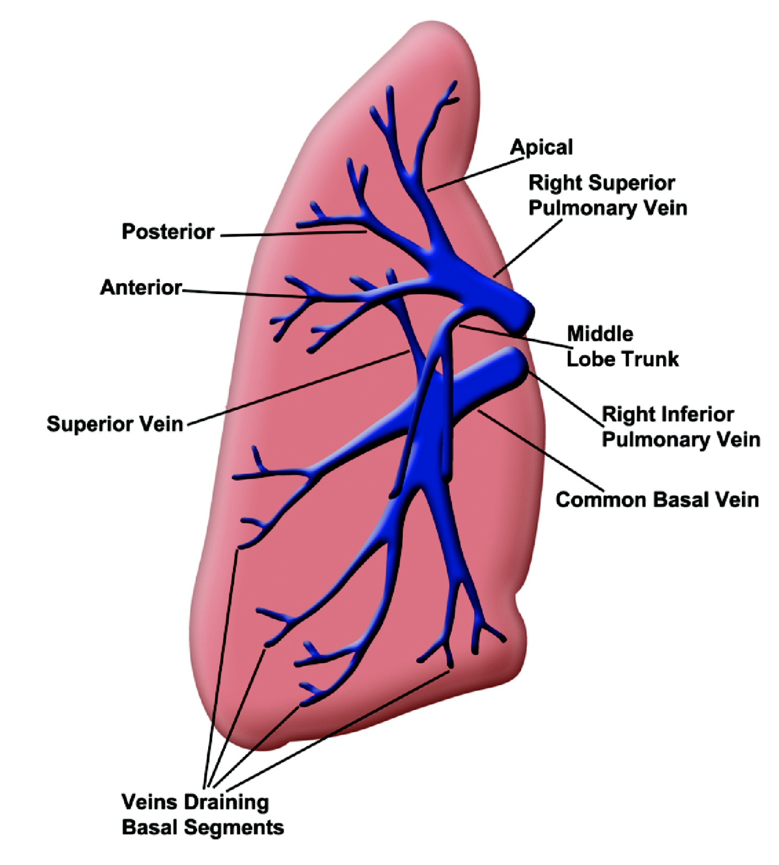
Question 50 of 200
The middle lobe vein is a tributary to which of the following structures?
- a) Directly to the left atrium
- b) Right superior pulmonary vein
- c) Right middle pulmonary vein
- d) Right inferior pulmonary vein
Correct Answer: c) Right middle pulmonary vein
Explanation:
「中葉静脈(middle lobe vein)」は「右中肺静脈(right middle pulmonary vein)」に流入します。他の選択肢は誤りです。
 |  |
| 項目 (Category) | 右肺 (Right Lung) | 左肺 (Left Lung) |
|---|---|---|
| 葉の数 (Number of lobes) | 3つの葉(上葉、中葉、下葉) 3 lobes (superior, middle, inferior) | 2つの葉(上葉、下葉) 2 lobes (superior, inferior) |
| 上肺静脈 (Superior pulmonary vein) | 右上肺静脈は上葉と中葉から血液を運ぶ Right superior pulmonary vein drains blood from the superior and middle lobes | 左上肺静脈は上葉全体から血液を運ぶ Left superior pulmonary vein drains blood from the entire superior lobe |
| 中葉の静脈 (Middle lobe vein) | 中葉静脈は右上肺静脈に流入 Middle lobe vein drains into the right superior pulmonary vein | 中葉は存在しない No middle lobe |
| 下肺静脈 (Inferior pulmonary vein) | 右下肺静脈は下葉から血液を運ぶ Right inferior pulmonary vein drains blood from the inferior lobe | 左下肺静脈は下葉から血液を運ぶ Left inferior pulmonary vein drains blood from the inferior lobe |
| 静脈の本数 (Number of veins) | 2本(右上肺静脈、右下肺静脈) 2 veins (right superior and right inferior pulmonary veins) | 2本(左上肺静脈、左下肺静脈) 2 veins (left superior and left inferior pulmonary veins) |
| 心臓への流入 (Drainage to heart) | 右上肺静脈と右下肺静脈はそれぞれ左心房へ流入 Right superior and right inferior pulmonary veins drain into the left atrium | 左上肺静脈と左下肺静脈はそれぞれ左心房へ流入 Left superior and left inferior pulmonary veins drain into the left atrium |

コメント