
Contents
- 1 教科書
- 1.1 卵巣 (Ovaries)
- 1.2 卵巣の初期発生 (Early Development of the Ovary)
- 1.3 卵胞 (Ovarian Follicles)
- 1.4 卵胞の成長と発達 (Follicular Growth & Development)
- 1.5 卵母細胞の成長 (Oocyte Growth)
- 1.6 卵胞細胞の成長 (Follicular Cell Growth)
- 1.7 透明帯 (Zona Pellucida) の形成
- 1.8 臨床応用 (Medical Application)
- 1.9 卵胞の成長 (Follicular Growth)
- 1.10 卵胞腔 (Antrum) の形成
- 1.11 卵胞液 (Follicular Fluid) の成分
- 1.12 卵丘 (Cumulus Oophorus) と放射冠 (Corona Radiata)
- 1.13 成熟卵胞 (Mature Follicle)
- 1.14 卵胞の閉鎖 (Follicular Atresia)
- 1.15 臨床応用 (Medical Application)
- 1.16 排卵とそのホルモン調節 (Ovulation & Its Hormonal Regulation)
- 1.17 減数分裂の進行 (Progression of Meiosis)
- 1.18 第二減数分裂の開始 (Onset of the Second Meiotic Division)
- 1.19 卵胞の成長とホルモン調節 (Follicular Development and Hormonal Regulation)
- 1.20 排卵時の主要な出来事 (Major Events During Ovulation)
- 1.21 卵胞の破裂 (Rupture of the Follicle)
- 1.22 黄体 (Corpus Luteum) の形成
- 1.23 黄体の構造と機能 (Structure and Function of the Corpus Luteum)
- 1.24 黄体の運命 (Fate of the Corpus Luteum)
- 1.25 白体 (Corpus Albicans) の形成
- 1.26 妊娠が起こった場合 (If Pregnancy Occurs)
- 1.27 卵管 (Uterine Tubes)
- 1.28 卵管の構造 (Structure of the Uterine Tube)
- 1.29 卵管の壁の構造 (Wall of the Uterine Tube)
- 1.30 卵管の粘膜 (Mucosa of the Uterine Tube)
- 1.31 排卵時の変化 (Changes During Ovulation)
- 1.32 卵の移動と受精 (Transport and Fertilization of the Oocyte)
- 1.33 受精の主な出来事 (Major Events of Fertilization)
- 1.34 1. 放射冠の貫通 (Penetration of the Corona Radiata)
- 1.35 2. 透明帯の通過 (Penetration of the Zona Pellucida)
- 1.36 3. 卵母細胞膜との融合 (Fusion with the Oocyte Plasmalemma)
- 1.37 4. 第二減数分裂の完了 (Completion of Meiosis II)
- 1.38 5. 雄性前核の形成 (Formation of the Male Pronucleus)
- 1.39 6. 前核の融合 (Fusion of Pronuclei)
- 1.40 胚の輸送 (Transport of the Embryo)
- 1.41 医療応用 (Medical Application)
- 1.42 子宮 (Uterus)
- 1.43 子宮壁の構造 (Structure of the Uterine Wall)
- 1.44 筋層 (Myometrium)
- 1.45 内膜 (Endometrium)
- 1.46 内膜の2つの層 (Two Layers of the Endometrium)
- 1.47 内膜への血液供給 (Blood Supply to the Endometrium)
- 1.48 月経周期 (Menstrual Cycle)
- 1.49 医療応用 (Medical Application)
- 1.50 月経周期の期間 (Duration of the Menstrual Cycle)
- 1.51 月経の開始 (Start of Menstruation)
- 1.52 月経周期の3つの主要な段階 (Three Major Phases of the Menstrual Cycle)
- 1.53 増殖期 (Proliferative Phase)
- 1.54 分泌期 (Secretory Phase)
- 1.55 受精が起こった場合 (If Fertilization Occurs)
- 1.56 月経期 (Menstrual Phase)
- 1.57 細胞の変化 (Cellular Changes)
- 1.58 機能層と基底層の変化 (Changes in the Functional and Basal Layers)
- 1.59 月経期の終わり (End of the Menstrual Phase)
- 1.60 胚の着床、脱落膜、および胎盤 (Embryonic Implantation, Decidua, & The Placenta)
- 1.61 胚盤胞の形成 (Formation of the Blastocyst)
- 1.62 着床 (Implantation)
- 1.63 栄養膜の分化 (Differentiation of the Trophoblast)
- 1.64 脱落膜 (Decidua)
- 1.65 脱落膜の領域 (Regions of the Decidua)
- 1.66 医療応用 (Medical Application)
- 1.67 胎盤の機能 (Function of the Placenta)
- 1.68 胎盤の発達 (Development of the Placenta)
- 1.69 胎盤の構造 (Structure of the Placenta)
- 1.70 物質交換の仕組み (Mechanism of Exchange)
- 1.71 胎盤の内分泌機能 (Endocrine Function of the Placenta)
- 1.72 子宮頸部 (Cervix)
- 1.73 子宮頸部の内腔 (Endocervical Mucosa)
- 1.74 子宮頸管外部 (Exocervical Mucosa)
- 1.75 扁平円柱移行帯 (Transformation Zone)
- 1.76 子宮頸部の粘液の変化 (Changes in Cervical Mucus)
- 1.77 子宮頸部の壁 (Wall of the Cervix)
- 1.78 子宮頸部の役割 (Role of the Cervix)
- 1.79 医療応用 (Medical Application)
- 1.80 膣 (Vagina)
- 1.81 膣の粘膜 (Mucosa of the Vagina)
- 1.82 膣の粘液 (Mucus in the Vagina)
- 1.83 膣の筋層 (Muscular Layer of the Vagina)
- 1.84 膣の外膜 (Adventitia of the Vagina)
- 1.85 医療応用 (Medical Application)
- 1.86 女性の外陰部 (Female External Genitalia, Vulva)
- 1.87 乳房の乳腺 (Mammary Glands of the Breasts)
- 1.88 乳腺の構造 (Structure of the Mammary Glands)
- 1.89 思春期の乳房の発達 (Breast Development During Puberty)
- 1.90 成人女性の乳腺 (Mammary Glands in Adult Women)
- 1.91 乳頭と乳輪 (Nipple and Areola)
- 1.92 乳頭 (Nipple)
- 1.93 妊娠および授乳中の乳房 (Breasts During Pregnancy & Lactation)
- 1.94 妊娠中の乳腺の変化 (Changes in the Mammary Glands During Pregnancy)
- 1.95 初乳 (Colostrum) の産生 (Production of Colostrum)
- 1.96 分娩後の授乳 (Lactation After Parturition)
- 1.97 乳腺の分泌活動 (Secretory Activities of the Mammary Glands)
- 1.98 医療応用 (Medical Application)
- 1.99 授乳後の乳腺の退行 (Postlactational Regression in the Mammary Glands)
- 1.100 医療応用 (Medical Application)
- 2 サマリー
- 3 教科書問題
- 4 BRS
- 4.1 二次卵胞の特徴(Secondary Ovarian Follicles)
- 4.2 母乳(初乳)の抗体(Antibodies in Colostrum)
- 4.3 黄体の変化(Corpus Luteum)
- 4.4 月経周期の相(Phases of Menstrual Cycle)
- 4.5 膣粘膜の特徴(Vaginal Mucosa)
- 4.6 卵管の特徴(Characteristics of the Oviduct)
- 4.7 未熟奇形腫(Immature Teratoma)
- 4.8 黄体形成ホルモン(LH)の作用(Function of Luteinizing Hormone, LH)
- 4.9 子宮内膜の基底層(Basal Layer of Endometrium)
- 4.10 月経周期の増殖期(Proliferative Phase of Menstrual Cycle)
- 5 重要箇所穴埋め問題
教科書
女性の生殖器系 (Female Reproductive System) は、一対の卵巣 (Ovaries) と卵管 (Oviducts または Uterine Tubes)、子宮 (Uterus)、膣 (Vagina)、および外陰部 (External Genitalia) で構成されています(図22–1参照)。このシステムは、女性の配偶子 (Gametes, 卵母細胞 Oocytes) を産生し、受精 (Fertilization) のための環境を提供し、胚 (Embryo) を胎児期 (Fetal Stage) を経て出産 (Birth) まで完全に発育させる機能を持っています。男性の性腺 (Male Gonads) と同様に、卵巣 (Ovaries) は、ステロイド性性ホルモン (Steroidal Sex Hormones) を産生し、生殖器系 (Reproductive System) の器官を制御し、他の器官にも影響を与えます。
初経 (Menarche)、すなわち最初の月経 (First Menses) が発生すると、神経ホルモン機構 (Neurohormonal Mechanisms) によって制御される月経周期 (Monthly Changes) に合わせて、生殖器系は構造 (Structure) と機能 (Function) に変化が生じます。閉経 (Menopause) は、周期的変化 (Cyclic Changes) が不規則になり、最終的には消失する期間であり、閉経後 (Postmenopausal Period) には、生殖器官は徐々に退縮 (Involution) します。乳腺 (Mammary Glands) は、生殖器系には含まれませんが、その機能的状態と密接に関係する変化を受けるため、ここに含まれます。
卵巣 (Ovaries)
卵巣 (Ovaries) はアーモンド (Almond)-形の器官で、長さ約3cm、幅1.5cm、厚さ1cm です。それぞれの卵巣は、単層立方上皮 (Simple Cuboidal Epithelium) に覆われており、この表面上皮 (Surface Epithelium または Germinal Epithelium) は中皮 (Mesothelium) に連続しています。その下には、密性結合組織の被膜 (Dense Connective Tissue Capsule)、すなわち白膜 (Tunica Albuginea) が存在し、これは精巣 (Testis) に見られるものと類似しています。
卵巣の大部分は、皮質 (Cortex) で構成されており、高度に細胞化された結合組織の間質 (Highly Cellular Connective Tissue Stroma) と、卵胞 (Ovarian Follicles) が多数存在しています。卵胞の大きさは、初経 (Menarche) 以降、大きく変化します(図22–1参照)。卵巣の最内部 (Innermost Part) は、髄質 (Medulla) であり、疎性結合組織 (Loose Connective Tissue) と、血管 (Blood Vessels) から構成されています。これらの血管は、間膜 (Mesenteries) を通して卵巣に入り、門 (Hilum) から進入します(図22–1および図22–2参照)。卵巣皮質 (Ovarian Cortex) と卵巣髄質 (Ovarian Medulla) の間には明確な境界 (Distinct Border) はありません。
卵巣の初期発生 (Early Development of the Ovary)
胚発生 (Embryonic Development) の第1月 (First Month) に、原始生殖細胞 (Primordial Germ Cells) は、卵黄嚢 (Yolk Sac) から生殖腺原基 (Gonadal Primordia) へと移動 (Migration) します。卵巣が発生する際、これらの原始生殖細胞は同期した有糸分裂 (Synchronized Mitotic Divisions) を行い、不完全な細胞質分裂 (Incomplete Cytokinesis) を伴って、数百万の卵母細胞 (Oogonia) を生成します。ヒト (Humans) では、妊娠11〜12週 (11-12 Weeks Gestation) に有糸分裂 (Mitotic Activity) が停止します。
その後、クラスター化 (Clustered) された卵母細胞の一部はアポトーシス (Apoptosis, プログラム細胞死) によって死滅しますが、他の細胞は第一減数分裂 (First Meiotic Division) の前期 (Prophase) に入り、接合 (Synapsis) や遺伝的組換え (Genetic Recombination) を完了し、さらに進行せず減数分裂の停止 (Meiotic Arrest) に入ります。この段階の細胞は一次卵母細胞 (Primary Oocyte) と呼ばれ、これに扁平な支持細胞 (Flattened Support Cells) が取り巻き、非成長性の卵胞 (Non-Growing Follicle) を形成します。
出生時には、約68万個 (680,000) の卵胞 (Follicles) があり、これを**「卵巣予備能 (Ovarian Reserve)」** と呼びますが、思春期 (Puberty) までに約46万個 (460,000) が残ります。残りは閉経 (Menopause) までに萎縮 (Atresia, 退化) して失われます。月経周期 (Menstrual Cycle) ごとに1つの卵母細胞 (Oocyte) が排卵 (Ovulation) によって放出されるため、生殖年齢 (Reproductive Life) 約30-35年 の間に排出される卵母細胞は約450個 だけです。他のすべての卵母細胞は萎縮 (Atresia) によって消失します。
卵胞 (Ovarian Follicles)
卵胞 (Ovarian Follicle) とは、卵母細胞 (Oocyte) が1層以上の上皮細胞 (Epithelial Cells) に囲まれ、基底膜 (Basal Lamina) 内に存在する構造を指します。
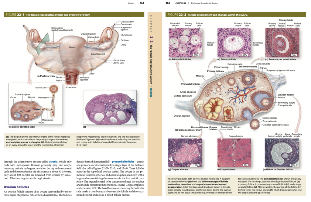
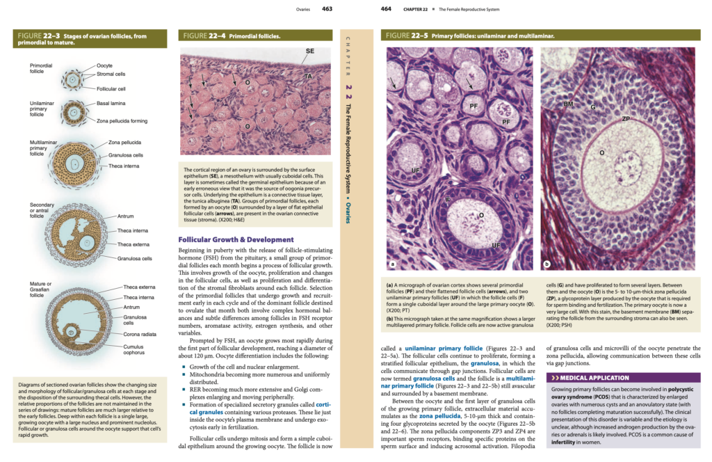
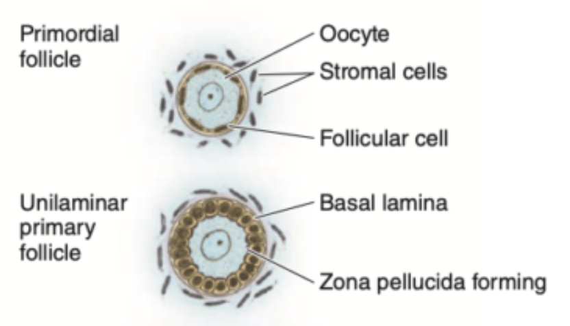
胎児期 (Fetal Life) に形成される卵胞は、原始卵胞 (Primordial Follicles) と呼ばれ、一次卵母細胞 (Primary Oocyte) が1層の扁平な卵胞細胞 (Flattened Follicular Cells) に包まれています(図22–2b、図22–3、図22–4参照)。これらの卵胞は、卵巣皮質 (Superficial Ovarian Cortex) に存在します。原始卵胞内の卵母細胞 (Oocyte in Primordial Follicle) は直径約25μmの球形 (Spherical Shape) で、第一減数分裂の前期 (First Meiotic Prophase) に染色体 (Chromosomes) を含む大きな核 (Large Nucleus) を有しています。
基底膜 (Basal Lamina) は、卵胞 (Follicle) と血管化された間質 (Vascularized Stroma) の間に明確な境界 (Clear Boundary) を形成し、血液-卵胞バリア (Blood-Follicle Barrier) の役割を果たしています。
卵胞の成長と発達 (Follicular Growth & Development)
思春期 (Puberty) が始まると、下垂体 (Pituitary) から卵胞刺激ホルモン (FSH, Follicle-Stimulating Hormone) が分泌され、毎月少数の原始卵胞 (Primordial Follicles) が卵胞成長 (Follicular Growth) のプロセスを開始します。この過程には、
卵母細胞 (Oocyte) の成長、
卵胞細胞 (Follicular Cells) の増殖 (Proliferation) と変化、
各卵胞を取り囲む間質の線維芽細胞 (Stromal Fibroblasts) の増殖 (Proliferation) および
分化 (Differentiation)
が含まれます。
各月経周期の早期 (Early Stage of Cycle) に成長 (Growth) を開始する原始卵胞の選択と、その月に排卵 (Ovulation) される優勢卵胞 (Dominant Follicle) の選択は、ホルモンバランス (Hormonal Balances) や卵胞間の微妙な違い (Subtle Differences Among Follicles) に依存しています。これには、FSH受容体の数 (FSH Receptor Numbers)、アロマターゼ活性 (Aromatase Activity)、エストロゲン合成 (Estrogen Synthesis) などの要素が関与しています。
卵母細胞の成長 (Oocyte Growth)
FSH (卵胞刺激ホルモン) によって刺激され、卵母細胞 (Oocyte) は、卵胞発達の最初の段階で急速に成長し、直径約120μm に達します。卵母細胞の分化 (Differentiation) には、以下のプロセスが含まれます。
- 細胞の成長 (Growth of the Cell) と核の肥大 (Nuclear Enlargement)
- ミトコンドリア (Mitochondria) が増加 (More Numerous) し、均等に分布 (Uniformly Distributed) する
- 粗面小胞体 (RER, Rough Endoplasmic Reticulum) が拡大 (More Extensive) し、ゴルジ体 (Golgi Complexes) が肥大 (Enlarging) し、細胞の周辺部へ移動 (Moving Peripherally)
- 特殊な分泌顆粒 (Specialized Secretory Granules) の形成。これらは皮質顆粒 (Cortical Granules) と呼ばれ、プロテアーゼ (Proteases) を含んでいます。これらの顆粒は、卵母細胞の細胞膜 (Oocyte’s Plasma Membrane) のすぐ内側に位置し、受精 (Fertilization) の初期段階でエキソサイトーシス (Exocytosis) を起こします。
卵胞細胞の成長 (Follicular Cell Growth)
卵胞細胞 (Follicular Cells) は有糸分裂 (Mitosis) を行い、成長する卵母細胞 (Growing Oocyte) を取り囲む単層立方上皮 (Simple Cuboidal Epithelium) を形成します。この時点で、一層性一次卵胞 (Unilaminar Primary Follicle) と呼ばれます(図22–3および図22–5a参照)。
卵胞細胞はさらに増殖 (Proliferation) を続け、多層性卵胞上皮 (Stratified Follicular Epithelium) を形成します。これを顆粒層 (Granulosa) と呼び、細胞間連絡 (Gap Junctions) を通じて相互に通信 (Communication) します。
この段階の卵胞細胞は顆粒膜細胞 (Granulosa Cells) と呼ばれ、卵胞は多層性一次卵胞 (Multilaminar Primary Follicle) として知られています(図22–3および図22–5b参照)。この時点でも、卵胞は無血管 (Avascular) であり、基底膜 (Basement Membrane) に囲まれています。
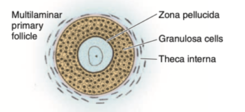
透明帯 (Zona Pellucida) の形成
成長する一次卵胞 (Growing Primary Follicle) の卵母細胞 (Oocyte) と最初の顆粒膜細胞の層 (First Layer of Granulosa Cells) の間に、細胞外マトリックス (Extracellular Material) が蓄積し、透明帯 (Zona Pellucida) が形成されます。透明帯の厚さは5~10μm で、卵母細胞 (Oocyte) によって分泌される4つの糖タンパク質 (Glycoproteins) が含まれています(図22–5bおよび図22–6参照)。

透明帯 (Zona Pellucida) の重要な構成要素は、ZP3 およびZP4 であり、これらは精子受容体 (Sperm Receptors) として機能します。精子 (Sperm) の表面にある特定のタンパク質 (Proteins) に結合し、先体反応 (Acrosomal Activation) を誘発します。顆粒膜細胞のフィロポディア (Filopodia of Granulosa Cells) と卵母細胞の微絨毛 (Microvilli of the Oocyte) は、透明帯 (Zona Pellucida) に入り込み、ギャップ結合 (Gap Junctions) を介して通信 (Communication) を行います。
臨床応用 (Medical Application)
成長する一次卵胞 (Growing Primary Follicles) は、多嚢胞性卵巣症候群 (PCOS, Polycystic Ovary Syndrome) に関与する可能性があります。PCOSは、卵巣の肥大 (Enlarged Ovaries) と多数の嚢胞 (Numerous Cysts) を伴う無排卵状態 (Anovulatory State)(すなわち、卵胞が成熟を完了しない (No Follicles Completing Maturation Successfully) 状態)を特徴としています。
PCOS (多嚢胞性卵巣症候群) の臨床症状 (Clinical Presentation) は変動性 (Variable) であり、その病因 (Etiology) は明確ではありません (Unclear)。ただし、卵巣 (Ovaries) または副腎 (Adrenals) によるアンドロゲン産生の増加 (Increased Androgen Production) が関与している可能性が高いと考えられています。
PCOS (多嚢胞性卵巣症候群) は、女性の不妊 (Infertility in Women) の一般的な原因 (Common Cause) の1つです。
卵胞の成長 (Follicular Growth)
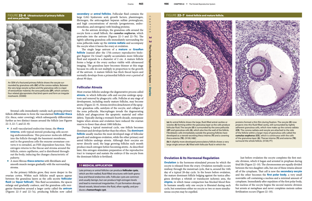
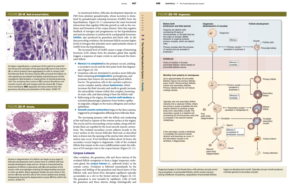
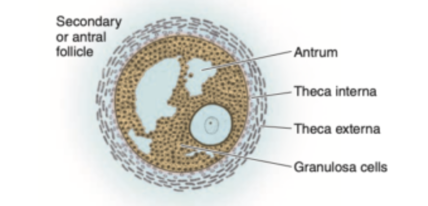
成長する一次卵胞 (Growing Primary Follicle) のすぐ外側にある間質細胞 (Stromal Cells) は分化 (Differentiate) し、血管化された卵胞の鞘 (Vascularized Follicular Theca, ギリシャ語のThecaは「外被」) を形成します。その後、卵胞の周囲に2つの異なる組織 (Two Distinct Tissues) が分化します(図22–3、図22–7、図22–8参照)。
- 内卵胞鞘 (Theca Interna)
- 血管が豊富な内分泌組織 (Well-Vascularized Endocrine Tissue) で、アンドロステンジオン (Androstenedione) を分泌する典型的なステロイド産生細胞 (Typical Steroid-Producing Cells) から構成されます。
- アンドロステンジオン (Androstenedione) は基底膜 (Basement Membrane) を通過して卵胞 (Follicle) 内に拡散し、顆粒膜細胞 (Granulosa Cells) によってアロマターゼ (Aromatase) によりエストラジオール (Estradiol) に変換されます。このプロセスはFSH (卵胞刺激ホルモン) に依存します。
- 生成されたエストロゲン (Estrogen) は、卵胞周囲の鞘 (Thecae) や間質 (Stroma) に戻り、毛細血管 (Capillaries) に入って体内に分布します。これにより、思春期 (Puberty) に特徴的な変化が誘発されます。
- 外卵胞鞘 (Theca Externa)
- 線維性の多い組織 (More Fibrous Tissue) で、線維芽細胞 (Fibroblasts) と平滑筋 (Smooth Muscle) が含まれています。
- 周囲の間質 (Surrounding Stroma) と徐々に融合します。
卵胞腔 (Antrum) の形成
一次卵胞 (Primary Follicles) が成長すると、卵巣皮質 (Ovarian Cortex) の深部 (Deeper Region) へと移動します。これらの卵胞内では、顆粒膜細胞 (Granulosa Cells) が卵胞液 (Follicular Fluid, 別名リクール・フォリクリ (Liquor Folliculi)) を分泌し、顆粒膜層 (Granulosa Layers) の間に小さな空間 (Small Spaces) が現れます。これらの空間は拡大 (Enlarge) し、最終的に結合 (Coalesce) して、大きな腔 (Larger Cavity)、すなわち卵胞腔 (Antrum) を形成します(図22–3、図22–7a参照)。この段階の卵胞は二次卵胞 (Secondary Follicles) または胞状卵胞 (Antral Follicles) と呼ばれます。
卵胞液 (Follicular Fluid) の成分
卵胞液 (Follicular Fluid) には、以下の成分が含まれています。
- ヒアルロン酸 (Hyaluronic Acid, GAG)
- 成長因子 (Growth Factors)
- プラスミノーゲン (Plasminogen)
- フィブリノーゲン (Fibrinogen)
- 抗凝固性のヘパラン硫酸プロテオグリカン (Heparan Sulfate Proteoglycan)
- 高濃度のステロイド (High Concentrations of Steroids)(
プロゲステロン (Progesterone)、
アンドロステンジオン (Androstenedione)、
エストロゲン (Estrogens))および
結合タンパク質 (Binding Proteins)
卵丘 (Cumulus Oophorus) と放射冠 (Corona Radiata)
卵胞腔 (Antrum) が発達すると、卵母細胞 (Oocyte) の周囲に顆粒膜細胞 (Granulosa Cells) が集まり、卵丘 (Cumulus Oophorus) と呼ばれる小さな丘 (Small Hillock) を形成します。この卵丘 (Cumulus Oophorus) は卵胞腔 (Antrum) に突き出ています(図22–3、図22–7b参照)。
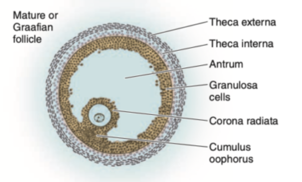
放射冠 (Corona Radiata) は、透明帯 (Zona Pellucida) のすぐ周囲にある密着した顆粒膜細胞 (Tightly Adhering Granulosa Cells) から構成されます。排卵 (Ovulation) の際には、放射冠 (Corona Radiata) は卵母細胞 (Oocyte) とともに卵巣から放出されます。
成熟卵胞 (Mature Follicle)
成熟卵胞 (Mature Follicle) またはグラーフ卵胞 (Graafian Follicle) は、17世紀の生殖生物学者 (17th-Century Reproductive Biologist) であるRegnier De Graaf にちなんで名付けられました。成熟卵胞 (Mature Follicle) の単一の大きな卵胞腔 (Single Large Antrum) は卵胞液 (Follicular Fluid) を急速に蓄積し、直径約2cm に拡大します。
この段階では、超音波画像 (Ultrasound Imaging) で確認できるように、卵巣の表面 (Ovary Surface) に膨らみ (Bulge) が現れます。顆粒膜層 (Granulosa Layer) は、卵胞の成長に対して顆粒膜細胞が増殖しないため (Do Not Multiply in Proportion to the Growth of the Antrum)、薄くなります (Becomes Thinner)。成熟卵胞 (Mature Follicle) は厚い卵胞鞘 (Thick Thecal Layers) を持ち、通常は約90日間 (About 90 Days) かけて原始卵胞 (Primordial Follicle) から発達します。
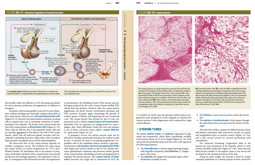
卵胞の閉鎖 (Follicular Atresia)
卵巣卵胞 (Ovarian Follicles) の大部分は、萎縮 (Atresia) と呼ばれる退化過程 (Degenerative Process) を経ます。萎縮 (Atresia) においては、卵胞細胞 (Follicular Cells) および卵母細胞 (Oocyte) がアポトーシス (Apoptosis) を起こし、貪食細胞 (Phagocytic Cells) によって除去 (Removal) されます。
アポトーシス (Apoptosis) に関する主要な出来事:
- 顆粒膜細胞 (Granulosa Cells) の剥離 (Detachment)
- 卵母細胞 (Oocyte) の自己融解 (Autolysis)
- 透明帯 (Zona Pellucida) の崩壊 (Collapse)
- マクロファージ (Macrophages) の侵入 (Invasion) と貪食 (Phagocytosis)
臨床応用 (Medical Application)
後期の一次卵胞 (Late Primary Follicles) や胞状卵胞 (Antral Follicles) では、卵胞嚢胞 (Follicular Cysts) が形成されることがあります。これらは薄い壁 (Thin-Walled) を持つ液体が充填された構造 (Fluid-Filled Structures) で、顆粒膜細胞 (Granulosa Cells) および内卵胞鞘の内分泌細胞 (Thecal Endocrine Cells) が含まれています。卵胞嚢胞 (Follicular Cysts) は一般的 (Common) で良性 (Benign) ですが、高エストロゲン (High Estrogen Levels) を引き起こす場合があります。
排卵とそのホルモン調節 (Ovulation & Its Hormonal Regulation)
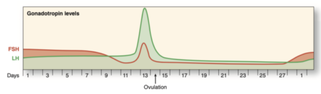
排卵 (Ovulation) は、ホルモンによって刺激 (Hormone-Stimulated Process) されるプロセスで、卵母細胞 (Oocyte) が卵巣 (Ovary) から放出される現象です。排卵 (Ovulation) は、通常、月経周期 (Menstrual Cycle) の中間、すなわち28日周期 (Typical 28-Day Cycle) では14日目 (Around the 14th Day) に発生します。
排卵の数時間前 (Hours Before Ovulation)、成熟した優勢卵胞 (Mature Dominant Follicle) が白膜 (Tunica Albuginea) に突き出し、白色または半透明の虚血性領域 (Whitish or Translucent Ischemic Area) が形成されます。この領域はスティグマ (Stigma) と呼ばれ、組織の圧縮 (Tissue Compaction) により血流が遮断 (Blocked Blood Flow) されます。
ヒトでは、通常1つの卵母細胞 (One Oocyte) が各周期 (Each Cycle) に放出 (Liberated) されますが、時には卵母細胞が放出されない (No Oocyte) こともあれば、同時に2つ以上の卵母細胞 (Two or More Simultaneous Oocytes) が放出されることもあります。
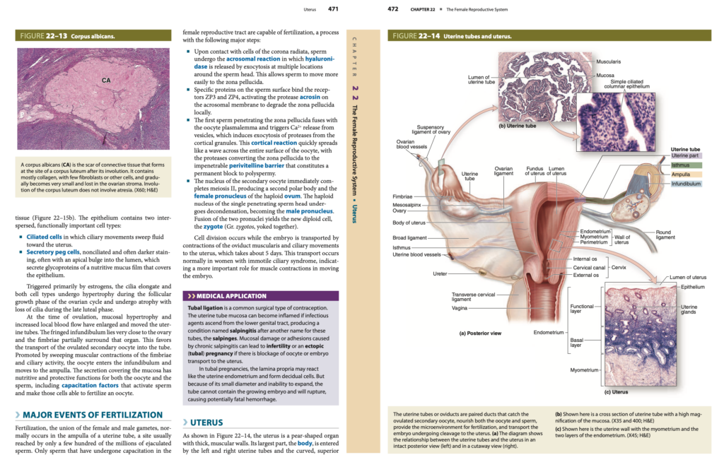
減数分裂の進行 (Progression of Meiosis)
排卵直前 (Just Before Ovulation)、卵母細胞 (Oocyte) は、胎児期 (Fetal Life) に前期 (Prophase) で開始 (Begun) し、その後停止 (Arrested) していた第一減数分裂 (First Meiotic Division) を完了 (Completes) します(図22–10参照)。
- 染色体 (Chromosomes) は、2つの娘細胞 (Two Daughter Cells) に等しく分配 (Equally Divided) されますが、そのうちの1つの細胞 (One of These Cells) はほとんどすべての細胞質 (Almost All of the Cytoplasm) を保持します。
- その細胞が二次卵母細胞 (Secondary Oocyte) となり、もう1つの細胞 (The Other Cell) は第一極体 (First Polar Body) となります。
- 第一極体 (First Polar Body) は、核 (Nucleus) と最小限の細胞質 (Minimal Amount of Cytoplasm) を含む非常に小さい生存不能な細胞 (Very Small Nonviable Cell) です。
第二減数分裂の開始 (Onset of the Second Meiotic Division)
第一極体の放出直後 (Immediately After Expulsion of the First Polar Body)、卵母細胞 (Oocyte) の核 (Nucleus) は第二減数分裂 (Second Meiotic Division) を開始します。
- しかし、中期 (Metaphase) で停止 (Arrests) し、受精 (Fertilization) が起こらない限り (Unless Fertilization Occurs)、減数分裂は完了しません (Never Completes Meiosis)(図22–10参照)。
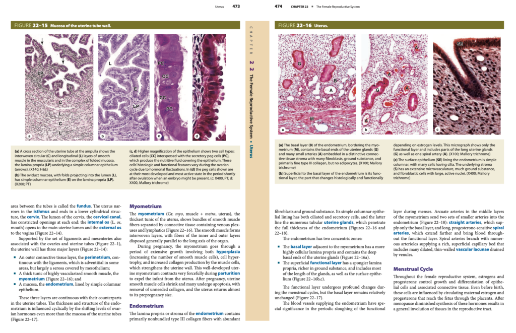
卵胞の成長とホルモン調節 (Follicular Development and Hormonal Regulation)
これまでに述べたように、卵胞の発達 (Follicular Development) は、下垂体のゴナドトロープ (Pituitary Gonadotrophs) からの卵胞刺激ホルモン (FSH, Follicle-Stimulating Hormone) に依存しています。このFSHの分泌 (Secretion of FSH) は、視床下部 (Hypothalamus) からのゴナドトロピン放出ホルモン (GnRH, Gonadotropin-Releasing Hormone) によって刺激されます。
図22–11 には、卵胞の成長 (Follicular Growth)、排卵 (Ovulation)、および黄体の形成 (Formation of the Corpus Luteum) を調節する主なホルモンの相互作用 (Main Hormonal Interactions) が示されています。
エストロゲン (Estrogen) およびプロゲステロン (Progesterone) は、視床下部 (Hypothalamus) と前葉下垂体 (Anterior Pituitary) に対して負のフィードバック (Negative Feedback) を行います。このフィードバックは、顆粒膜細胞 (Granulosa Cells) や黄体細胞 (Luteal Cells) によって産生されるポリペプチドホルモン (Polypeptide Hormone) であるインヒビン (Inhibin) によって強化されます。
排卵 (Ovulation) に先立つ数日間、優勢卵胞 (Dominant Follicle) はより高いレベルのエストロゲン (Higher Levels of Estrogen) を分泌し、これが視床下部 (Hypothalamus) からのGnRHのパルス状の放出 (Pulsatile Release of GnRH) を促進します。
排卵時の主要な出来事 (Major Events During Ovulation)
GnRHの増加 (Increased Level of GnRH) によって、下垂体 (Pituitary Gland) からの黄体形成ホルモン (LH, Luteinizing Hormone) のサージ (Surge) が引き起こされます。これにより、優勢卵胞 (Dominant Follicle) 内外で一連の主要な出来事が急速に引き起こされます。
- 減数分裂の再開 (Resumption of Meiosis)
- 一次卵母細胞 (Primary Oocyte) が第一減数分裂 (Meiosis I) を完了します。
- 二次卵母細胞 (Secondary Oocyte) と第一極体 (First Polar Body) が形成されます。
- 第一極体 (First Polar Body) は退化 (Degenerates) します(図22–10参照)。
- 顆粒膜細胞の活動 (Granulosa Cell Activity)
- 顆粒膜細胞 (Granulosa Cells) は、卵胞液 (Follicular Fluid) の産生を増加させます。
- この卵胞液 (Follicular Fluid) には、プロスタグランジン (Prostaglandins)、プロテオグリカン (Proteoglycans)、およびプロテアーゼ (Proteases) が含まれ、血液-卵胞バリア (Blood-Follicle Barrier) を除去します。
- 卵丘-卵母細胞複合体 (Cumulus Oophorus-Oocyte Complex) における顆粒膜細胞 (Granulosa Cells) は、ヒアルロン酸 (Hyaluronan) を放出し、体液の粘度 (Fluid Viscosity) を増加させます。これにより、複合体の細胞外体積 (Extracellular Volume of the Complex) が大幅に増加し、外側の細胞が緩む (Loosening of the Outer Cells) ことで、卵胞壁 (Follicle Wall) からの分離 (Dissociation) が促されます。
- 卵巣壁の破壊 (Disruption of the Ovarian Wall)
- スティグマ (Stigma) において、卵巣の壁 (Ovarian Wall) は膨らみ (Ballooning)、活性化されたプラスミノーゲン (Activated Plasminogen, Plasmin) によって、白膜 (Tunica Albuginea) および表面上皮 (Surface Epithelium) のコラーゲン (Collagen) が分解されます。
- 平滑筋の収縮 (Smooth Muscle Contraction)
- 外卵胞鞘 (Theca Externa) 内の平滑筋 (Smooth Muscle) は、卵胞液 (Follicular Fluid) から拡散するプロスタグランジン (Prostaglandins) によって収縮 (Contraction) します。
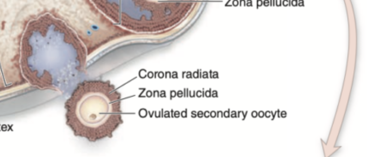
卵胞の破裂 (Rupture of the Follicle)
卵胞内の圧力の増加 (Increasing Pressure within the Follicle) と卵巣壁の弱体化 (Weakening of the Wall) により、スティグマ (Stigma) での卵胞の破裂 (Rupture of the Follicle) が発生します。
- 卵母細胞 (Oocyte) とその放射冠 (Corona Radiata) は、卵胞液 (Follicular Fluid) と共に卵巣 (Ovary) から放出 (Expelled) されます。
- 排出 (Expulsion) は、平滑筋の局所収縮 (Local Smooth Muscle Contractions) によって促進されます。
- 排卵された二次卵母細胞 (Ovulated Secondary Oocyte) は、粘性の卵胞液 (Viscous Follicular Fluid) にゆるく付着 (Adheres Loosely) し、卵管 (Uterine Tube) の開口部に引き込まれ (Drawn In)、受精 (Fertilization) が起こる可能性があります。
- 受精が24時間以内に起こらない場合 (If Not Fertilized Within 24 Hours)、二次卵母細胞 (Secondary Oocyte) は退化 (Degenerates) し始めます。
黄体 (Corpus Luteum) の形成
排卵後 (After Ovulation)、排卵された卵胞 (Ovulated Follicle) の顆粒膜細胞 (Granulosa Cells) と内卵胞鞘 (Theca Interna) は再分化 (Redifferentiate) し、卵巣皮質 (Ovarian Cortex) に一時的な内分泌腺 (Temporary Endocrine Gland) である黄体 (Corpus Luteum, 黄色い体の意) を形成します。
- 排卵後 (After Ovulation)、顆粒膜 (Granulosa) および卵胞壁の卵胞鞘 (Thecal Layers of the Follicle Wall) は崩壊 (Collapse) および折りたたまれます (Folding)。
- 破れた毛細血管 (Disrupted Capillaries) からの血液 (Blood) が以前の卵胞腔 (Former Antrum) に凝血塊 (Clot) として蓄積 (Accumulates) します(図22–12参照)。
- 顆粒膜細胞 (Granulosa Cells) と内卵胞鞘 (Theca Interna) の細胞は、黄体形成ホルモン (LH, Luteinizing Hormone) の影響を受けて、エストロゲン (Estrogens) に加えて、プロゲステロン (Progesterone) をより多量に産生する (More Extensive Production) 細胞に変化 (Change) します。
黄体の構造と機能 (Structure and Function of the Corpus Luteum)
顆粒膜細胞 (Granulosa Cells) は、分裂 (Dividing) することなく大きさが大幅に増加 (Increase Greatly in Size) し、直径20~35μm に達します。これらの細胞は黄体の約80% (About 80% of the Corpus Luteum) を構成します。この時点で、これらの細胞は顆粒膜黄体細胞 (Granulosa Lutein Cells) と呼ばれるようになります(図22–12参照)。
- 顆粒膜黄体細胞 (Granulosa Lutein Cells) は、タンパク質分泌細胞 (Protein-Secreting Cells) に特有の多くの特徴を失い、アロマターゼ (Aromatase) によるアンドロステンジオン (Androstenedione) のエストラジオール (Estradiol) への変換の役割が拡大 (Expand) します。
一方、内卵胞鞘 (Theca Interna) は、黄体の残りの部分 (Rest of the Corpus Luteum) を形成し、卵胞鞘黄体細胞 (Theca Lutein Cells) と呼ばれます(図22–12参照)。
- 卵胞鞘黄体細胞 (Theca Lutein Cells) は、顆粒膜黄体細胞 (Granulosa Lutein Cells) の半分の大きさ (Half the Size) で、黄体壁 (Wall of the Corpus Luteum) の折り目 (Folds) に集まる (Aggregated) 傾向があります。
- すべての内分泌腺と同様に、黄体 (Corpus Luteum) も血管が豊富 (Well Vascularized) です。
- 黄体形成ホルモン (LH, Luteinizing Hormone) によって、プロゲステロン (Progesterone) およびアンドロステンジオン (Androstenedione) の大量の産生 (Production of Large Amounts) が促進されます。
黄体の運命 (Fate of the Corpus Luteum)
黄体 (Corpus Luteum) の短期的な運命 (Short-Term Fate) は、妊娠 (Pregnancy) が発生するかどうか (Depends on Whether Pregnancy Occurs) によって決まります。
- 妊娠が起こらない場合 (If Pregnancy Does Not Occur)
- 排卵時のLHサージ (Ovulatory LH Surge) によって、黄体 (Corpus Luteum) は10~12日間 (10-12 Days) プロゲステロン (Progesterone) を分泌 (Secrete) します。
- さらなるLHの刺激がない (Without Further LH Stimulation)、および妊娠が起こらない (Absence of Pregnancy) 状態では、黄体の2つの主要な細胞型 (Both Major Cell Types of the Corpus Luteum) はステロイド産生 (Steroid Production) を停止 (Cease) し、アポトーシス (Apoptosis) を経て組織が退縮 (Regression of the Tissue) します。
- プロゲステロン (Progesterone) の分泌の減少は、月経 (Menstruation)、すなわち子宮粘膜 (Uterine Mucosa) の一部の剥離 (Shedding) を引き起こします。
- アクティブな黄体 (Active Corpus Luteum) によって産生されるエストロゲン (Estrogen) は、下垂体 (Pituitary) からのFSH (卵胞刺激ホルモン) の分泌を抑制 (Inhibit) します。
- 黄体が退化 (Degenerate) すると、血中のステロイド濃度 (Blood Steroid Concentration) は減少 (Decreases) し、FSHの分泌 (Secretion of FSH) は再び増加 (Increases Again) し、新しい卵胞群 (Another Group of Follicles) の成長が刺激 (Stimulated) されます。これが次の月経周期 (Next Menstrual Cycle) を開始させる要因です。
- このような黄体 (Corpus Luteum) は、月経の黄体 (Corpus Luteum of Menstruation) と呼ばれます。
白体 (Corpus Albicans) の形成
黄体 (Corpus Luteum) の退化 (Regression) によって残存した細胞残渣 (Remnants) は、マクロファージ (Macrophages) によって貪食 (Phagocytosed) されます。その後、線維芽細胞 (Fibroblasts) がその領域に侵入 (Invade) し、高密度結合組織 (Dense Connective Tissue) の瘢痕 (Scar) を生成 (Produce) します。これが白体 (Corpus Albicans, 白い体の意) です(図22–13参照)。
妊娠が起こった場合 (If Pregnancy Occurs)
妊娠が起こった場合 (If Pregnancy Occurs)、子宮粘膜 (Uterine Mucosa) は月経 (Menstruation) を起こしてはならない (Must Not Undergo Menstruation) ため、胚 (Embryo) が失われるのを防ぐ必要があります。
- 循環するプロゲステロン (Circulating Progesterone) の減少 (Drop) を防ぐために、着床した胚 (Implanted Embryo) の栄養膜細胞 (Trophoblast Cells) は、糖タンパク質ホルモン (Glycoprotein Hormone) であるヒト絨毛性ゴナドトロピン (hCG, Human Chorionic Gonadotropin) を産生します。
- hCG (ヒト絨毛性ゴナドトロピン) は、LH (黄体形成ホルモン) と同様の標的 (Targets) と活性 (Activity) を持っています。
- hCG (ヒト絨毛性ゴナドトロピン) は、黄体 (Corpus Luteum) のさらなる成長 (Further Growth) を維持 (Maintains) し、プロゲステロン (Progesterone) の分泌を促進 (Stimulate Secretion) します。これにより、子宮粘膜 (Uterine Mucosa) が維持 (Maintain) されます。
妊娠黄体 (Corpus Luteum of Pregnancy) は、非常に大きく (Very Large) なり、hCG (ヒト絨毛性ゴナドトロピン) によって4~5か月間 (4-5 Months) 維持されます。
- 4~5か月後 (By 4-5 Months)、胎盤 (Placenta) 自体が、プロゲステロン (Progesterone)(およびエストロゲン (Estrogens))を十分なレベル (Levels Adequate) で産生するようになります。
- その後、黄体 (Corpus Luteum) は退化 (Degenerates) し、大きな白体 (Large Corpus Albicans) に置き換えられます。
卵管 (Uterine Tubes)
一対の卵管 (Paired Uterine Tubes)、別名卵管 (Oviducts) は、靭帯 (Ligaments) および間膜 (Mesenteries) によって支えられており、これによりかなりの可動性 (Considerable Mobility) を持つようになっています。卵管 (Uterine Tube) はそれぞれ約10〜12cm (About 10-12 cm) の長さがあり(図22-14参照)、卵巣 (Ovary) 付近の腹腔 (Peritoneal Cavity) に開口 (Opens) しています。
卵管の構造 (Structure of the Uterine Tube)
卵管は、卵巣側 (Ovary Side) から子宮 (Uterus) に向かって、次の4つの領域 (Four Regions) に分かれています。
- 卵管采 (Infundibulum)
- 漏斗状の開口部 (Funnel-Shaped Opening) であり、繊毛のような突起 (Fingerlike Extensions) が付いています。
- これらの突起 (Extensions) は卵管采 (Fimbriae, ラテン語で「フリンジ」) と呼ばれ、卵巣 (Ovary) の近くに位置します。
- 膨大部 (Ampulla)
- 卵管の最も長い部分 (Longest Region) であり、拡張した部分 (Expanded Region) です。
- 通常、受精 (Fertilization) はこの膨大部 (Ampulla) で発生します。
- 峡部 (Isthmus)
- 子宮の近く (Near the Uterus) に位置するより狭い部分 (Narrow Portion) です。
- 子宮内部分 (Uterine or Intramural Part)
- 子宮の壁 (Wall of the Uterus) を通過し、子宮の内部 (Interior of the Uterus) に開口します。
卵管の壁の構造 (Wall of the Uterine Tube)
卵管 (Oviduct) の壁は、以下の3つの層 (Three Layers) で構成されています。
- 粘膜 (Mucosa)
- 折り重なった粘膜 (Folded Mucosa) であり、縦方向の分岐 (Branching, Longitudinal Folds) を形成しています。
- これらの粘膜の縦方向のヒダ (Longitudinal Mucosal Folds) は、膨大部 (Ampulla) で最も顕著であり、断面図 (Cross Section) では迷路のように見える (Resembles a Labyrinth)(図22-14b参照)。
- 子宮に近い領域 (Regions Closer to the Uterus) では、これらの粘膜のヒダ (Mucosal Folds) は小さくなり、子宮内部分 (Intramural Portion of the Tube) では存在しなくなる (Absent) ことが多いです。
- 筋層 (Muscularis)
- 厚く (Thick)、明確に定義された (Well-Defined) 筋層 (Muscularis) を持ち、円形 (Circular または Spiral) と縦方向 (Longitudinal Layers) の平滑筋 (Smooth Muscle) が交差 (Interwoven) しています(図22-15a参照)。
- 漿膜 (Serosa)
- 内臓腹膜 (Visceral Peritoneum) によって覆われ、中皮 (Mesothelium) を持つ薄い漿膜 (Thin Serosa) です。
卵管の粘膜 (Mucosa of the Uterine Tube)
- 粘膜 (Mucosa) は、疎性結合組織 (Loose Connective Tissue) からなる固有層 (Lamina Propria) 上の単層円柱上皮 (Simple Columnar Epithelium) によって裏打ちされています(図22-15b参照)。
- 上皮 (Epithelium) には、2種類の機能的に重要な細胞 (Two Functionally Important Cell Types) が含まれています。
- 線毛細胞 (Ciliated Cells)
- 線毛運動 (Ciliary Movements) により、液体 (Fluid) を子宮方向 (Toward the Uterus) に移動 (Sweep) させます。
- 分泌細胞 (Secretory Peg Cells)
- 線毛を持たない (Nonciliated) 細胞であり、暗く染色される (Darker Staining) ことが多いです。
- 腔 (Lumen) に向かって頂部が突出 (Apical Bulge) している場合があり、糖タンパク質 (Glycoproteins) を含む栄養性の粘液膜 (Nutritive Mucus Film) を分泌 (Secrete) します。
- この粘液 (Mucus) は、上皮 (Epithelium) を覆い、卵母細胞 (Oocyte) および精子 (Sperm) に対して栄養 (Nutritive) および保護機能 (Protective Functions) を提供します。
排卵時の変化 (Changes During Ovulation)
エストロゲン (Estrogen) によって引き起こされる変化 (Triggered by Estrogens):
- 線毛 (Cilia) は伸長 (Elongate) し、卵胞成長期 (Follicular Growth Phase) において、両方の細胞型 (Both Cell Types) が肥大 (Hypertrophy) します。
- 黄体期後期 (Late Luteal Phase) では、線毛が消失 (Loss of Cilia) し、細胞の萎縮 (Atrophy of Cells) が起こります。
排卵時 (At the Time of Ovulation):
- 粘膜の肥大 (Mucosal Hypertrophy) と局所的な血流の増加 (Increased Local Blood Flow) によって、卵管 (Uterine Tubes) は拡大 (Enlarged) し、卵巣 (Ovary) のすぐ近くに移動 (Moved Close) します。
- 卵管采 (Fimbriae) は卵巣 (Ovary) を部分的に取り囲む (Partially Surround) ようになります。
卵の移動と受精 (Transport and Fertilization of the Oocyte)
- 卵 (Oocyte) は、卵管采 (Fimbriae) の掃引運動 (Sweeping Movements) と線毛運動 (Ciliary Activity) によって、漏斗部 (Infundibulum) に取り込まれ (Enters the Infundibulum)、膨大部 (Ampulla) へと移動します。
- 粘膜を覆う分泌物 (Secretions Covering the Mucosa) は、卵母細胞 (Oocyte) と精子 (Sperm) に対して栄養 (Nutritive) および保護 (Protective) の機能を提供します。
- 分泌物 (Secretions) には、容量化因子 (Capacitation Factors) が含まれています。
- 容量化因子 (Capacitation Factors) は、精子 (Sperm) を活性化 (Activate) し、卵母細胞 (Oocyte) を受精 (Fertilize) する能力を持たせる (Make Able) 役割を果たします。
受精の主な出来事 (Major Events of Fertilization)
受精 (Fertilization) とは、女性と男性の配偶子 (Female and Male Gametes) が結合 (Union) するプロセスで、通常、卵管の膨大部 (Ampulla of a Uterine Tube) で発生します。射精 (Ejaculation) された数百万の精子 (Millions of Sperm) のうち、卵管の膨大部 (Ampulla) に到達するのは数百の精子 (A Few Hundred Sperm) に過ぎません。
受精 (Fertilization) を行う能力を持つのは、女性の生殖器官 (Female Reproductive Tract) 内で容量化 (Capacitation) を受けた精子 (Sperm) のみです。受精には、以下の主なステップ (Major Steps) が含まれます。
1. 放射冠の貫通 (Penetration of the Corona Radiata)
- 放射冠 (Corona Radiata) の細胞に接触 (Upon Contact with Cells of the Corona Radiata) すると、精子 (Sperm) は先体反応 (Acrosomal Reaction) を引き起こします。
- この反応では、精子の頭部 (Sperm Head) の周囲の複数の部位から、エキソサイトーシス (Exocytosis) によってヒアルロニダーゼ (Hyaluronidase) が放出されます。
- これにより、精子 (Sperm) は透明帯 (Zona Pellucida) へとより容易に移動 (Move More Easily) できるようになります。
2. 透明帯の通過 (Penetration of the Zona Pellucida)
- 精子の表面 (Surface of the Sperm) にある特定のタンパク質 (Specific Proteins) が、透明帯 (Zona Pellucida) 上の受容体 (Receptors) ZP3 およびZP4 に結合 (Bind) します。
- これにより、先体膜 (Acrosomal Membrane) 上のプロテアーゼ (Protease) アクロシン (Acrosin) が活性化 (Activated) され、局所的に透明帯 (Locally Degrades the Zona Pellucida) を分解 (Degrade) します。
3. 卵母細胞膜との融合 (Fusion with the Oocyte Plasmalemma)
- 透明帯 (Zona Pellucida) を最初に貫通した精子 (First Sperm to Penetrate the Zona Pellucida) は、卵母細胞の原形質膜 (Oocyte Plasmalemma) と融合 (Fuses) します。
- この融合 (Fusion) により、小胞 (Vesicles) からCa²⁺ (カルシウムイオン, Calcium Ions) が放出 (Release) されます。
- カルシウムイオンの放出 (Release of Ca²⁺) によって、卵母細胞 (Oocyte) の皮質顆粒 (Cortical Granules) からプロテアーゼ (Proteases) がエキソサイトーシス (Exocytosis) によって放出されます。
- 皮質反応 (Cortical Reaction) は、卵母細胞 (Oocyte) の表面全体 (Entire Surface) に波のように広がり (Spreads Like a Wave)、透明帯 (Zona Pellucida) を侵入不可能な周囲胚膜障壁 (Impenetrable Perivitelline Barrier) に変換 (Converts) します。これにより、多精子受精 (Polyspermy) が永久に防止 (Permanent Block) されます。
4. 第二減数分裂の完了 (Completion of Meiosis II)
- 二次卵母細胞 (Secondary Oocyte) の核 (Nucleus) は、減数分裂 II (Meiosis II) を直ちに完了 (Immediately Completes) します。
- これにより、第二極体 (Second Polar Body) と卵子 (Haploid Ovum) の雌性前核 (Female Pronucleus) が形成されます。
5. 雄性前核の形成 (Formation of the Male Pronucleus)
- 精子の頭部 (Head of the Penetrating Sperm) の半数体の核 (Haploid Nucleus) は脱凝縮 (Decondensation) を受け、雄性前核 (Male Pronucleus) に変換 (Becomes) されます。
6. 前核の融合 (Fusion of Pronuclei)
- 雌性前核 (Female Pronucleus) と雄性前核 (Male Pronucleus) が融合 (Fusion) し、新しい二倍体細胞 (New Diploid Cell) である接合子 (Zygote, ラテン語の “結合する” の意) が形成されます。
胚の輸送 (Transport of the Embryo)
- 接合子 (Zygote) は、分裂 (Cell Division) を続けながら、卵管の筋層 (Muscularis of the Oviduct) の収縮 (Contractions) と線毛運動 (Ciliary Movements) によって子宮 (Uterus) へと輸送 (Transport) されます。
- 胚の輸送 (Transport of the Embryo) には約5日間 (About 5 Days) かかります。
- 不動性線毛症 (Immotile Ciliary Syndrome) の女性 (Women) においても、この輸送 (Transport) が正常に行われることから、筋層の収縮 (Muscle Contractions) が胚の輸送において重要 (More Important Role) であることが示唆されています。
医療応用 (Medical Application)
- 卵管結紮術 (Tubal Ligation)
- 避妊 (Contraception) のための一般的な外科手術 (Common Surgical Procedure) です。
- 卵管炎 (Salpingitis)
- 卵管の別名 (Another Name for Uterine Tubes) はサルピンゲス (Salpinges) です。
- 下部生殖管 (Lower Genital Tract) から感染因子 (Infectious Agents) が上行 (Ascend) すると、卵管の粘膜 (Mucosa of the Uterine Tube) が炎症 (Inflamed) を起こし、これが卵管炎 (Salpingitis) です。
- 慢性卵管炎 (Chronic Salpingitis) によって生じる粘膜損傷 (Mucosal Damage) や癒着 (Adhesions) は、不妊症 (Infertility) や異所性妊娠 (Ectopic Pregnancy, Tubal Pregnancy) を引き起こす可能性があります。
- 異所性妊娠 (Ectopic Pregnancy)
- 卵管妊娠 (Tubal Pregnancy) では、固有層 (Lamina Propria) が子宮内膜 (Uterine Endometrium) のように脱落細胞 (Decidual Cells) を形成する場合があります。
- しかし、卵管 (Uterine Tube) は拡張 (Expansion) できないため、胚 (Embryo) が成長すると、卵管が破裂 (Rupture of the Tube) し、潜在的に致命的な出血 (Potentially Fatal Hemorrhage) を引き起こす可能性があります。
子宮 (Uterus)
子宮 (Uterus) は、図22-14 に示されているように、洋ナシ形 (Pear-Shaped) の器官であり、厚い筋肉の壁 (Thick, Muscular Walls) を持っています。
- 子宮の最大の部分 (Largest Part of the Uterus) は体部 (Body) であり、左および右の卵管 (Left and Right Uterine Tubes) がここに接続します。
- 卵管の間の上部の湾曲した領域 (Curved, Superior Area Between the Tubes) は子宮底 (Fundus) と呼ばれます。
- 子宮 (Uterus) は峡部 (Isthmus) で細くなり (Narrows)、下方の円筒形の構造 (Lower Cylindrical Structure) である子宮頸部 (Cervix) で終わります。
- 子宮頸部 (Cervix) の腔 (Lumen) は子宮頸管 (Cervical Canal) であり、両端に狭い開口部 (Constricted Openings) を持ちます。
- 内子宮口 (Internal Os, “口”を意味するラテン語のOs) は子宮の主腔 (Main Uterine Lumen) に開口 (Opens) し、
- 外子宮口 (External Os) は膣 (Vagina) に開口 (Opens) します(図22-14参照)。
子宮壁の構造 (Structure of the Uterine Wall)
子宮壁 (Uterine Wall) は、卵巣 (Ovaries) や卵管 (Uterine Tubes) に関連する靭帯 (Ligaments) および間膜 (Mesenteries) によって支えられています(図22-1参照)。子宮壁 (Uterine Wall) には、次の3つの主要な層 (Three Major Layers) があります(図22-14参照)。
- 外膜 (Perimetrium)
- 結合組織の層 (Connective Tissue Layer) であり、靭帯 (Ligaments) と連続 (Continuous) しています。
- 一部の領域では外膜 (Adventitia) ですが、ほとんどは中皮 (Mesothelium) で覆われた漿膜 (Serosa) です。
- 筋層 (Myometrium)
- 高度に血管化された平滑筋 (Highly Vascularized Smooth Muscle) からなる厚い外套 (Thick Tunic) です(図22-16参照)。
- 内膜 (Endometrium)
- 粘膜 (Mucosa) であり、単層円柱上皮 (Simple Columnar Epithelium) で裏打ちされています。
これらの3つの層 (Three Layers) は、卵管 (Uterine Tubes) の対応する層と連続 (Continuous) しています。内膜 (Endometrium) の厚さ (Thickness) と構造 (Structure) は、卵巣ホルモン (Ovarian Hormones) の周期的な変動 (Cyclic Changes) の影響を受け、これは卵管の粘膜 (Mucosa of the Uterine Tubes) に見られる変化よりも顕著です(図22-17参照)。
筋層 (Myometrium)
筋層 (Myometrium)(ギリシャ語のmyo = 筋肉 (Muscle), metra = 子宮 (Uterus))は、子宮の最も厚い外套 (Thickest Tunic of the Uterus) です。
- 平滑筋繊維 (Smooth Muscle Fibers) は、静脈叢 (Venous Plexuses) やリンパ管 (Lymphatics) を含む結合組織 (Connective Tissue) によって分離されています(図22-16参照)。
- 平滑筋 (Smooth Muscle) は、交差する層 (Interwoven Layers) を形成し、内層および外層の繊維 (Fibers of the Inner and Outer Layers) は子宮の長軸 (Long Axis of the Organ) に沿って平行 (Parallel) に配置されています。
妊娠中 (During Pregnancy):
- 筋層 (Myometrium) は、過形成 (Hyperplasia, 平滑筋細胞の数の増加)、細胞肥大 (Cell Hypertrophy, 細胞サイズの増加)、およびコラーゲン産生 (Increased Collagen Production) による大規模な成長 (Extensive Growth) を経ます。
- 子宮の壁 (Uterine Wall) が強化 (Strengthens) されます。
出産時 (During Parturition):
- 子宮の平滑筋 (Uterine Smooth Muscle) は非常に強力に収縮 (Contracts Very Forcefully) し、胎児 (Infant) を子宮から排出 (Expel from the Uterus) します。
出産後 (After Pregnancy):
- 平滑筋細胞 (Smooth Muscle Cells) は縮小 (Shrink) し、多くの細胞はアポトーシス (Apoptosis) を受け、不要なコラーゲン (Unneeded Collagen) は除去 (Removal) されます。
- 子宮 (Uterus) は妊娠前のサイズ (Prepregnancy Size) にほぼ戻ります (Returns Almost)。
内膜 (Endometrium)
内膜 (Endometrium) の固有層 (Lamina Propria) または間質 (Stroma) は、主に束化されていないIII型コラーゲン繊維 (Nonbundled Type III Collagen Fibers)、豊富な線維芽細胞 (Abundant Fibroblasts)、および基質 (Ground Substance) からなります。
- 単層円柱上皮 (Simple Columnar Epithelial Lining) には、線毛細胞 (Ciliated Cells) と分泌細胞 (Secretory Cells) が含まれます。
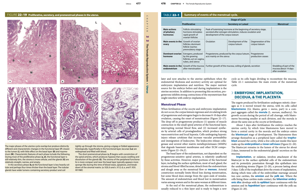
- 子宮腺 (Uterine Glands) は、内膜の全層を貫通 (Penetrate the Full Thickness of the Endometrium) します(図22-16および図22-18参照)。
内膜の2つの層 (Two Layers of the Endometrium)
- 基底層 (Basal Layer)
- 筋層 (Myometrium) に隣接し、高密度の固有層 (Highly Cellular Lamina Propria) があります。
- 子宮腺 (Uterine Glands) の深部の基底部 (Deep Basal Ends) を含んでいます。
- 機能層 (Functional Layer)
- 基質が豊富なスポンジ状の固有層 (Spongier Lamina Propria, Richer in Ground Substance) を持っています。
- 子宮腺 (Uterine Glands) のほとんどの長さ (Most of the Length of the Glands) と表面上皮 (Surface Epithelium) を含みます。
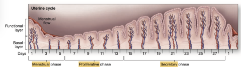
月経周期 (Menstrual Cycle) 中、機能層 (Functional Layer) は深い変化 (Profound Changes) を受けますが、基底層 (Basal Layer) はほぼ変化しません (Remains Relatively Unchanged)(図22-17参照)。
内膜への血液供給 (Blood Supply to the Endometrium)
- 筋層の中間層 (Middle Layers of the Myometrium) にある弓状動脈 (Arcuate Arteries) は、内膜 (Endometrium) に2つの小さな動脈 (Two Smaller Arteries) を送ります。
- 直動脈 (Straight Arteries) は基底層 (Basal Layer) へ血液を供給します。
- らせん動脈 (Spiral Arteries) は機能層 (Functional Layer) に血液を供給 (Bring Blood) します。
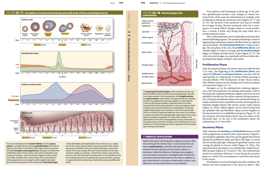
月経周期 (Menstrual Cycle)
女性の生殖器系 (Female Reproductive System) においては、エストロゲン (Estrogens) とプロゲステロン (Progesterone) が、上皮細胞 (Epithelial Cells) およびそれに関連する結合組織 (Connective Tissue) の成長 (Growth) や分化 (Differentiation) を制御します。
出生前 (Before Birth) でさえ、これらの細胞は胎盤 (Placenta) を通じて胎児 (Fetus) に届く母体のエストロゲンとプロゲステロン (Maternal Estrogen and Progesterone) の循環 (Circulation) の影響を受けます。
閉経後 (After Menopause) には、これらのホルモンの合成 (Synthesis of These Hormones) が減少 (Diminished) し、生殖管 (Reproductive Tract) の組織は一般的な退縮 (General Involution) を示します。
医療応用 (Medical Application)
子宮内膜 (Endometrial Cells) は頻繁に (Frequently) 月経の逆流 (Menstrual Reflux) を起こし、卵管 (Uterine Tubes) へ、あるいは卵管を通じて (Through the Uterine Tubes) 流れ込みます。
一部の女性では、これが子宮内膜症 (Endometriosis) を引き起こす可能性があります。子宮内膜症 (Endometriosis) とは、卵巣 (Ovaries)、卵管 (Oviducts)、またはその他の部位 (Elsewhere) に子宮内膜組織 (Endometrial Tissue) が増殖 (Growing) する障害 (Disorder) です。
- エストロゲン (Estrogen) およびプロゲステロン (Progesterone) の影響下で、異所性の子宮内膜組織 (Ectopic Endometrial Tissue) は毎月増殖 (Grows) および退縮 (Degenerates) しますが、この組織は体外に効果的に排出されることはありません (Cannot Be Removed Effectively from the Body)。
- 痛み (Pain) に加えて、炎症 (Inflammation)、卵巣嚢胞 (Ovarian Cysts)、癒着 (Adhesions)、および瘢痕組織 (Scar Tissue) を引き起こす (Produce) ことがあり、これが不妊症 (Infertility) の原因になることもあります。
月経周期の期間 (Duration of the Menstrual Cycle)
思春期 (Puberty) から閉経 (Menopause)(約45~50歳 (About Age 45-50))まで、下垂体のゴナドトロピン (Pituitary Gonadotropins) によって卵巣ホルモン (Ovarian Hormone) の周期的な変動 (Cyclic Changes) が引き起こされます。
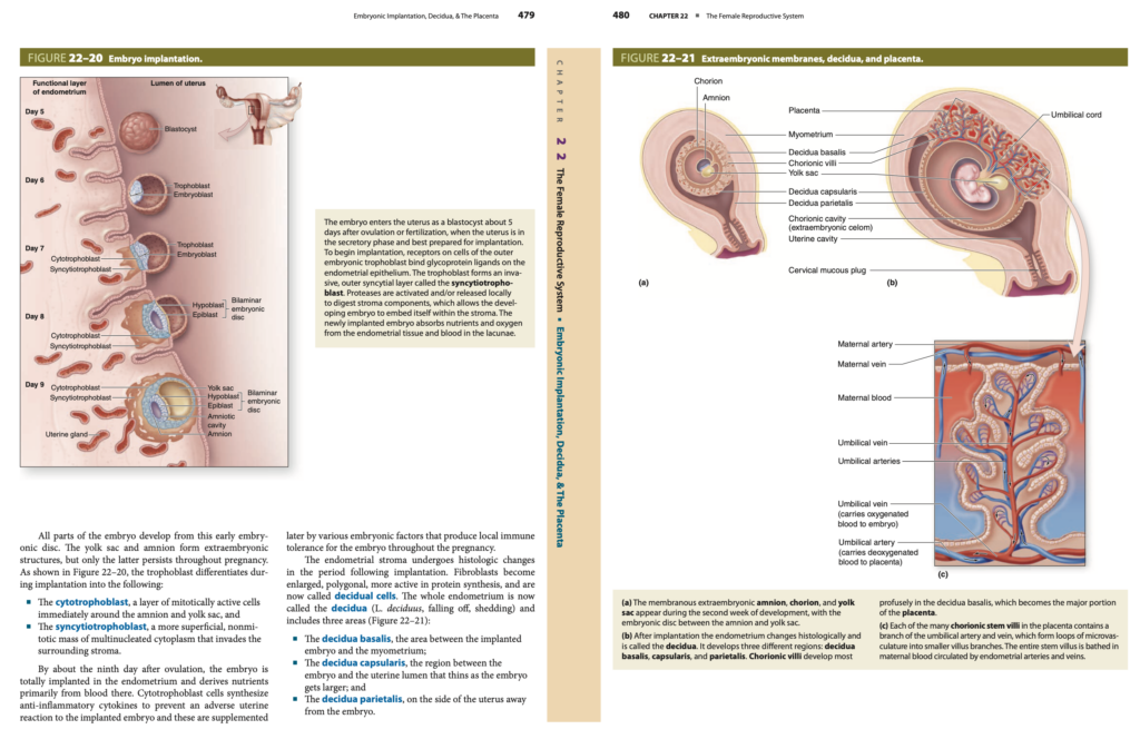
これにより、子宮内膜 (Endometrium) は月経周期 (Menstrual Cycle) 中に周期的な変化 (Cyclic Modifications) を示します(図22-17および図22-19参照)。月経周期 (Menstrual Cycle) の期間 (Duration) は変動 (Variable) する可能性がありますが、平均28日 (Average 28 Days) です。
- 月経周期 (Menstrual Cycles) は、卵母細胞 (Oocyte) の産生 (Production) に関連する卵胞の変化 (Changes in Ovarian Follicles) に依存しているため、月経周期が続く間 (During the Years of Menstrual Cycles) のみ、女性は妊娠可能 (Fertile) です。
月経の開始 (Start of Menstruation)
月経周期の1日目 (Day 1 of the Menstrual Cycle) は、通常、月経出血 (Menstrual Bleeding) が出現する日 (Day When Bleeding Appears) として考えられます。
- 月経の分泌物 (Menstrual Discharge) には、変性した子宮内膜 (Degenerating Endometrium) と、破裂した微小血管 (Ruptured Microvasculature) からの血液 (Blood) が混ざっています (Mixed)。
- 月経期間 (Menstrual Period) は、平均して3〜4日間 (3-4 Days on Average) 続きます。
月経周期の3つの主要な段階 (Three Major Phases of the Menstrual Cycle)
- 増殖期 (Proliferative Phase)
- 期間 (Duration): 8〜10日間 (8-10 Days on Average)
- 別名 (Other Names): 卵胞期 (Follicular Phase) またはエストロゲン期 (Estrogenic Phase)
- 排卵 (Ovulation) の直前に終わります。
- 分泌期 (Secretory Phase)
- 期間 (Duration): 約14日間 (About 14 Days)
- 排卵 (Ovulation) の後 (After Ovulation) に始まります。
- 月経期 (Menstrual Phase)
- 期間 (Duration): 平均3~4日 (3-4 Days on Average)
- 機能層 (Functional Layer) の剥離 (Shedding) が起こります。
増殖期 (Proliferative Phase)
月経期 (Menstrual Phase) の後 (After the Menstrual Phase)、子宮粘膜 (Uterine Mucosa) は比較的薄い (Relatively Thin)(約0.5 mm)。増殖期 (Proliferative Phase) の開始 (Beginning) は、卵胞期 (Follicular Phase) またはエストロゲン期 (Estrogenic Phase) とも呼ばれます。
- 卵胞 (Ovarian Follicles) が小胞状卵胞 (Vesicular Follicles) として急速に成長 (Rapid Growth) するのと一致 (Coincides) します。
- 内卵胞鞘 (Theca Interna) の発達 (Development) に伴い、これらの卵胞 (These Follicles) は活発にエストロゲン (Estrogen) を分泌 (Secrete) し、血漿濃度 (Plasma Concentration) を増加 (Increase) させます(図22-17参照)。
エストロゲン (Estrogens) は子宮内膜 (Endometrium) に作用し、月経中に失われた機能層 (Functional Layer Lost During Menstruation) の再生 (Regeneration) を誘発 (Induce) します。
- 腺の基底部の細胞 (Cells in the Basal Ends of Glands) が増殖 (Proliferate) し、移動 (Migrate) し、月経中に露出した表面 (Surface Exposed During Menstruation) に新しい上皮の覆い (New Epithelial Covering) を形成 (Form) します。
- 増殖期 (Proliferative Phase) の間、子宮内膜 (Endometrial Lining) は、単層円柱上皮 (Simple Columnar Surface Epithelium) です。
- 子宮腺 (Uterine Glands) は比較的まっすぐな管 (Relatively Straight Tubules) で、狭い (Narrow) 空の腔 (Nearly Empty Lumens) を持ちます。
- 機能層 (Functional Layer) が成長 (Grows) し、らせん動脈 (Spiral Arteries) が長くなります (Lengthen)。
増殖期の終わり (End of Proliferative Phase) には、子宮内膜 (Endometrium) は2〜3 mmの厚さ (2-3 mm Thick) になります。
分泌期 (Secretory Phase)
排卵 (Ovulation) の後、分泌期 (Secretory Phase) または黄体期 (Luteal Phase) が始まります。これは、黄体 (Corpus Luteum) から分泌 (Secreted) されるプロゲステロン (Progesterone) の影響 (As a Result) によるものです。
- プロゲステロン (Progesterone) は、増殖期 (Proliferative Phase) に形成された子宮腺 (Uterine Glands Formed) の上皮細胞 (Epithelial Cells) を刺激 (Stimulate) します。
- 子宮腺 (Uterine Glands) の細胞は、グリコーゲン (Glycogen) を分泌 (Secrete) し始め、細胞内に蓄積 (Accumulate) されます。
- これにより、腺の内腔 (Glandular Lumens) が拡張 (Dilating) し、腺 (Glands) はらせん状 (Coiled) になります(図22–19b、図22–19e参照)。
- 表層の微小血管系 (Superficial Microvasculature) には、薄壁の血液で満たされたラグーナ (Thin-Walled, Blood-Filled Lacunae) が含まれる (Includes) ようになります(図22–18および図22–19e参照)。
分泌期 (Secretory Phase) の間、子宮内膜 (Endometrium) は最大の厚さ (Maximum Thickness) に達します(5 mm)。これは、腺の分泌物の蓄積 (Accumulation of Secretions) および間質の浮腫 (Edema in the Stroma) によるものです。
受精が起こった場合 (If Fertilization Occurs)
- 受精 (Fertilization) が排卵後の翌日に起こった場合 (If Fertilization Occurred During the Day After Ovulation)、胚 (Embryo) は約5日後 (About 5 Days Later) に子宮 (Uterus) に輸送 (Transported) されます。
- この時、子宮内膜 (Endometrial Thickness) の厚さ (Thickness) と分泌活性 (Secretory Activity) は、胚の着床 (Embryonic Implantation) と栄養 (Nutrition) に最適な状態になります。
- 着床前および着床中 (Before and During Implantation)、胚 (Embryo) に対する主な栄養源 (Major Nutrient Source) は子宮の分泌物 (Uterine Secretion) です。
- プロゲステロン (Progesterone) は、この分泌 (Secretion) を促進 (Promote) すると同時に、胚の着床 (Embryo Implantation) に支障を与える可能性のある筋層 (Myometrium) の強い収縮 (Strong Contractions) を抑制 (Inhibits) します。
月経期 (Menstrual Phase)
受精 (Fertilization) が起こらず (Does Not Occur)、胚の着床 (Embryonic Implantation) が発生しない (Does Not Occur) 場合、黄体 (Corpus Luteum) は退縮 (Regresses) します。
- 黄体 (Corpus Luteum) の退縮により、プロゲステロン (Progesterone) およびエストロゲン (Estrogens) の血中濃度 (Circulating Levels) は排卵後8〜10日で (8-10 Days After Ovulation) 減少 (Decrease) し、月経の開始 (Onset of Menstruation) を引き起こします (Causing the Onset of Menstruation)(図22–17参照)。
プロゲステロンの減少 (Drop-off in Progesterone) により、以下の2つの重要な変化 (Two Major Changes) が発生します。
- らせん動脈の収縮 (Contraction of the Spiral Arteries)
- 機能層 (Functional Layer) の小さならせん動脈 (Small Spiral Arteries) において、筋肉の収縮 (Muscle Contraction) がけいれん (Spasms) を引き起こし、正常な血流 (Normal Blood Flow) が遮断されます (Interrupting Normal Blood Flow)。
- 血管の強い血管収縮 (Vasoconstriction) と低酸素 (Hypoxia)
- 動脈の細胞 (Arterial Cells) によるプロスタグランジン (Prostaglandins) の合成 (Synthesis) が増加 (Increased) し、強い血管収縮 (Strong Vasoconstriction) と局所的な低酸素状態 (Local Hypoxia) を引き起こします。
細胞の変化 (Cellular Changes)
- 低酸素状態 (Hypoxic Injury) にさらされた細胞 (Cells) は、サイトカイン (Cytokines) を放出 (Release) し、血管透過性 (Vascular Permeability) の増加 (Increase) と白血球 (Leukocytes) の移動 (Immigration) を誘導 (Induce) します。
- 白血球 (Leukocytes) は、コラゲナーゼ (Collagenase) およびマトリックスメタロプロテアーゼ (MMPs, Matrix Metalloproteinases) を放出 (Release) し、基底膜 (Basement Membranes) や細胞外マトリックス (ECM, Extracellular Matrix) 成分を分解 (Degrade) します(図22–19c、図22–19f参照)。
機能層と基底層の変化 (Changes in the Functional and Basal Layers)
- 機能層 (Functional Layer):
- 表面上皮 (Surface Epithelium)、子宮腺の大部分 (Most of Each Gland)、間質 (Stroma)、および血液で満たされたラグーナ (Blood-Filled Lacunae) は、子宮内膜 (Endometrium) から分離 (Detach) し、剥がれ落ち (Slough Away) ます。これが月経の流出 (Menstrual Flow or Menses) を形成します。
- らせん動脈 (Spiral Arteries) の収縮 (Constriction) により、月経中の出血量 (Amount of Blood Loss During Menstruation) が制限 (Limits) されますが、一部の血液 (Blood) は小静脈 (Venules) の開放端 (Open Ends) から排出 (Emerge) されます。
- 失われる子宮内膜 (Amount of Endometrium Lost) および血液の量 (Amount of Blood Lost) は、女性ごとに異なり (Varies Among Women)、同じ女性の周期によっても異なります (Varies in the Same Woman at Different Times)。
- 基底層 (Basal Layer):
- らせん動脈 (Spiral Arteries) に依存しない (Not Dependent) ため、これらの活動の影響を受けにくい (Relatively Unaffected) です。
月経期の終わり (End of the Menstrual Phase)
月経期 (Menstrual Phase) の終わり (End of the Phase) では、子宮内膜 (Endometrium) は薄い層 (Thin Layer) に減少 (Reduced) します。
- 粘膜 (Mucosa) を再構成 (Reconstitute) するために、細胞 (Cells) は分裂 (Divide) を開始し、新しい周期 (New Cycle) が始まります。
表22–1 には、月経周期の主要な出来事 (Main Events of the Menstrual Cycle) がまとめられています。
胚の着床、脱落膜、および胎盤 (Embryonic Implantation, Decidua, & The Placenta)
受精 (Fertilization) によって生成された接合子 (Zygote) は、子宮 (Uterus) に向かって移動する間、有糸分裂による卵割 (Mitotic Cleavages) を行います。卵割 (Cleavage) の間、接合子の細胞 (Cells of the Zygote) は割球 (Blastomeres, ギリシャ語の “blastos” = 芽, “meros” = 部分) と呼ばれます。
- 割球 (Blastomeres) は緊密に凝集 (Compact Aggregate) し、桑実胚 (Morula, ラテン語の “morum” = 桑の実) として知られる集合体 (Aggregate) を形成します。
- 卵割 (Cell Cleavage) の期間中は成長 (Growth) は起こりません (No Growth Occurs)。したがって、分裂ごとに割球 (Blastomeres) は小さくなり (Becoming Smaller)、桑実胚 (Morula) の大きさ (Size) は、受精時の卵母細胞 (Oocyte at Fertilization) とほぼ同じ (About the Same Size) です。
胚盤胞の形成 (Formation of the Blastocyst)
受精 (Fertilization) から約5日後 (About 5 Days Later)、胚 (Embryo) は子宮腔 (Uterine Cavity) に到達します。
- この時までに、割球 (Blastomeres) は移動 (Moved) し、桑実胚 (Morula) 内に中心腔 (Central Cavity) を形成 (Form) します。
- これにより、胚 (Embryo) は胚盤胞 (Blastocyst) の発生段階 (Stage of Development) に入ります (Enters)。
- 割球 (Blastomeres) は、空洞の周囲 (Around the Cavity) に周辺の層 (Peripheral Layer) を形成し、これを栄養膜 (Trophoblast) と呼びます。
- 栄養膜 (Trophoblast) のすぐ内側には、胚芽 (Embryoblast) または内部細胞塊 (Inner Cell Mass) を構成する細胞群 (Few Cells) が存在します(図22–20参照)。
- 胚盤胞 (Blastocyst) は、子宮の内腔 (Lumen of the Uterus) に約2日間 (About 2 Days) 留まります。
- 子宮粘膜 (Endometrial Mucosa) 上の子宮腺の分泌物 (Secretions of the Endometrial Glands) に浸される (Immersed) 形で存在します。
着床 (Implantation)
着床 (Implantation, 別名 “Nidation”) とは、胚盤胞 (Blastocyst) が子宮内膜の上皮細胞 (Surface Epithelial Cells of the Endometrium) に付着 (Attachment) し、この上皮を通って下の間質 (Stroma) に侵入 (Proteolytic Penetration) するプロセスです(図22–20参照)。このプロセスは約3日間 (About 3 Days) 続きます。
- 着床 (Implantation) においては、栄養膜 (Trophoblast) の細胞 (Cells) が主導的役割 (Drive the Events) を果たします。
- 同時に、胚芽 (Embryoblast) の細胞 (Cells) は、羊膜 (Amnion) および卵黄嚢 (Yolk Sac) という2つの新しい腔 (Two New Cavities) の周囲に再配置 (Rearrange) します。
- これらの腔 (These Cavities) を裏打ちする細胞が接触する場所 (Where the Cells Make Contact) に、二層性胚盤 (Bilaminar Embryonic Disc) が発生 (Develops) します。
- 上層 (Epiblast Layer) は羊膜 (Amnion) と連続 (Continuous) し、
- 下層 (Hypoblast Layer) は卵黄嚢 (Yolk Sac) と連続 (Continuous) します(図22–20参照)。
栄養膜の分化 (Differentiation of the Trophoblast)
着床 (Implantation) 中、栄養膜 (Trophoblast) は次の2つの異なる部分 (Two Distinct Parts) に分化 (Differentiates) します。
- 細胞性栄養膜 (Cytotrophoblast)
- 羊膜 (Amnion) および卵黄嚢 (Yolk Sac) のすぐ周囲 (Immediately Around) にある有糸分裂が活発な細胞の層 (Layer of Mitotically Active Cells) です。
- 合胞体性栄養膜 (Syncytiotrophoblast)
- より外側 (More Superficial) に存在し、有糸分裂しない (Non-Mitotic)、多核の細胞質 (Multinucleated Cytoplasm) からなる塊 (Mass) です。
- 周囲の間質 (Surrounding Stroma) に侵入 (Invades) します。
脱落膜 (Decidua)
排卵後9日 (About the Ninth Day After Ovulation) までに、胚 (Embryo) は子宮内膜 (Endometrium) に完全に着床 (Totally Implanted) します。これにより、胚 (Embryo) は栄養 (Nutrients) を子宮内膜の血液 (Blood in the Endometrium) から供給される (Derives) ようになります。
- 細胞性栄養膜 (Cytotrophoblast Cells) は、抗炎症性サイトカイン (Anti-Inflammatory Cytokines) を合成 (Synthesize) し、子宮 (Uterus) が着床した胚 (Implanted Embryo) に対して免疫反応 (Adverse Uterine Reaction) を引き起こさないようにします (Prevent an Adverse Reaction)。
- 胚 (Embryo) からの様々な因子 (Various Embryonic Factors) によって、局所的な免疫寛容 (Local Immune Tolerance) が妊娠 (Pregnancy) を通して維持されます (Maintained)。
脱落膜の領域 (Regions of the Decidua)
子宮内膜の間質 (Endometrial Stroma) は、着床後の期間 (Period Following Implantation) に組織学的変化 (Histologic Changes) を受けます。
- 線維芽細胞 (Fibroblasts) は、拡大 (Enlarged) し、多角形 (Polygonal) になり、タンパク質合成 (Protein Synthesis) が活性化 (More Active) します。
- これらの線維芽細胞 (Fibroblasts) は脱落細胞 (Decidual Cells) と呼ばれます。
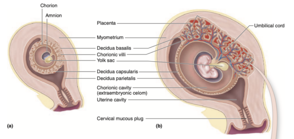
子宮内膜全体 (Whole Endometrium) は、脱落膜 (Decidua, ラテン語の「剥がれ落ちる」を意味する “deciduus”) として知られるようになります。
- 基底脱落膜 (Decidua Basalis): 着床した胚 (Implanted Embryo) と筋層 (Myometrium) の間の領域。
- 包絡脱落膜 (Decidua Capsularis): 胚 (Embryo) と子宮腔 (Uterine Lumen) の間の領域。
- 壁側脱落膜 (Decidua Parietalis): 胚 (Embryo) から離れた子宮の側面 (Side of the Uterus Away from the Embryo) に位置します。
医療応用 (Medical Application)
胚 (Embryo) の最初の付着 (Initial Attachment) は、通常、子宮体部 (Body of the Uterus) の腹側 (Ventral Wall) または背側 (Dorsal Wall) に発生 (Occurs) します。
- ただし、時折、内子宮口 (Internal Os) 付近に付着 (Attaches) することがあります。
- この場合、胎盤 (Placenta) は胎児 (Fetus) と膣 (Vagina) の間 (Interposed) に位置 (Positioned) するため、分娩時 (At Parturition) に胎児の通過 (Passage of the Fetus) が妨げられます (Obstructing the Passage)。
- この状態は、前置胎盤 (Placenta Previa) と呼ばれます。
- 前置胎盤 (Placenta Previa) は、医師 (Physician) によって認識される必要があり (Must Be Recognized)、胎児 (Fetus) は帝王切開 (Cesarean Section) によって分娩 (Delivered) される必要があります。
- 胎児の通過が妨げられたまま (Obstructed Parturition) だと、胎児の死亡 (Death of the Fetus) につながる可能性があります。
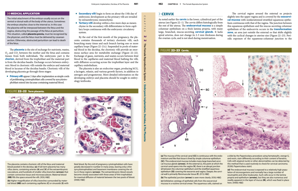
胎盤の機能 (Function of the Placenta)
胎盤 (Placenta) は、母体 (Mother) と胎児 (Fetus) の間での栄養 (Nutrients)、老廃物 (Wastes)、酸素 (O₂)、および二酸化炭素 (CO₂) の交換 (Exchange) の場 (Site) です。
- 胎盤 (Placenta) には、母体と胎児の両方の組織 (Tissues from Both Individuals) が含まれています。
- 胎児の部分 (Embryonic Part) は、絨毛膜 (Chorion) であり、これは栄養膜 (Trophoblast) に由来します。
- 母体の部分 (Maternal Part) は、基底脱落膜 (Decidua Basalis) から派生しています。
- 物質交換 (Exchange) は、胎児の血液 (Embryonic Blood) が胎児の外側 (Outside the Embryo) にある絨毛 (Chorionic Villi) で循環 (Circulating) し、母体の血液 (Maternal Blood) が基底脱落膜 (Decidua Basalis) のラグーナ (Lacunae, プールのような構造) に流れ込む (Flows In) ことで行われます (Occurs)。
胎盤の発達 (Development of the Placenta)
胎盤 (Placenta) の発達 (Development) において、絨毛 (Chorionic Villi) は3つの段階 (Three Stages) を経ます。
1. 一次絨毛 (Primary Villi)
- 着床 (Implantation) から2日後 (2 Days After Implantation) に出現 (Appear) します。
- 増殖中の細胞性栄養膜 (Proliferating Cytotrophoblast Cells) の単純な索状の構造 (Simple Cords) であり、合胞体栄養膜 (Syncytiotrophoblast) に覆われ (Covered) ています。
- 母体の血液 (Maternal Blood) を含むラグーナ (Lacunae) に突き出します (Extend Into)。
2. 二次絨毛 (Secondary Villi)
- 胚の発生15日目 (About the 15th Day of Embryonic Development) に形成 (Begin to Form) されます。
- 一次絨毛 (Primary Villi) が、胚外中胚葉 (Extraembryonic Mesenchyme) に侵入され (Invaded)、二次絨毛 (Secondary Villi) が形成 (Formed) されます。
3. 三次絨毛 (Tertiary Villi)
- 数日後 (A Few Days Later) に発達 (Develop) します。
- 二次絨毛 (Secondary Villi) 内の間充織 (Mesenchyme) が分化 (Differentiates) し、毛細血管のループ (Capillary Loops) が形成 (Formed) されます。
- これらの毛細血管のループ (Capillary Loops) は胎児の循環系 (Embryonic Circulatory System) に連続 (Continuous) しています。
胎盤の構造 (Structure of the Placenta)
妊娠の1か月後 (End of the First Month of Pregnancy) までに、胎盤 (Placenta) には数千の三次絨毛 (Thousands of Tertiary Chorionic Villi) が含まれる (Contains) ようになります。
- 各三次絨毛 (Tertiary Villi) は、多数の枝 (Many Branches) を持ち、各枝 (Each Branch) には1つまたは複数の毛細血管ループ (One or More Capillary Loops) が含まれます(図22–21c参照)。
- 母体の血液 (Maternal Blood) のプール (Pools) に懸濁 (Suspended) されている基底脱落膜 (Decidua Basalis) のラグーナ (Lacunae) に囲まれています。
- これにより (This Arrangement)、代謝物の交換 (Metabolite Exchange) に非常に大きな表面積 (Enormous Surface Area) が提供 (Provides) されます(図22–22参照)。
物質交換の仕組み (Mechanism of Exchange)
- ガス (Gases)(O₂ (酸素, Oxygen)、CO₂ (二酸化炭素, Carbon Dioxide))、栄養素 (Nutrients)、および老廃物 (Wastes) の交換 (Exchange) は、胎児の血液 (Fetal Blood) が毛細血管 (Capillaries) 内を流れ (Flows)、母体の血液 (Maternal Blood) が絨毛 (Villi) を洗浄 (Bathes) することによって行われます (Occurs)。
- 拡散 (Diffusion) は、栄養膜の層 (Trophoblast Layer) および毛細血管内皮 (Capillary Endothelium) を横切って発生 (Occurs Across) します。
胎盤の内分泌機能 (Endocrine Function of the Placenta)
胎盤 (Placenta) は、内分泌器官 (Endocrine Organ) としても機能 (Functions) します。
- hCG (ヒト絨毛性ゴナドトロピン, Human Chorionic Gonadotropin)
- 乳腺刺激ホルモン (Lactogen)
- リラキシン (Relaxin)
- 成長因子 (Growth Factors)
- エストロゲン (Estrogen)
- プロゲステロン (Progesterone)
これらのホルモンは、胎児の成長 (Growth of the Fetus) と妊娠の維持 (Maintenance of Pregnancy) に不可欠 (Essential) です。
子宮頸部 (Cervix)
子宮頸部 (Cervix) は、子宮 (Uterus) の下部 (Lower Part) に位置する円筒形の構造 (Cylindrical Part) です(図22–1参照)。
- 子宮頸部 (Cervix) は、子宮の他の部分 (Rest of the Uterus) とは組織学的に異なります (Differs Histologically)。
子宮頸部の内腔 (Endocervical Mucosa)
- 子宮頸部の内腔 (Endocervical Mucosa) は、厚い固有層 (Thick Lamina Propria) 上の単層円柱上皮 (Simple Columnar Epithelium) で裏打ち (Lined) されています。
- 多くの大きな分岐した (Many Large, Branched) 粘液分泌性の子宮頸腺 (Mucus-Secreting Cervical Glands) が存在 (Present) しています。
- 子宮頸部の内腔 (Endocervical Mucosa) には、らせん動脈 (Spiral Arteries) が存在しません (Lacks Spiral Arteries)。
- 卵巣周期 (Ovarian Cycle) の間も、2-3 mmの厚さ (Thickness of 2-3 mm) は変化しません (Does Not Change)。
- 月経 (Menstruation) の際に剥がれ落ちる (Shed) こともありません (Is Not Shed)。
子宮頸管外部 (Exocervical Mucosa)
- 子宮頸管外部 (Exocervical Mucosa) は、外子宮口 (External Os) 周辺の膣の上部 (Upper Vagina) にわずかに突出 (Projects Slightly) しています。
- 子宮頸管外部 (Exocervical Mucosa) は、非角化重層扁平上皮 (Nonkeratinized Stratified Squamous Epithelium) で覆われており (Covered)、膣 (Vagina) の上皮と連続 (Continuous) しています。
扁平円柱移行帯 (Transformation Zone)
- 扁平上皮 (Squamous Epithelium) と、内子宮頸部の粘液分泌性円柱上皮 (Mucus-Secreting Columnar Epithelium of the Endocervix) の接合部 (Junction) は、変換帯 (Transformation Zone) と呼ばれます。
- 変換帯 (Transformation Zone) は、外子宮口 (External Os) のすぐ外側 (Just Outside) にあり、子宮の大きさの周期的な変化 (Cyclical Changes in Uterine Size) に伴ってわずかに移動 (Shifts Slightly) します(図22–23参照)。
- 膣環境 (Vaginal Environment) に周期的にさらされる (Periodic Exposure) ことで、上皮幹細胞 (Epithelial Stem Cells) の再プログラミング (Reprogramming) が刺激 (Stimulated) される場合があります。
- この再プログラミング (Reprogramming) は、扁平上皮内腫瘍 (Intraepithelial Neoplasia) の原因となる (Occasionally Leads) 可能性があります。
子宮頸部の粘液の変化 (Changes in Cervical Mucus)
- プロゲステロン (Progesterone) の影響下 (Under the Influence) で、子宮頸部の粘液 (Cervical Mucus) の粘稠度 (Consistency) が周期的に変化 (Changes Cyclically) し、受精 (Fertilization) および初期の妊娠 (Early Pregnancy) に重要な役割 (Significant Role) を果たします。
- 排卵時 (At Ovulation) には、粘液分泌 (Mucous Secretion) が豊富 (Abundant) になり、水様性 (Watery) になります。
- これにより、精子 (Sperm) の子宮内への移動 (Movement into the Uterus) が促進されます (Facilitating)。
- 黄体期 (Luteal Phase) では、粘液 (Mucus) はより粘性 (More Viscous) になり、精子の通過 (Passage of Sperm) が妨げられます (Hinders Passage)。
- 妊娠中 (During Pregnancy) では、子宮頸腺 (Cervical Glands) が増殖 (Proliferate) し、非常に粘稠な粘液 (Highly Viscous Mucus) を分泌 (Secrete) します。
- この粘液 (Mucus) は子宮頸管 (Cervical Canal) に栓 (Plug) を形成 (Forms) します(図22–21b参照)。
子宮頸部の壁 (Wall of the Cervix)
- 子宮頸部の深部の壁 (Deeper Wall of the Cervix) は、高密度の結合組織 (Dense Connective Tissue) から主に構成 (Consists Mainly) されています。
- 平滑筋 (Smooth Muscle) の量は、子宮の他の部分 (Rest of the Uterus) に比べてはるかに少ない (Much Less) です(図22–23参照)。
子宮頸部の役割 (Role of the Cervix)
- 妊娠中 (During Pregnancy)、子宮頸部 (Cervix) は比較的硬くなる (Becomes Relatively Rigid) ため、胎児 (Fetus) が子宮内 (Uterus) にとどまるのを助けます (Helps Retain the Fetus)。
- 分娩前 (Before Parturition) には、子宮頸部の熟化 (Cervical Effacement) と呼ばれるプロセス (Process) が発生 (Occurs) します。
- 結合組織 (Connective Tissue) は広範なリモデリング (Extensive Remodeling) を受け、大幅なコラーゲンの除去 (Significant Collagen Removal) が行われます。
- この変化 (Change) は、マクロファージ (Macrophages) によって仲介 (Mediated) されます。
- 子宮頸部 (Cervix) は軟化 (Softens) し、子宮頸管 (Cervical Canal) が拡張 (Dilates) するため、分娩 (Birth) が容易に (More Easily) 行えるようになります。
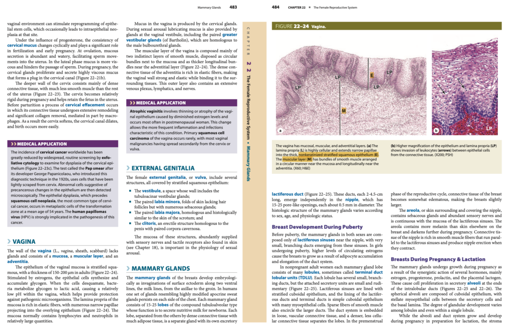
医療応用 (Medical Application)
子宮頸がん (Cervical Cancer) の発生率 (Incidence) は、世界的に大幅に減少 (Greatly Reduced Worldwide) しています。これは、子宮頸部上皮 (Cervical Epithelium) の異形成 (Dysplasia) を検査 (Examine) するための剥離細胞診 (Exfoliative Cytology) による広範かつ定期的なスクリーニング (Widespread, Routine Screening) の実施 (Implementation) によるものです(図22-23c参照)。
- この検査 (Test) は、開発者 (Developer) であるジョージ・パパニコラウ (George Papanicolaou) にちなんでパパニコロウ塗抹検査 (Pap Smear) と呼ばれます (Called)。
- 1920年代 (1920s) に導入 (Introduced) されたこの診断技術 (Diagnostic Technique) では、子宮頸部 (Cervix) から軽くこすり取られた (Lightly Scraped) 細胞 (Cells) を使用します。
- 前がん状態の変化 (Precancerous Changes) が示唆される異常な細胞 (Abnormal Cells Suggestive of Changes) は、その後顕微鏡で検出 (Detected Microscopically) されます。
扁平上皮異形成 (Epithelial Dysplasia) は、子宮頸がん (Cervical Cancer) の最も一般的なタイプ (Most Common Type) である扁平上皮性腫瘍 (Squamous Cell Neoplasia) に先行 (Precedes) します。
- この異形成 (Dysplasia) は、変換帯 (Transformation Zone) の化生細胞 (Metaplastic Cells) に発生することが多く、平均54歳 (Mean Age of 54 Years) で発生 (Occurs) します。
- ヒトパピローマウイルス (HPV, Human Papillomavirus) は、このがん (This Cancer) の病因 (Pathogenesis) に強く関与 (Strongly Implicated) しています。
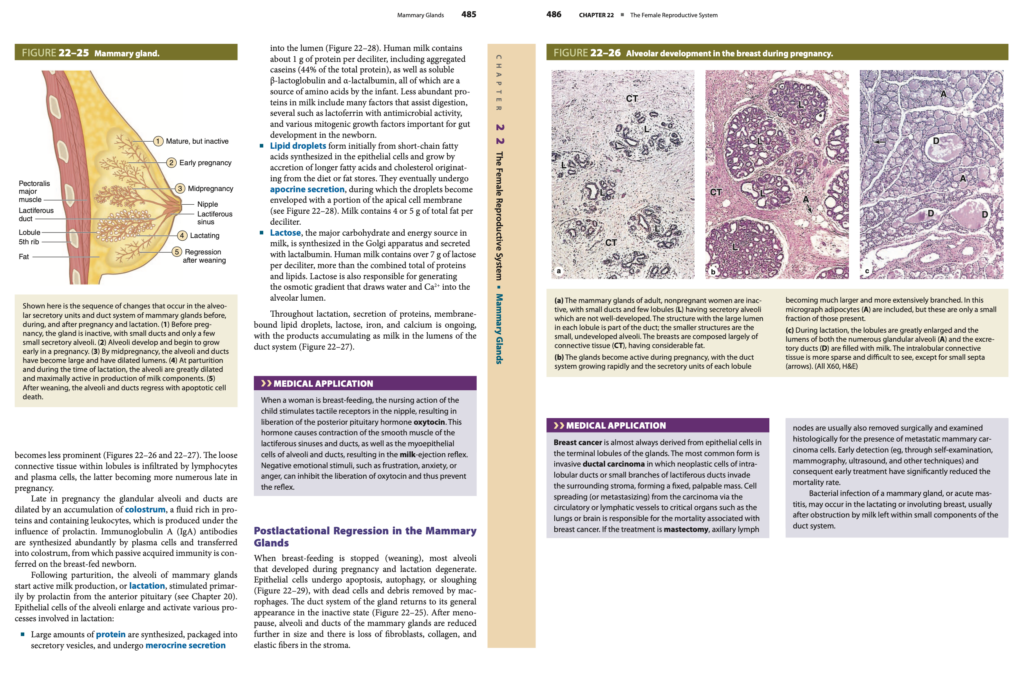
膣 (Vagina)
膣 (Vagina, ラテン語で「さや、鞘」) の壁 (Wall) は、腺 (Glands) を欠いている (Lacks) ため、粘膜 (Mucosa)、筋層 (Muscular Layer)、および外膜 (Adventitia) で構成されています。
膣の粘膜 (Mucosa of the Vagina)
- 膣の粘膜 (Vaginal Mucosa) の上皮 (Epithelium) は、重層扁平上皮 (Stratified Squamous Epithelium) です。
- 成人 (Adults) では、150-200μmの厚さ (Thickness of 150-200 μm) があります(図22–24参照)。
- エストロゲン (Estrogens) に刺激される (Stimulated) と、上皮細胞 (Epithelial Cells) はグリコーゲン (Glycogen) を合成 (Synthesize) して蓄積 (Accumulate) します。
- 細胞が脱落 (Desquamate, 脱落) すると、細菌 (Bacteria) がグリコーゲン (Glycogen) を代謝 (Metabolize) して乳酸 (Lactic Acid) を生成 (Produce) します。
- これにより、膣内 (Within the Vagina) は比較的低いpH (Relatively Low pH) になり、病原微生物 (Pathogenic Microorganisms) に対する保護 (Protection) に役立ちます (Helps Provide Protection)。
- 粘膜の固有層 (Lamina Propria of the Mucosa) には、豊富な弾性繊維 (Rich in Elastic Fibers) が含まれ、細い乳頭 (Narrow Papillae) が上皮 (Epithelium) に**突き出しています (Projecting Into)。
- 粘膜 (Mucosa) には、リンパ球 (Lymphocytes) や好中球 (Neutrophils) が比較的大量に (Relatively Large Quantities) 含まれています(図22–24参照)。
膣の粘液 (Mucus in the Vagina)
- 膣内の粘液 (Mucus in the Vagina) は、子宮頸腺 (Cervical Glands) によって産生されます (Produced)。
- 性的興奮時 (During Sexual Arousal) には、膣前庭 (Vaginal Vestibule) にある腺 (Glands) も、潤滑粘液 (Lubricating Mucus) を分泌 (Provide) します。
- 大前庭腺 (Greater Vestibular Glands, バルトリン腺とも呼ばれる Bartholin’s Glands) は、男性の球尿道腺 (Male Bulbourethral Glands) に相同 (Homologous) です。
膣の筋層 (Muscular Layer of the Vagina)
- 膣の筋層 (Muscular Layer of the Vagina) は、主に2つの不明瞭な層 (Two Indistinct Layers) から構成されています。
- 粘膜のすぐ隣 (Next to the Mucosa) には円形の平滑筋束 (Circular Bundles of Smooth Muscle) があり、
- 外膜の近く (Near the Adventitial Layer) には、厚い縦方向の平滑筋束 (Thicker Longitudinal Bundles) があります(図22–24参照)。
膣の外膜 (Adventitia of the Vagina)
- 膣の外膜 (Adventitia of the Vagina) は、高密度の結合組織 (Dense Connective Tissue) から構成 (Composed) されています。
- 弾性繊維 (Elastic Fibers) が豊富 (Rich) であるため、膣壁 (Vaginal Wall) は強靭 (Strong) かつ弾力性がある (Elastic) 構造を持ち、周囲の組織 (Surrounding Tissues) に結合 (Binding) されています。
- 外膜 (Outer Layer) には、広範な静脈叢 (Extensive Venous Plexus)、リンパ管 (Lymphatics)、および神経 (Nerves) が**含まれています (Contains)。
医療応用 (Medical Application)
- 萎縮性膣炎 (Atrophic Vaginitis) は、エストロゲン濃度の低下 (Diminished Estrogen Levels) によって引き起こされる膣上皮 (Vaginal Epithelium) の菲薄化 (Thinning) または萎縮 (Atrophy) です。
- 閉経後の女性 (Postmenopausal Women) に最も頻繁に発生 (Occurs Most Often) します。
- 変化 (Change) により、炎症 (Inflammation) や感染 (Infections) の頻度が増加 (More Frequent) し、この状態の特徴 (Characteristic of This Condition) となります。
- 原発性の扁平上皮がん (Primary Squamous Cell Carcinoma of the Vagina) はまれに発生 (Occurs Rarely) しますが、多くは子宮頸部 (Cervix) や外陰部 (Vulva) から二次的に広がる (Spread Secondarily) ものです。
女性の外陰部 (Female External Genitalia, Vulva)
女性の外陰部 (Female External Genitalia, または Vulva) には、いくつかの構造 (Several Structures) が含まれており、すべて重層扁平上皮 (Stratified Squamous Epithelium) で覆われています (Covered)。
- 前庭 (Vestibule)
- 管状胞状の前庭腺 (Tubuloacinar Vestibular Glands) を含む壁 (Wall Including Glands) を持つ空間 (Space) です。
- 小陰唇 (Labia Minora)
- 対をなすヒダ (Paired Folds of Skin) であり、毛包 (Hair Follicles) がない (Lacking) ものの、多数の皮脂腺 (Numerous Sebaceous Glands) を含んでいます。
- 大陰唇 (Labia Majora)
- 対をなすヒダ (Paired Folds of Skin) であり、陰嚢 (Scrotum) に相同 (Homologous) であり、組織学的にも類似 (Histologically Similar) しています。
- 陰核 (Clitoris)
- 勃起性の構造 (Erectile Structure) であり、陰茎 (Penis) に相同 (Homologous) です。
- 対をなす陰茎海綿体 (Paired Corpora Cavernosa) を含んでいます。
これらの構造の粘膜 (Mucosa of These Structures) には、感覚神経 (Sensory Nerves) および皮膚に見られる触覚受容体 (Tactile Receptors Found in Skin, 第18章参照) が豊富に供給 (Abundantly Supplied) されています。
これらの神経および受容体 (Nerves and Receptors) は、性的興奮 (Sexual Arousal) の生理学において重要 (Important in Physiology) な役割を果たします (Play a Significant Role)。
乳房の乳腺 (Mammary Glands of the Breasts)
乳房の乳腺 (Mammary Glands of the Breasts) は、発生学的 (Embryologically) には、表面外胚葉 (Surface Ectoderm) の陥入 (Invaginations) として発達 (Develop) します。
- 乳腺 (Mammary Glands) は、脇の下 (Axillae) から鼠径部 (Groin) までの2本の腹側線 (Two Ventral Lines)、すなわち乳腺線 (Milk Lines) に沿って発生します。
- ヒト (Humans) では、左右の胸部 (Each Side of the Chest) に一対の乳腺 (One Set of Glands) が残ります (Persists)。
- これらの乳腺 (Mammary Glands) は、高度に修飾された頂泌汗腺 (Highly Modified Apocrine Sweat Glands) に類似 (Resembling) しています。
乳腺の構造 (Structure of the Mammary Glands)
- 乳腺 (Mammary Glands) は、15〜25の小葉 (15-25 Lobes) から構成されており、複合管胞状腺 (Compound Tubuloalveolar Type) に分類されます。
- その機能 (Function) は、新生児 (Newborns) に栄養豊富な乳 (Nutritive Milk) を分泌 (Secrete) することです。
- 各小葉 (Lobe) は、多くの脂肪組織 (Much Adipose Tissue) を含む高密度の結合組織 (Dense Connective Tissue) によって他の小葉と分離 (Separated) されており、それぞれが独立した腺 (Separate Gland) です。
- 各小葉 (Lobe) には排泄乳管 (Excretory Lactiferous Duct) が1本 (One Duct) 存在し、これが独立して (Independently) 乳頭 (Nipple) に開口 (Emerge) します(図22–25参照)。
- 排泄乳管 (Lactiferous Ducts) は、2〜4.5 cmの長さ (2-4.5 cm Long) があり、乳頭 (Nipple) には15〜25個の小孔 (15-25 Pore-Like Openings) が開いており (Open)、各開口 (Each Opening) の直径は約0.5 mm (About 0.5 mm in Diameter) です。
思春期の乳房の発達 (Breast Development During Puberty)
- 思春期前 (Before Puberty) では、男女 (Both Sexes) の乳腺 (Mammary Glands) は、乳頭 (Nipple) の近くにある乳管洞 (Lactiferous Sinuses) のみ (Only) で構成されており、これらの洞 (Sinuses) から分岐する非常に小さな管 (Very Small, Branching Ducts) が出現 (Emerging) します。
- 思春期の少女 (Girls Undergoing Puberty) では、循環中のエストロゲン (Circulating Estrogens) の高レベル (Higher Levels) によって乳房 (Breasts) が成長 (Grow) します。
- これは、脂肪細胞 (Adipocytes) の蓄積 (Accumulation) および管系の伸長 (Elongation of the Duct System) によるものです。
成人女性の乳腺 (Mammary Glands in Adult Women)
- 妊娠していない成人女性 (Nonpregnant Adult Women) では、各小葉 (Each Mammary Gland Lobe) は、多くの小葉 (Many Lobules) で構成されています。
- 小葉 (Lobules) は、時折末端乳管小葉単位 (Terminal Duct Lobular Units, TDLU) と呼ばれます。
- 各小葉 (Lobule) には、いくつかの小さな分岐管 (Several Small, Branching Ducts) がありますが、付随する分泌単位 (Attached Secretory Units) は小さく (Small)、原始的 (Rudimentary) です(図22–25参照)。
乳頭と乳輪 (Nipple and Areola)
- 乳輪 (Areola) は、乳頭 (Nipple) を囲む皮膚 (Skin Surrounding the Nipple) です。
- 乳輪 (Areola) には皮脂腺 (Sebaceous Glands) が含まれており、感覚神経 (Abundant Sensory Nerves) も豊富 (Abundant) です。
- 乳輪 (Areola) は、乳管洞の粘膜 (Mucosa of the Lactiferous Sinuses) と連続 (Continuous) しています。
- 妊娠中 (During Pregnancy)、乳輪 (Areola) のメラニン (Melanin) は増加 (Increases) し、さらに暗くなります (Darkens Further)。
乳頭 (Nipple)
- 乳頭 (Nipple) の結合組織 (Connective Tissue) には、平滑筋繊維 (Smooth Muscle Fibers) が豊富 (Rich) です。
- これらの平滑筋 (Smooth Muscle) は、乳管洞 (Lactiferous Sinuses) に平行 (Parallel) に走行し、収縮 (Contraction) によって乳頭の勃起 (Nipple Erection) を引き起こします (Produce)。
妊娠および授乳中の乳房 (Breasts During Pregnancy & Lactation)
妊娠中 (During Pregnancy)、乳腺 (Mammary Glands) は、いくつかのホルモン (Several Hormones) の相乗効果 (Synergistic Action) によって成長 (Growth) します。主要なホルモン (Main Hormones) には、以下が含まれます。
- エストロゲン (Estrogen)
- プロゲステロン (Progesterone)
- プロラクチン (Prolactin)
- 胎盤性ラクトゲン (Placental Lactogen)
これらのホルモンは、小葉内乳管 (Intralobular Ducts) の末端 (Ends) にある分泌性肺胞 (Secretory Alveoli) における細胞増殖 (Cell Proliferation) を促進 (Cause) します(図22–25および図22–26参照)。
- 球形の肺胞 (Spherical Alveoli) は、立方上皮 (Cuboidal Epithelium) で構成 (Composed) され、
- 分泌細胞 (Secretory Cells) と基底膜 (Basal Lamina) の間 (Between) には、星状の筋上皮細胞 (Stellate Myoepithelial Cells) が存在します。
- 腺の発達の程度 (Degree of Glandular Development) は、小葉間 (Among Lobules)、さらには単一の小葉内 (Within a Single Lobule) でも異なります (Varies)。
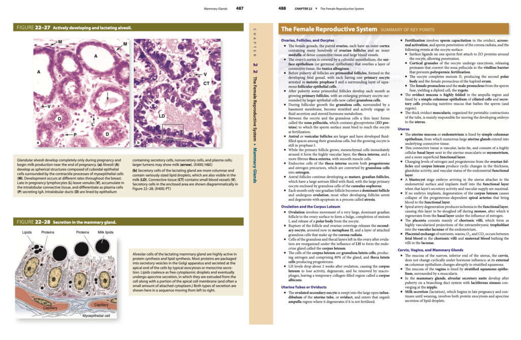
妊娠中の乳腺の変化 (Changes in the Mammary Glands During Pregnancy)
- 肺胞 (Alveoli) および管系 (Duct System) が成長 (Grow) し、授乳 (Lactation) に備えて発達 (Develop in Preparation) する一方で、間質 (Stroma) は目立たなくなります (Becomes Less Prominent)(図22–26および図22–27参照)。
- 小葉内の疎な結合組織 (Loose Connective Tissue within Lobules) には、リンパ球 (Lymphocytes) や形質細胞 (Plasma Cells) が浸潤 (Infiltrated) し、
- 妊娠後期 (Late in Pregnancy) には、形質細胞 (Plasma Cells) の数が増加 (Become More Numerous) します。
初乳 (Colostrum) の産生 (Production of Colostrum)
- 妊娠後期 (Late in Pregnancy) には、プロラクチン (Prolactin) の影響下 (Under the Influence) で、腺胞 (Glandular Alveoli) および乳管 (Ducts) が拡張 (Dilated) し、初乳 (Colostrum) が蓄積 (Accumulation) します。
- 初乳 (Colostrum) は、タンパク質が豊富 (Rich in Proteins) で、白血球 (Leukocytes) を含む (Containing) 液体です。
- 形質細胞 (Plasma Cells) は、免疫グロブリンA (IgA, Immunoglobulin A) 抗体を豊富に合成 (Synthesized Abundantly) し、初乳 (Colostrum) に移行 (Transferred) させます。
- 初乳 (Colostrum) からは、母乳を与えられた新生児 (Breast-Fed Newborn) に受動的獲得免疫 (Passive Acquired Immunity) が付与 (Conferred) されます。
分娩後の授乳 (Lactation After Parturition)
- 分娩後 (Following Parturition)、乳腺の肺胞 (Alveoli of Mammary Glands) は活発な乳の産生 (Active Milk Production)、すなわち授乳 (Lactation) を開始 (Start) します。
- 授乳 (Lactation) は、下垂体前葉 (Anterior Pituitary) からのプロラクチン (Prolactin) によって主に刺激 (Stimulated Primarily) されます(第20章参照)。
乳腺の分泌活動 (Secretory Activities of the Mammary Glands)
乳腺の上皮細胞 (Epithelial Cells of the Alveoli) は肥大 (Enlarge) し、以下の授乳に関わるプロセス (Processes Involved in Lactation) を活性化 (Activate) します。
- タンパク質の分泌 (Protein Secretion)
- 大量のタンパク質 (Large Amounts of Protein) が合成 (Synthesized) され、分泌小胞 (Secretory Vesicles) にパッケージ (Packaged) され、
- 分泌 (Merocrine Secretion) によって腔内 (Lumen) に放出 (Released) されます(図22-28参照)。
- 脂肪の分泌 (Lipid Secretion)
- 脂質滴 (Lipid Droplets) は、上皮細胞 (Epithelial Cells) 内で短鎖脂肪酸 (Short-Chain Fatty Acids) から形成 (Form) され、
- 食事 (Diet) や脂肪貯蔵 (Fat Stores) に由来する長鎖脂肪酸 (Longer Fatty Acids) やコレステロール (Cholesterol) によって成長 (Grow) します。
- 脂質 (Lipids) は頂泌分泌 (Apocrine Secretion) によって放出 (Released) され、
- 乳 (Milk) には1デシリットルあたり4〜5gの総脂肪 (4-5 g of Total Fat per Deciliter) が含まれています (Contains)。
- 乳糖の分泌 (Lactose Secretion)
- 乳 (Milk) の主な炭水化物 (Major Carbohydrate) であり、エネルギー源 (Energy Source) である乳糖 (Lactose) は、ゴルジ装置 (Golgi Apparatus) 内で合成 (Synthesized) され、
- ラクトアルブミン (Lactalbumin) と共に分泌 (Secreted) されます。
医療応用 (Medical Application)
- 授乳中 (During Breast-Feeding)、乳頭 (Nipple) にある触覚受容体 (Tactile Receptors) が刺激される (Stimulated) と、
- 後葉ホルモン (Posterior Pituitary Hormone) であるオキシトシン (Oxytocin) が放出 (Liberated) されます。
- オキシトシン (Oxytocin) は、乳管洞 (Lactiferous Sinuses)、乳管 (Ducts)、および肺胞 (Alveoli) の平滑筋 (Smooth Muscle) を収縮 (Contract) させ、
- 乳射出反射 (Milk-Ejection Reflex) を引き起こします (Results in the Reflex)。
授乳後の乳腺の退行 (Postlactational Regression in the Mammary Glands)
- 授乳が終了 (When Breast-Feeding Stops) すると、妊娠中および授乳中に発達した (Developed During Pregnancy and Lactation) ほとんどの肺胞 (Alveoli) が退縮 (Degenerate) します。
- 乳腺の導管系 (Duct System of the Gland) は非活動状態 (Inactive State) に戻ります(図22-25参照)。
医療応用 (Medical Application)
乳がん (Breast Cancer) は、ほぼ常に腺の末端小葉 (Terminal Lobules of the Glands) にある上皮細胞 (Epithelial Cells) に由来します。
- 最も一般的な形態は浸潤性乳管がん (Invasive Ductal Carcinoma) で、小葉内乳管 (Intralobular Ducts) や乳管の小さな分枝 (Small Branches of Lactiferous Ducts) に存在する腫瘍細胞 (Neoplastic Cells) が周囲の間質 (Surrounding Stroma) に浸潤 (Invade) します。
- これにより、固定された (Fixed) かつ触知可能な腫瘤 (Palpable Mass) が形成 (Form) されます。
がん細胞 (Cancer Cells) は、循環系 (Circulatory Vessels) またはリンパ管 (Lymphatic Vessels) を介して肺 (Lungs) や脳 (Brain) などの重要な臓器 (Critical Organs) へ転移 (Metastasize) します。
- 乳がん (Breast Cancer) における死亡率 (Mortality) は、これらの転移 (Metastasis) によるものです。
治療 (Treatment) に乳房切除術 (Mastectomy) が含まれる場合、通常は腋窩リンパ節 (Axillary Lymph Nodes) も外科的に切除 (Removed Surgically) されます。
- 腋窩リンパ節 (Axillary Lymph Nodes) は組織学的に検査 (Examined Histologically) され、転移性の乳がん細胞 (Metastatic Mammary Carcinoma Cells) が存在するかどうか (Presence of Cells) を確認します。
早期発見 (Early Detection) と早期治療 (Early Treatment) は、死亡率 (Mortality Rate) の大幅な低下 (Significantly Reduced) に貢献 (Contribute) しています。
- 早期発見の方法 (Methods of Early Detection) には以下が含まれます。
- 自己検診 (Self-Examination)
- マンモグラフィ (Mammography)
- 超音波検査 (Ultrasound)
- その他の技術 (Other Techniques)
乳腺の細菌感染症 (Bacterial Infection of a Mammary Gland)、すなわち急性乳腺炎 (Acute Mastitis) は、授乳中 (Lactating) または退縮期 (Involuting Breast) に発生する可能性があります (May Occur)。
- 急性乳腺炎 (Acute Mastitis) は、乳管系 (Duct System) の小さな部分 (Small Components) に残留した乳 (Milk Left Within) による閉塞 (Obstruction) の後 (After) に通常発生 (Usually Occurs) します。
サマリー
卵巣、卵胞、および卵母細胞 (Ovaries, Follicles, and Oocytes)
- 女性の生殖腺 (Female Gonads) である一対の卵巣 (Paired Ovaries) には、
- 数百の卵胞 (Many Hundreds of Ovarian Follicles) を含む外側の皮質 (Outer Cortex) と、
- 高密度の結合組織 (Dense Connective Tissue) および大きな血管 (Large Blood Vessels) を含む内側の髄質 (Inner Medulla) があります。
- 卵巣の皮質 (Ovary’s Cortex) は、立方上皮の中皮 (Cuboidal Mesothelium) に覆われ (Covered) ており、これを表面上皮 (Surface Epithelium または Germinal Epithelium) と呼びます。
- この表面上皮 (Surface Epithelium) は、結合組織の層 (Layer of Connective Tissue) である白膜 (Tunica Albuginea) の上に位置 (Overlies) しています。
- 思春期前 (Before Puberty) のすべての卵胞 (All Follicles) は、胎児の性腺 (Developing Fetal Gonad) において形成 (Formed) される原始卵胞 (Primordial Follicles) です。
- 各原始卵胞 (Primordial Follicle) には、減数分裂の第一分裂前期 (Meiotic Prophase I) で停止 (Arrested) した一次卵母細胞 (Primary Oocyte) が1つと、
- 周囲に存在する (Surrounding) 扁平な卵胞上皮細胞 (Squamous Follicular Epithelial Cells) から構成されます。
- 思春期以降 (After Puberty)、一部の原始卵胞 (Primordial Follicles) は毎月 (Each Month)成長中の一次卵胞 (Growing Primary Follicles) へと発達 (Develop) します。
- 一次卵母細胞 (Primary Oocyte) は拡大 (Enlarging) し、大きな上皮細胞 (Larger Epithelial Cells) に囲まれ (Surrounded)、これらの上皮細胞は顆粒細胞 (Granulosa Cells) と呼ばれます (Called)。
- 卵胞の成長 (Follicular Growth) の間、顆粒細胞 (Granulosa Cells) は基底膜 (Basement Membrane) に囲まれ (Surrounded)、
- 重層構造 (Stratified Structure) を形成 (Form) し、液体の分泌 (Fluid Secretion) およびステロイドホルモンの代謝 (Steroid Hormone Metabolism) に積極的に関与 (Actively Engage) します。
- 卵母細胞 (Oocyte) と顆粒細胞 (Granulosa Cells) の間には、透明帯 (Zona Pellucida) と呼ばれる薄い層 (Thin Layer) が形成 (Forms) されます。
- 透明帯 (Zona Pellucida) には糖タンパク質 (Glycoproteins, ZO Proteins) が含まれ (Contains) れており、
- 受精時 (At Fertilization) に精子の表面 (Sperm Surface) が結合 (Bind) する必要があります。
- 胞状卵胞 (Antral or Vesicular Follicles) は、より大きな (Larger) 卵胞であり、顆粒細胞 (Granulosa Cells) の間に液体で満たされた空間 (Fluid-Filled Spaces) が発達 (Developed) しています。
- ただし、成長中の卵母細胞 (Growing Oocyte) は依然として第一分裂前期 (Prophase I) に停止 (Arrested) しています。
- 一次卵胞 (Primary Follicle) が成長 (Grows) する間、その周囲の間充織細胞 (Mesenchymal Cells) は、
- 高血管層 (Highly Vascular Layer) である内卵胞膜 (Theca Interna) と、
- 平滑筋細胞 (Smooth Muscle Cells) を含む (Containing) より線維性の外卵胞膜 (More Fibrous Theca Externa) を形成 (Form) します。
- 内卵胞膜の内分泌細胞 (Endocrine Cells of the Theca Interna) は、プロゲステロン (Progesterone) およびエストロゲンの前駆体 (Estrogen Precursors) を分泌 (Secrete) します。
- これらの前駆体は、顆粒細胞 (Granulosa Cells) によってエストロゲン (Estrogen) に変換 (Converted) されます。
- 胞状卵胞 (Antral Follicles) は、成熟卵胞 (Mature Follicles)、すなわちグラーフ卵胞 (Graafian Follicles) として成長を続け (Continue Developing) します。
- これらの卵胞は、液体で満たされた大きな卵胞腔 (Large Antrum Filled with Fluid) を持つ (Have) ほか、
- 大きな一次卵母細胞 (Large Primary Oocyte) が卵丘顆粒細胞 (Granulosa Cells of the Cumulus Oophorus) に囲まれ (Enclosed) ています。
- 毎月 (Each Month)、グラーフ卵胞 (Graafian Follicle) のうち1つのみ (Only One) が優勢な卵胞 (Dominant Follicle) になり、排卵 (Ovulation) します。
- それ以外の発達中の卵胞 (Other Developing Follicles) は停止 (Arrest) し、アポトーシス (Apoptosis) による退化 (Degenerate) を経験 (Experience) します。
- このプロセス (Process) は、閉鎖 (Atresia) と呼ばれます。
排卵および黄体 (Ovulation and the Corpus Luteum)
- 排卵 (Ovulation) では、非常に大きな (Very Large)優勢なグラーフ卵胞 (Dominant Graafian Follicle) が卵巣の表面 (Ovary Surface) へ移動 (Movement) し、
- 膨らみ (Bulge) を形成 (Form) し、第一減数分裂 (Meiosis I) が完了 (Completion) し、
- 卵母細胞 (Oocyte) から極体 (Polar Body) が放出 (Release) されます。
- 卵胞の破裂 (Rupture of the Follicle) および卵巣被膜 (Ovarian Coverings) により、
- 二次卵母細胞 (Secondary Oocyte) と、顆粒細胞 (Granulosa Cells) の層 (Layer) からなる放線冠 (Corona Radiata) が放出 (Releases) されます。
- 黄体 (Corpus Luteum) の細胞 (Cells) には、
- 顆粒黄体細胞 (Granulosa Lutein Cells)(腺の80% (80% of the Gland))および卵胞膜黄体細胞 (Theca Lutein Cells) が含まれます (Contained)。
卵管または輸卵管 (Uterine Tubes or Oviducts)
- 排卵された二次卵母細胞 (Ovulated Secondary Oocyte) は、卵管 (Uterine Tube または Oviduct) の大きく開いた漏斗部 (Large Open Infundibulum) に取り込まれ (Swept Into)、
- 膨大部 (Ampulla Region) に入り (Enters) ます。
- もし受精 (Fertilization) しなければ、卵母細胞 (Oocyte) は退化 (Degenerates) します。
受精 (Fertilization)
受精 (Fertilization) には、以下の一連のプロセス (Series of Processes) が含まれます。
- 1. 精子の受精能獲得 (Sperm Capacitation)
- 卵管 (Oviduct) で精子 (Sperm) が活性化 (Capacitation) します。
- 2. 先体反応 (Acrosomal Activation)
- 精子 (Sperm) における先体の活性化 (Acrosomal Activation) が誘発 (Induced) されます。
- 3. 放線冠への精子の侵入 (Sperm Penetration of the Corona Radiata)
- 精子 (Sperm) は、卵母細胞 (Oocyte) の放線冠 (Corona Radiata) を通過 (Penetrate) します。
- 4. 卵母細胞の表面でのイベント (Events at the Oocyte Surface)
- 精子の表面リガンド (Surface Ligands on Sperm) が、卵母細胞 (Oocyte) を取り囲む (Surrounding) 透明帯 (Zona Pellucida) に存在するZOタンパク質 (ZO Proteins) に最初に付着 (Attach) します。
- 卵母細胞の皮質顆粒 (Cortical Granules of the Oocyte) が開口分泌 (Exocytosis) を起こし (Undergo)、
- プロテアーゼ (Proteases) を放出 (Release) します。
- このプロテアーゼ (Proteases) は、透明帯 (Zona Pellucida) をビテリナバリア (Vitelline Barrier) に変換 (Convert) します。
- ビテリナバリア (Vitelline Barrier) によって、多精子受精 (Polyspermic Fertilization) が防止 (Prevents) されます。
- 卵母細胞 (Oocyte) は、第二減数分裂 (Meiosis II) を完了 (Completes) し、
- 第二極体 (Second Polar Body) および半数体の雌性前核 (Haploid Female Pronucleus) を生成 (Produce) します。
- 雌性前核 (Female Pronucleus) と精子由来の雄性前核 (Male Pronucleus from the Sperm) が融合 (Fuse) し、
- 二倍体細胞 (Diploid Cell) である接合子 (Zygote) が形成 (Yield) されます。
卵管の構造 (Structure of the Oviduct)
- 卵管の粘膜 (Oviduct Mucosa) は、膨大部 (Ampulla Region) において高度に折りたたまれた (Highly Folded) 構造を持っています (Have)。
- 単層円柱上皮 (Simple Columnar Epithelium) で覆われ (Lined) ており、
- 線毛細胞 (Ciliated Cells) および分泌細胞 (Secretory Cells) を含んでいます (Contain)。
- 分泌細胞 (Secretory Cells) は、栄養粘液 (Nutritive Mucus) を生成 (Produce) し、
- 精子 (Sperm) や接合子 (Zygote) を覆います (Bathe)。
- 卵管の筋層 (Oviduct Muscularis) は厚い (Thick) 構造を持ち (Have)、
- 蠕動運動 (Peristaltic Contractions) を促進するように編成 (Organized for) されています。
- この筋層の収縮 (Contraction of the Muscularis) によって、
- 発生中の胚 (Developing Embryo) が子宮 (Uterus) へ移動 (Moves) します。
子宮 (Uterus)
- 子宮粘膜 (Uterine Mucosa または Endometrium) は、単層円柱上皮 (Simple Columnar Epithelium) で裏打ち (Lined) されており、
- 多数の大きな子宮腺 (Numerous Large Uterine Glands) が基底の結合組織 (Underlying Connective Tissue) に伸びています (Extend Into)。
- この結合組織 (This Connective Tissue) は、
- 血管が豊富 (Vascular) で、脂肪を欠き (Lacks Fat)、
- 子宮の筋層 (Uterine Muscularis または Myometrium) に隣接する (Next To) 高い細胞密度の基底層 (Highly Cellular Basal Layer) と、
- より表層の機能層 (More Superficial Functional Layer) から構成 (Consists) されています。
- 卵巣の卵胞 (Ovarian Follicles) および黄体 (Corpus Luteum) から分泌される (Produced)エストロゲン (Estrogen) およびプロゲステロン (Progesterone) の濃度の変化 (Changing Levels) によって、
- 子宮内膜の機能層 (Endometrial Functional Layer) の厚さ (Thickness)、腺の活動 (Glandular Activity)、および血管の状態 (Vascular Status) に周期的な変化 (Cyclic Changes) が引き起こされます (Produce)。
- 胚盤胞 (Blastocyst Stage Embryo) が子宮に到達 (Arriving in the Uterus) すると、
- 子宮内膜の表面 (Endometrial Surface) に付着 (Attaches) し、
- 機能層 (Functional Layer) に埋め込まれ (Implants Itself) ます。
- このとき、分泌活動 (Secretory Activity) および血管供給 (Vascular Supply) が最大 (Maximal) になります。
- 胚 (Embryo) が着床 (Implants) しなかった場合、
- 黄体 (Corpus Luteum) の退化 (Degeneration) によって、
- 機能層 (Functional Layer) に血液を供給する (Bring Blood) プロゲステロン依存性のらせん動脈 (Progesterone-Dependent Spiral Arteries) が崩壊 (Collapse) します。
- らせん動脈の退縮 (Spiral Artery Degeneration) によって、
- 機能層 (Functional Layer) に虚血 (Ischemia) が発生 (Produces) し、
- 機能層 (Functional Layer) は剥がれ落ち (Sloughed Off)、
- 月経 (Menses) として排出 (Discharged) されます。
- その後、基底層 (Basal Layer) から再生 (Regenerates) し、
- エストロゲン (Estrogen) の影響下 (Under the Influence) で新たな機能層が形成 (New Functional Layer Forms) されます。
- 胎盤 (Placenta) は、
- 栄養膜 (Extraembryonic Trophoblast) から子宮内膜 (Endometrium) の血管性の小腔 (Vascular Lacunae) に突き出した (Project Into)、
- 高度に血管化された (Highly Vascularized) 絨毛 (Chorionic Villi) から主に構成 (Consists Mainly) されています。
- 胎盤 (Placenta) では、
- 栄養素 (Nutrients)、老廃物 (Wastes)、酸素 (O₂)、および二酸化炭素 (CO₂) の交換 (Exchange) が、
- 絨毛の胎児血 (Fetal Blood in the Chorionic Villi) と、
- 小腔内の母体血 (Maternal Blood Bathing the Villi in the Lacunae) の間 (Between) で行われます (Occurs)。
子宮頸部、膣、および乳腺 (Cervix, Vagina, and Mammary Glands)
子宮頸部 (Cervix)
- 子宮の下部 (Inferior End of the Uterus) に位置する子宮頸部 (Cervix) の粘膜 (Mucosa) は、
- ホルモンの影響下 (Under Hormone Influence) で周期的に変化しません (Does Not Change Cyclically)。
- 外子宮口 (External Os) では、
- 円柱上皮 (Columnar Epithelium) が急激に (Abruptly) 重層扁平上皮 (Stratified Squamous Epithelium) に変化 (Changes) します。
膣 (Vagina)
- 膣 (Vagina) の粘膜 (Mucosa) は、
- 重層扁平上皮 (Stratified Squamous Epithelium) で裏打ち (Lined) され、
- 筋層 (Muscularis) に囲まれています (Surrounded)。
乳腺 (Mammary Glands)
- 乳腺 (Mammary Glands) では、
- 分岐乳管系 (Branching Duct System) に乳腺洞 (Lactiferous Sinuses) が合流 (Converging) し、
- 乳頭 (Nipple) に開口 (Open) します。
- アルベオラー分泌単位 (Alveolar Secretory Units) は、
- 思春期後 (After Puberty) に発達 (Develop) します。
- 母乳の分泌 (Milk Secretion または Lactation) は、
- 妊娠後期 (Late Pregnancy) に始まり (Begins)、離乳 (Weaning) まで続きます (Continues)。
- 乳の分泌 (Milk Secretion) には、
- タンパク質 (Protein) の開口分泌 (Exocytosis) と、
- 脂肪滴 (Lipid Droplets) の頂泌分泌 (Apocrine Secretion) が関与 (Involves) します。
教科書問題
1. 卵胞発達段階での卵胞液蓄積の開始
Q: Which stage of ovarian follicle development is characterized by an initial period of follicular fluid accumulation?
a. Graafian follicle
b. Mature follicle
c. Primordial follicle
d. Oocyte
e. Secondary follicle
Answer: e. Secondary follicle
Explanation:
二次卵胞(secondary follicle)は、卵胞液が初めて形成され始める段階です。卵胞腔(antrum)が発達し、顆粒膜細胞に囲まれた構造が見られます。
他の選択肢について:
- Graafian follicle: 最終的に排卵する成熟卵胞。
- Primordial follicle: 初期の卵胞で液体の蓄積は見られない。
- Oocyte: 卵胞の中の卵細胞そのもの。
- Mature follicle: 卵胞液は既に十分に蓄積している。
2. 顆粒膜黄体細胞の特徴
Q: Which of the following is characteristic of granulosa lutein cells?
a. Are a minor cell type in the corpus luteum
b. Derive from the theca interna
c. Contain abundant rough endoplasmic reticulum
d. Are small and dark-staining
e. Secrete progesterone
Answer: e. Secrete progesterone
Explanation:
顆粒膜黄体細胞(granulosa lutein cells)は、黄体で主要なプロゲステロン(progesterone)分泌細胞です。他の選択肢について:
- a: 顆粒膜黄体細胞は黄体の主要細胞。
- b: 内卵胞膜細胞ではなく、顆粒膜細胞由来。
- c: 粗面小胞体ではなく、滑面小胞体が豊富。
- d: 大きく、淡染性。
3. 排卵を引き起こすホルモン
Q: Which of the following hormones is primarily responsible for inducing ovulation?
a. Relaxin
b. LH
c. Progesterone
d. FSH
e. Estrogen
Answer: b. LH
Explanation:
黄体形成ホルモン(LH)は、急激なサージが起こることで排卵を誘導します。他の選択肢について:
- Relaxin: 子宮筋弛緩や分娩時の骨盤弛緩に関与。
- Progesterone: 排卵後に黄体が分泌するホルモン。
- FSH: 卵胞の成熟を助けるが排卵の直接的な誘因ではない。
- Estrogen: LHサージを引き起こす要因の1つ。
4. 白体の特徴
Q: Which feature is characteristic of the corpora albicans but not of atretic follicles?
a. May contain degenerating granulosa cells floating in remnants of follicular fluid
b. Resemble large collagenous scars
c. Eventually removed by macrophages and replaced by stroma
d. Are remnants of follicles that degenerate before maturation
e. May contain degenerating oocytes
Answer: b. Resemble large collagenous scars
Explanation:
白体(corpora albicans)は、排卵後の黄体が退化してできる大きな膠原性瘢痕に似た構造です。他の選択肢について:
- a: これは退化卵胞(atretic follicles)の特徴。
- c: 白体にも適用されるが、選択肢bがより明確な特徴。
- d: 退化卵胞に該当。
- e: 退化卵胞に見られる場合がある。
5. 子宮内膜腺の発達と分泌物の充満
Q: Endometrial glands are typically most fully developed and filled with product during which day(s) or phase of a woman’s menstrual cycle?
a. Menstrual phase
b. Days 1-4
c. The day ovulation occurs
d. Proliferative phase
e. Days 15-28
Answer: e. Days 15-28
Explanation:
子宮内膜腺は、分泌期(days 15-28)に最も発達し、分泌物で満たされます。他の選択肢について:
- a, b: 月経期には分泌腺は壊れる。
- c: 排卵日は腺がまだ十分に発達していない。
- d: 増殖期には腺が伸長するが、分泌物は少ない。
6. 基底層の特徴
Q: Which feature is characteristic of the endometrium’s basal layer but not of its functional layer?
a. Includes the uterine surface epithelium
b. Includes connective tissue
c. Contains cells that replace the surface epithelium after menstruation
d. Relies solely on spiral arteries for its blood supply
e. Undergoes cyclic thickening and shedding
Answer: c. Contains cells that replace the surface epithelium after menstruation
Explanation:
基底層(basal layer)は、月経後の子宮内膜再生に必要な細胞を含んでいます。他の選択肢について:
- a: 表層上皮は機能層の一部。
- b: 両層とも結合組織を含む。
- d: 基底層はらせん動脈だけでなく基底動脈からも供給される。
- e: 周期的な変化は機能層に限られる。
7. 母乳中の脂質分泌機構
Q: Most lipid in milk is released from cells by which mechanism?
a. Apocrine secretion
b. Paracrine secretion
c. Holocrine secretion
d. Merocrine secretion
e. Autocrine secretion
Answer: a. Apocrine secretion
Explanation:
母乳中の脂質はアポクリン分泌(apocrine secretion)により細胞から放出されます。他の選択肢について:
- b: 細胞間情報伝達で使われる。
- c: 細胞全体が崩壊して分泌する方法(例:皮脂腺)。
- d: 膜を通過して分泌する方式(例:タンパク質)。
- e: 自己分泌でホルモン調整に使われる。
8. 基礎体温とホルモン値の異常
Q: A 33-year-old woman with an average menstrual cycle of 28 days comes in for a routine Pap smear. It has been 35 days since the start of her last menstrual period, and a vaginal smear reveals clumps of basophilic cells. As her physician you suspect which of the following?
a. She will begin menstruating in a few days.
b. She will ovulate within a few days.
c. Her serum progesterone levels will be found to be very low.
d. There will be detectable levels of hCG in her serum and urine.
e. She is undergoing early menopause.
Answer: d. There will be detectable levels of hCG in her serum and urine.
Explanation:
月経が遅れ、塗抹検査で基底好性細胞が見られることは妊娠の可能性を示唆し、hCGの測定が推奨されます。他の選択肢について:
- a: 月経前には基底好性細胞は通常見られない。
- b: 排卵後35日以上経過している。
- c: 妊娠ではプロゲステロン値が高い。
- e: 初期閉経は他の症状と併発する。
9. 異所性妊娠が見られる卵管部位
Q: The surgeon removes her right uterine tube that is inflamed, scarified, and contains the implanted embryonic tissue in the region where fertilization normally occurs. Where is this?
a. The uterine part of the oviduct
b. The ampulla region with highly folded mucosa
c. The only oviduct region attached to the mesosalpinx
d. The infundibulum region with fimbriae
e. The isthmus region
Answer: b. The ampulla region with highly folded mucosa
Explanation:
受精は卵管膨大部(ampulla)で最も頻繁に起こり、異所性妊娠もこの部位で発生することが多いです。他の選択肢について:
- a: 子宮内に近い部分。
- c: 卵管全体が間接的に中間膜に接続される。
- d: 卵管采(fimbriae)を持つ開口部。
- e: 子宮に近い狭窄部位。
10. 膣内の低pH維持のメカニズム
Q: The normally low pH in the vagina is maintained by which of the following?
a. A proton pump in the epithelial cells similar to that in osteoclasts and parietal cells
b. Secretions derived from intracellular carbonic acid
c. Secretion of lactic acid by cells of the stratified squamous epithelium
d. Bacterial metabolism of glycogen to produce an organic acid
e. Synthesis and accumulation of acid hydrolases in the epithelium
Answer: d. Bacterial metabolism of glycogen to produce an organic acid
Explanation:
膣内の低pHは、乳酸菌が上皮細胞のグリコーゲンを代謝し乳酸を生成することで維持されます。他の選択肢について:
- a, b: プロトンポンプや炭酸は関与しない。
- c: 上皮細胞そのものは乳酸を分泌しない。
- e: 酸性加水分解酵素の生成は関係ない。
BRS
二次卵胞の特徴(Secondary Ovarian Follicles)
Q1: Which of the following statements about secondary ovarian follicles is true?
a. They lack liquor folliculi.
b. They contain a secondary oocyte.
c. Their continued maturation requires follicle-stimulating hormone (FSH).
d. They have a single layer of cuboidal follicular cells surrounding the oocyte.
Answer: c. Their continued maturation requires follicle-stimulating hormone (FSH).
Explanation:
二次卵胞(secondary ovarian follicles)の成熟には卵胞刺激ホルモン(FSH:follicle-stimulating hormone)が必要です。FSHは視床下部-下垂体軸から分泌され、卵巣内の卵胞の成長を促進します。
他の選択肢が間違っている理由は以下の通りです:
- a. They lack liquor folliculi.
→ 誤り。二次卵胞は「液胞(liquor folliculi)」を持っています。これは顆粒膜細胞(granulosa cells)間に液体がたまったもので、一次卵胞には存在しません。 - b. They contain a secondary oocyte.
→ 誤り。二次卵胞には「一次卵母細胞(primary oocyte)」が含まれています。二次卵母細胞(secondary oocyte)は排卵時に形成されます。 - d. They have a single layer of cuboidal follicular cells surrounding the oocyte.
→ 誤り。一次卵胞には一層の立方形の顆粒膜細胞が存在しますが、二次卵胞では顆粒膜細胞の層が複数になります。
母乳(初乳)の抗体(Antibodies in Colostrum)
Q2: Which antibody is present in colostrum?
a. IgA
b. IgD
c. IgE
d. IgM
Answer: a. IgA
Explanation:
初乳(colostrum)にはIgA抗体が多く含まれています。IgAは新生児の消化管の粘膜を保護する役割を果たし、免疫防御を助けます。
他の選択肢が間違っている理由は以下の通りです:
- b. IgD
→ 誤り。IgDは主にB細胞の表面に見られ、母乳や初乳には含まれません。 - c. IgE
→ 誤り。IgEはアレルギー反応に関与する抗体であり、初乳や母乳には含まれません。 - d. IgM
→ 誤り。IgMは免疫系の初期応答に関与する抗体であり、母乳の主要な抗体ではありません。
黄体の変化(Corpus Luteum)
Q3: What happens to the corpus luteum after it ceases to function?
a. It produces luteinizing hormone (LH).
b. It produces follicle-stimulating hormone (FSH).
c. It becomes the corpus albicans.
d. It is derived from atretic follicles.
Answer: c. It becomes the corpus albicans.
Explanation:
黄体(corpus luteum)が機能を停止すると、**白体(corpus albicans)**になります。これは、退縮した黄体が結合組織に変化したもので、卵巣内に一時的に残ります。
他の選択肢が間違っている理由は以下の通りです:
- a. It produces luteinizing hormone (LH).
→ 誤り。LHは下垂体前葉から分泌されるホルモンであり、黄体が生成するものではありません。 - b. It produces follicle-stimulating hormone (FSH).
→ 誤り。FSHも下垂体前葉から分泌され、黄体が生成するホルモンではありません。 - d. It is derived from atretic follicles.
→ 誤り。黄体は排卵後のグラーフ卵胞から生じるもので、閉鎖卵胞(atretic follicles)からは形成されません。
月経周期の相(Phases of Menstrual Cycle)
Q4: Which of the following is a recognized phase of the menstrual cycle?
a. Gestational phase
b. Active phase
c. Follicular phase
d. Anaphase
Answer: c. Follicular phase
Explanation:
月経周期は卵胞期(follicular phase)、黄体期(luteal phase)、および月経期(menstrual phase)の3つの相で構成されています。卵胞期は卵胞の成熟とエストロゲンの分泌が促進される期間です。
他の選択肢が間違っている理由は以下の通りです:
- a. Gestational phase
→ 誤り。妊娠期(gestational phase)は妊娠に関する用語であり、月経周期には含まれません。 - b. Active phase
→ 誤り。月経周期には「active phase」という名称は使用されません。 - d. Anaphase
→ 誤り。「anaphase(中期)」は細胞分裂の一部を指す用語であり、月経周期の一部ではありません。
膣粘膜の特徴(Vaginal Mucosa)
Q5: Which of the following statements about the vaginal mucosa is true?
a. It is lined by stratified columnar epithelium.
b. It is lined by stratified squamous nonkeratinized epithelium.
c. It contains no elastic fibers.
d. It is lubricated by glands in the cervix.
Answer: b. It is lined by stratified squamous nonkeratinized epithelium.
Explanation:
膣の粘膜は重層扁平非角化上皮(stratified squamous nonkeratinized epithelium)で覆われています。この上皮は物理的な摩擦に耐えながらも柔軟性を維持する特徴があります。
他の選択肢が間違っている理由は以下の通りです:
- a. It is lined by stratified columnar epithelium.
→ 誤り。重層円柱上皮(stratified columnar epithelium)は膣の内壁には存在せず、他の器官(気管など)で見られます。 - c. It contains no elastic fibers.
→ 誤り。膣の粘膜下層には弾性線維(elastic fibers)が存在し、伸縮性を提供しています。 - d. It is lubricated by glands in the cervix.
→ 部分的に正しいが不十分です。膣自体には腺(glands)が存在しませんが、子宮頸部(cervix)の腺からの分泌物が膣を潤滑します。
卵管の特徴(Characteristics of the Oviduct)
Q6: Which of the following statements about the oviduct is true?
a. It is lined by a simple cuboidal epithelium.
b. Its epithelium contains peg cells.
c. Fertilization most often occurs in its fimbriated portion.
d. Its epithelium contains goblet cells.
Answer: b. Its epithelium contains peg cells.
Explanation:
卵管(oviduct)の上皮は単層円柱上皮(simple columnar epithelium)で構成され、繊毛細胞(ciliated cells)とペグ細胞(peg cells)からなります。ペグ細胞は、栄養物や分泌液を生成して、卵管内の環境を整える役割を果たします。
他の選択肢が間違っている理由は以下の通りです:
- a. It is lined by a simple cuboidal epithelium.
→ 誤り。卵管は**単層円柱上皮(simple columnar epithelium)**で覆われており、単層立方上皮(simple cuboidal epithelium)ではありません。 - c. Fertilization most often occurs in its fimbriated portion.
→ 誤り。受精は**卵管の膨大部(ampulla)**で最も頻繁に起こります。繊毛のついた卵管采(fimbriated portion)は、卵子を卵管に取り込む役割を果たしますが、受精の主な場ではありません。 - d. Its epithelium contains goblet cells.
→ 誤り。卵管の上皮にはゴブレット細胞(goblet cells)は存在しません。ゴブレット細胞は主に消化管(特に小腸や大腸)の粘液分泌に関与しています。
未熟奇形腫(Immature Teratoma)
Q7: Which one of the following teratomas is a malignant, fast-growing tumor?
a. Mature teratoma
b. Monodermal teratoma
c. Immature teratoma
d. Struma ovarii
Answer: c. Immature teratoma
Explanation:
未熟奇形腫(immature teratoma)は悪性腫瘍(malignant tumor)で、成長が速いのが特徴です。未熟な胚性組織を含むため、悪性度が高く、他の奇形腫と異なり、化学療法が必要になることが多いです。
他の選択肢が間違っている理由は以下の通りです:
- a. Mature teratoma
→ 誤り。**成熟奇形腫(mature teratoma)は良性腫瘍(benign tumor)**であり、しばしば皮様嚢腫(dermoid cyst)と呼ばれます。これには皮膚、毛髪、脂肪組織などが含まれます。 - b. Monodermal teratoma
→ 誤り。単一胚葉性奇形腫(monodermal teratoma)は一種類の細胞型で構成される腫瘍で、通常は良性です。 - d. Struma ovarii
→ 誤り。ストルーマ卵巣腫(struma ovarii)は、甲状腺組織を含むまれな種類の奇形腫で、ほとんどの場合、良性です。
黄体形成ホルモン(LH)の作用(Function of Luteinizing Hormone, LH)
Q8: Which of the following is a function of luteinizing hormone (LH)?
a. It triggers the completion of the second meiotic division in secondary oocytes.
b. It triggers ovulation.
c. It suppresses the release of estrogens.
d. It induces primary follicles to become secondary follicles.
Answer: b. It triggers ovulation.
Explanation:
黄体形成ホルモン(LH)は排卵(ovulation)を引き起こす重要なホルモンです。月経周期の中頃(約14日目)にLHの急上昇(LHサージ)が起こり、それが卵胞の破裂と卵子の放出を促進します。
他の選択肢が間違っている理由は以下の通りです:
- a. It triggers the completion of the second meiotic division in secondary oocytes.
→ 誤り。第二減数分裂(second meiotic division)の完了は受精時にのみ起こります。LHは第二減数分裂の完了を促進しません。 - c. It suppresses the release of estrogens.
→ 誤り。エストロゲンの放出はLHによって抑制されません。むしろ、エストロゲン濃度の上昇がLHサージを引き起こします。 - d. It induces primary follicles to become secondary follicles.
→ 誤り。一次卵胞から二次卵胞への成長はFSH(卵胞刺激ホルモン)によって促進されます。LHはこの過程には関与しません。
子宮内膜の基底層(Basal Layer of Endometrium)
Q9: Which of the following statements about the basal layer of the endometrium is true?
a. It is sloughed during menstruation.
b. It is supplied by straight arteries.
c. It has no glands.
d. It is supplied by coiled arteries.
Answer: b. It is supplied by straight arteries.
Explanation:
子宮内膜の基底層(basal layer)は直動脈(straight arteries)によって供給されます。この基底層は、機能層が月経によって剥がれた後、子宮内膜を再生するための細胞源となります。
他の選択肢が間違っている理由は以下の通りです:
- a. It is sloughed during menstruation.
→ 誤り。基底層は月経時には剥がれ落ちません。月経時に剥がれるのは機能層(functional layer)です。 - c. It has no glands.
→ 誤り。基底層には腺(glands)が存在します。これらの腺は月経後に機能層の再生に関与します。 - d. It is supplied by coiled arteries.
→ 誤り。らせん動脈(coiled arteries)は機能層を供給する血管であり、基底層は直動脈によって供給されます。
月経周期の増殖期(Proliferative Phase of Menstrual Cycle)
Q10: Which of the following changes occur in the endometrium during the proliferative phase?
a. Blood vessels become ischemic.
b. The epithelium is renewed.
c. The stroma swells due to edema.
d. Glands become coiled.
Answer: b. The epithelium is renewed.
Explanation:
増殖期(proliferative phase)では、子宮内膜の機能層が再生され、表面の上皮と腺の上皮が新たに形成されます。この過程はエストロゲンの影響を受けます。
他の選択肢が間違っている理由は以下の通りです:
- a. Blood vessels become ischemic. → これは月経期の特徴です。
- c. The stroma swells due to edema. → これは**分泌期(secretory phase)**の特徴です。
- d. Glands become coiled. → これも分泌期の特徴です。
重要箇所穴埋め問題
Question 1: Ovaries (卵巣)
Q: Fill in the blanks with the correct terms.
The ovaries are shaped like an _________, measuring about 3 cm in length, 1.5 cm in width, and 1 cm in thickness. They are covered by a single layer of _________ epithelium, also known as germinal epithelium, which is continuous with the _________, a membrane lining the abdominal cavity.
Answer: almond / simple cuboidal / mesothelium
Explanation:
卵巣はアーモンドのような形をしており、長さは約3cm、幅は1.5cm、厚さは1cmです。卵巣は、単層立方上皮 (simple cuboidal epithelium) で覆われており、これは胚上皮 (germinal epithelium) とも呼ばれます。この上皮は、腹腔を裏打ちする膜である中皮 (mesothelium) と連続しています。
Question 2: Ovarian Follicles (卵胞)
Q: Fill in the blanks with the correct terms.
A primordial follicle consists of a primary oocyte surrounded by a single layer of _________ follicular cells. The primary oocyte is arrested in the _________ stage of the first meiotic division. These follicles are found in the _________ of the ovarian cortex.
Answer: flattened / prophase / superficial region
Explanation:
原始卵胞 (primordial follicle) は、一次卵母細胞 (primary oocyte) が一層の扁平 (flattened) な卵胞細胞 (follicular cells) に包まれた構造です。一次卵母細胞は、第一減数分裂の前期 (prophase) に停止しており、この段階の卵母細胞は発育を中断します。原始卵胞は、卵巣皮質の浅い部分 (superficial region) に存在します。
Question 3: Menstrual Cycle (月経周期)
Q: Fill in the blanks with the correct terms.
The first menstrual period is known as _________, and it marks the beginning of monthly cyclic changes controlled by _________. As women age, the cyclic changes become irregular, eventually leading to _________, marking the end of reproductive capacity.
Answer: menarche / neurohormonal mechanisms / menopause
Explanation:
初経 (menarche) は、最初の月経を意味し、月経周期の開始を示します。月経周期は、神経ホルモン機構 (neurohormonal mechanisms) によって制御されており、これにより生殖器系の機能的変化が引き起こされます。女性の加齢とともに、月経の周期的な変化は不規則になり、最終的に閉経 (menopause) となり、生殖能力が終わります。
Question 4: Early Development of the Ovary (卵巣の初期発生)
Q: Fill in the blanks with the correct terms.
During the first month of embryonic development, _________ germ cells migrate from the yolk sac to the _________. These cells undergo mitotic division, forming numerous _________, which later develop into primary oocytes.
Answer: primordial / gonadal primordia / oogonia
Explanation:
発生第1月 (first month) に、原始生殖細胞 (primordial germ cells) は卵黄嚢 (yolk sac) から生殖腺原基 (gonadal primordia) に移動します。これらの細胞は有糸分裂 (mitotic division) を行い、多数の卵母細胞 (oogonia) を生成します。これらの卵母細胞の一部は後に一次卵母細胞 (primary oocyte) になります。
Question 5: Ovarian Reserve (卵巣予備能)
Q: Fill in the blanks with the correct terms.
At birth, the number of follicles in the ovaries is approximately _________. This number decreases to around _________ by puberty, and only about _________ oocytes are ovulated during a woman’s reproductive life.
Answer: 680,000 / 460,000 / 450
Explanation:
出生時には、約68万個 (680,000) の卵胞 (follicles) が卵巣内に存在しますが、思春期 (puberty) までにその数は約46万個 (460,000) に減少します。生殖年齢 (reproductive life) の約30〜35年の間に排卵 (ovulation) によって放出される卵母細胞 (oocytes) の数は、約450個にすぎません。
Question 6: Follicular Development (卵胞の発育)
Q: Fill in the blanks with the correct terms.
With the onset of puberty, the pituitary gland releases _________, which stimulates the growth of primordial follicles. As follicles develop, follicular cells form a stratified layer called the _________. The oocyte also develops a structure known as the _________, which plays a role in fertilization.
Answer: follicle-stimulating hormone (FSH) / granulosa / zona pellucida
Explanation:
思春期が始まると、下垂体 (pituitary) から卵胞刺激ホルモン (FSH, follicle-stimulating hormone) が分泌され、原始卵胞 (primordial follicle) が成長を開始します。発育する卵胞では、卵胞細胞 (follicular cells) が増殖し、顆粒層 (granulosa) を形成します。さらに、卵母細胞 (oocyte) の周囲には透明帯 (zona pellucida) が形成され、これは受精 (fertilization) において重要な役割を果たします。
Question 7: Zona Pellucida (透明帯)
Q: Fill in the blanks with the correct terms.
The zona pellucida is an extracellular matrix surrounding the oocyte. It contains the glycoproteins _________ and _________, which act as sperm receptors. They bind to proteins on the surface of sperm and induce the _________ reaction.
Answer: ZP3 / ZP4 / acrosomal
Explanation:
透明帯 (zona pellucida) は、卵母細胞 (oocyte) を取り囲む細胞外マトリックス (extracellular matrix) です。この帯には、糖タンパク質のZP3およびZP4が含まれており、これらは精子受容体 (sperm receptors) として機能します。ZP3およびZP4は、精子 (sperm) の表面にある特定のタンパク質に結合し、先体反応 (acrosomal reaction) を誘発します。
Question 8: Polycystic Ovary Syndrome (多嚢胞性卵巣症候群, PCOS)
Q: Fill in the blanks with the correct terms.
PCOS is a condition where the ovaries become _________ and contain numerous _________. It is associated with increased production of _________, which may contribute to the symptoms of the disorder.
Answer: enlarged / cysts / androgens
Explanation:
多嚢胞性卵巣症候群 (PCOS, polycystic ovary syndrome) は、卵巣 (ovary) が肥大 (enlarged) し、無数の嚢胞 (cysts) が形成される疾患です。PCOSの症状には、アンドロゲン (androgens) の産生増加が関連していると考えられています。これにより、無排卵状態 (anovulation) や不妊 (infertility) が引き起こされる可能性があります。
Question 1: Follicular Growth (卵胞の成長)
Q: Fill in the blanks with the correct terms.
As the primary follicle grows, stromal cells differentiate to form the _________, which produces androgens like _________ that are converted to estradiol by _________ in granulosa cells.
Answer: theca interna / androstenedione / aromatase
Explanation:
一次卵胞 (primary follicle) が成長するにつれて、周囲の間質細胞 (stromal cells) が分化し、内卵胞鞘 (theca interna) を形成します。内卵胞鞘はアンドロゲン (androgens)、特にアンドロステンジオン (androstenedione) を分泌します。このアンドロステンジオンは基底膜を通過し、顆粒膜細胞 (granulosa cells) によってアロマターゼ (aromatase) によりエストラジオール (estradiol) に変換されます。
Question 2: Antrum Formation (卵胞腔の形成)
Q: Fill in the blanks with the correct terms.
During follicular development, granulosa cells secrete _________, which accumulates in small spaces that eventually coalesce to form the _________. At this stage, the follicle is called a _________.
Answer: follicular fluid / antrum / secondary follicle
Explanation:
卵胞の発育中、顆粒膜細胞 (granulosa cells) は卵胞液 (follicular fluid) を分泌し、これが小さな空間 (small spaces) に蓄積します。これらの空間は次第に結合 (coalesce) し、大きな腔 (antrum) を形成します。この段階の卵胞は二次卵胞 (secondary follicle) または胞状卵胞 (antral follicle) として知られています。
Question 3: Theca Layers (卵胞鞘の構造)
Q: Fill in the blanks with the correct terms.
The follicle is surrounded by two distinct thecal layers: the _________, which is rich in blood vessels, and the _________, which is composed of fibroblasts and smooth muscle. The thecal layers are derived from the surrounding _________.
Answer: theca interna / theca externa / stroma
Explanation:
卵胞は、2つの異なる卵胞鞘 (theca layers) に囲まれています。内卵胞鞘 (theca interna) は血管が豊富 (rich in blood vessels) であり、ステロイド産生細胞が含まれています。外卵胞鞘 (theca externa) は、線維芽細胞 (fibroblasts) と平滑筋 (smooth muscle) から構成され、周囲の間質 (stroma) に由来しています。
Question 4: Cumulus Oophorus (卵丘)
Q: Fill in the blanks with the correct terms.
As the antrum enlarges, granulosa cells surround the oocyte to form the _________. The tightly adhering cells surrounding the zona pellucida are known as the _________. During ovulation, the oocyte, along with the corona radiata, is released from the _________.
Answer: cumulus oophorus / corona radiata / ovary
Explanation:
卵胞腔 (antrum) が拡大すると、顆粒膜細胞 (granulosa cells) が卵母細胞 (oocyte) の周囲に集まり、卵丘 (cumulus oophorus) を形成します。透明帯 (zona pellucida) のすぐ外側にある密着した顆粒膜細胞 (tightly adhering granulosa cells) は放射冠 (corona radiata) と呼ばれます。排卵 (ovulation) の際には、卵母細胞 (oocyte) は放射冠 (corona radiata) とともに卵巣 (ovary) から放出されます。
Question 5: Follicular Fluid (卵胞液)
Q:
Follicular fluid contains several important components, including the glycosaminoglycan _________, the growth factor _________, and steroid hormones like _________.
Answer: hyaluronic acid / plasminogen / estrogen
Explanation:
卵胞液 (follicular fluid) には、ヒアルロン酸 (hyaluronic acid)、成長因子 (growth factor) であるプラスミノーゲン (plasminogen)、およびステロイドホルモン (steroid hormones) であるエストロゲン (estrogen) などが含まれています。これらの成分は、卵母細胞 (oocyte) の成熟および卵胞の成長において重要な役割を果たします。
Question 6: Ovulation (排卵)
Q: Fill in the blanks with the correct terms.
Ovulation occurs around day 14 of a 28-day cycle and is triggered by a surge in _________. The follicle ruptures at a specific site called the _________. The oocyte, surrounded by the _________, is released from the ovary.
Answer: luteinizing hormone (LH) / stigma / corona radiata
Explanation:
排卵 (ovulation) は、28日周期の月経周期 (menstrual cycle) の14日目ごろに発生し、黄体形成ホルモン (luteinizing hormone, LH) の急増 (surge) によって引き起こされます。卵胞は、スティグマ (stigma) と呼ばれる特定の部位で破裂し、卵母細胞 (oocyte) は放射冠 (corona radiata) に囲まれて卵巣 (ovary) から放出されます。
Question 7: Meiosis (減数分裂)
Q: Fill in the blanks with the correct terms.
Before ovulation, the oocyte resumes meiosis and completes the _________ meiotic division. It produces a secondary oocyte and a small nonviable cell called the _________. The secondary oocyte begins meiosis II but arrests at the _________ stage.
Answer: first / first polar body / metaphase
Explanation:
排卵前 (before ovulation) に、卵母細胞 (oocyte) は減数分裂 (meiosis) を再開し、第一減数分裂 (first meiotic division) を完了します。これにより、二次卵母細胞 (secondary oocyte) と第一極体 (first polar body) が形成されます。第一極体は、核 (nucleus) を含む小さな生存不能な細胞 (nonviable cell) です。二次卵母細胞は第二減数分裂 (meiosis II) を開始しますが、中期 (metaphase) で停止 (arrest) します。
Question 8: Follicular Atresia (卵胞の萎縮)
Q: Fill in the blanks with the correct terms.
Most ovarian follicles undergo atresia, a degenerative process that involves _________ of granulosa cells, _________ of the oocyte, and the phagocytosis of cellular debris by _________.
Answer: apoptosis / autolysis / macrophages
Explanation:
卵巣の卵胞 (ovarian follicles) の大部分は、萎縮 (atresia) という退化過程 (degenerative process) を経ます。この過程では、顆粒膜細胞 (granulosa cells) のアポトーシス (apoptosis)、卵母細胞 (oocyte) の自己融解 (autolysis)、およびマクロファージ (macrophages) による細胞の破片 (cellular debris) の貪食 (phagocytosis) が含まれます。
Question 1: Corpus Luteum (黄体)
Q: Fill in the blanks with the correct terms.
After ovulation, granulosa cells and _________ cells of the follicle wall undergo differentiation to form the _________, a temporary endocrine gland. This structure primarily secretes the hormones _________ and progesterone.
Answer: theca interna / corpus luteum / estrogen
Explanation:
排卵後 (after ovulation)、顆粒膜細胞 (granulosa cells) および内卵胞鞘 (theca interna) の細胞は分化 (differentiation) し、一時的な内分泌腺 (temporary endocrine gland) である黄体 (corpus luteum) を形成します。この黄体は、エストロゲン (estrogen) およびプロゲステロン (progesterone) を主に分泌し、子宮内膜 (endometrium) の維持を助けます。
Question 2: Granulosa Lutein Cells (顆粒膜黄体細胞)
Q: Fill in the blanks with the correct terms.
Granulosa cells transform into _________ cells after ovulation. These cells increase in size and make up about _________ percent of the corpus luteum. They play a critical role in the production of _________ by converting androstenedione into estradiol.
Answer: granulosa lutein / 80 / estrogen
Explanation:
排卵後、顆粒膜細胞 (granulosa cells) は顆粒膜黄体細胞 (granulosa lutein cells) に変化します。これらの細胞は大きさが大幅に増加し、黄体 (corpus luteum) の約80% (80 percent) を構成します。顆粒膜黄体細胞は、アンドロステンジオン (androstenedione) をエストラジオール (estradiol) に変換し、エストロゲン (estrogen) の産生に関与します。
Question 3: Theca Lutein Cells (卵胞鞘黄体細胞)
Q: Fill in the blanks with the correct terms.
Theca interna cells transform into _________ cells after ovulation. These cells are located in the _________ of the corpus luteum and are about _________ the size of granulosa lutein cells.
Answer: theca lutein / folds / half
Explanation:
排卵後、内卵胞鞘 (theca interna) の細胞は、卵胞鞘黄体細胞 (theca lutein cells) に変化します。これらの細胞は黄体 (corpus luteum) の折り目 (folds) に位置し、顆粒膜黄体細胞 (granulosa lutein cells) の約半分 (half) の大きさです。
Question 4: Fate of the Corpus Luteum (黄体の運命)
Q: Fill in the blanks with the correct terms.
If pregnancy does not occur, the corpus luteum functions for about _________ days before undergoing _________. The remnants of the corpus luteum are phagocytosed by macrophages and replaced by _________ to form the corpus albicans.
Answer: 10-12 / regression / dense connective tissue
Explanation:
妊娠が起こらない場合、黄体 (corpus luteum) は約10〜12日間 (10-12 days) 機能した後、退縮 (regression) します。その後、マクロファージ (macrophages) によって黄体の細胞残渣が貪食 (phagocytosis) され、線維芽細胞 (fibroblasts) によって高密度結合組織 (dense connective tissue) に置き換えられ、白体 (corpus albicans) が形成されます。
Question 5: Corpus Luteum of Pregnancy (妊娠黄体)
Q: Fill in the blanks with the correct terms.
If pregnancy occurs, trophoblast cells of the embryo secrete _________, which maintains the corpus luteum. This hormone mimics the activity of _________, stimulating the continued production of _________.
Answer: human chorionic gonadotropin (hCG) / luteinizing hormone (LH) / progesterone
Explanation:
妊娠が起こると、胚 (embryo) の栄養膜細胞 (trophoblast cells) はヒト絨毛性ゴナドトロピン (hCG, human chorionic gonadotropin) を分泌します。hCGは、黄体形成ホルモン (luteinizing hormone, LH) に似た働きをし、黄体 (corpus luteum) を維持 (maintain) し続け、プロゲステロン (progesterone) の分泌を刺激 (stimulate) します。
Question 6: Formation of the Corpus Albicans (白体の形成)
Q: Fill in the blanks with the correct terms.
When the corpus luteum regresses, macrophages phagocytose the residual cells, and _________ invade the area. These cells produce _________, forming a scar known as the _________.
Answer: fibroblasts / dense connective tissue / corpus albicans
Explanation:
黄体 (corpus luteum) が退縮 (regression) すると、マクロファージ (macrophages) が残存する細胞を貪食 (phagocytose) し、線維芽細胞 (fibroblasts) がその領域に侵入 (invade) します。これらの線維芽細胞は、高密度結合組織 (dense connective tissue) を生成し、白体 (corpus albicans) として知られる瘢痕 (scar) を形成します。
Question 7: Function of the Corpus Luteum (黄体の機能)
Q: Fill in the blanks with the correct terms.
The main function of the corpus luteum is to secrete _________ and _________, which help maintain the endometrium. If pregnancy occurs, the corpus luteum is maintained for about _________ months before the placenta takes over hormone production.
Answer: progesterone / estrogen / 4-5
Explanation:
黄体 (corpus luteum) の主な機能は、プロゲステロン (progesterone) およびエストロゲン (estrogen) を分泌することです。これにより、子宮内膜 (endometrium) が維持されます。妊娠が成立した場合、黄体は約4〜5か月 (4-5 months) 維持され、その後、胎盤 (placenta) がホルモン産生の役割を引き継ぎます。
Question 8: Hormonal Control of the Corpus Luteum (黄体のホルモン制御)
Q: Fill in the blanks with the correct terms.
The formation of the corpus luteum is stimulated by a surge of _________. If fertilization occurs, the trophoblast secretes _________, which maintains the corpus luteum and promotes the secretion of _________ to sustain the endometrium.
Answer: luteinizing hormone (LH) / human chorionic gonadotropin (hCG) / progesterone
Explanation:
黄体 (corpus luteum) の形成は、黄体形成ホルモン (luteinizing hormone, LH) のサージ (surge) によって刺激されます。受精が起こると、胚 (embryo) の栄養膜細胞 (trophoblast cells) はヒト絨毛性ゴナドトロピン (hCG, human chorionic gonadotropin) を分泌し、黄体の維持 (maintenance) を促します。hCGはプロゲステロン (progesterone) の分泌を促進し、子宮内膜 (endometrium) の維持に必要な環境を作ります。
Question 1: Fertilization (受精)
Q: Fill in the blanks with the correct terms.
Fertilization typically occurs in the _________ of the uterine tube. The sperm must undergo _________ to acquire the ability to fertilize the oocyte. The first step of fertilization involves the sperm penetrating the _________.
Answer: ampulla / capacitation / corona radiata
Explanation:
受精 (fertilization) は通常、卵管 (uterine tube) の膨大部 (ampulla) で発生します。精子 (sperm) は、受精能力を得るために、女性の生殖器内で容量化 (capacitation) を受ける必要があります。受精の最初のステップは、精子が放射冠 (corona radiata) を貫通することです。
Question 2: Acrosomal Reaction (先体反応)
Q: Fill in the blanks with the correct terms.
The acrosomal reaction occurs when sperm bind to _________ receptors on the zona pellucida. This triggers the release of _________, which helps degrade the zona pellucida. The first sperm to penetrate the zona pellucida fuses with the _________ of the oocyte.
Answer: ZP3 / acrosin / plasmalemma
Explanation:
先体反応 (acrosomal reaction) は、精子 (sperm) が透明帯 (zona pellucida) 上のZP3受容体 (ZP3 receptors) に結合することで引き起こされます。この反応は、先体膜 (acrosomal membrane) からアクロシン (acrosin) というプロテアーゼが放出され、透明帯 (zona pellucida) を分解 (degrade) します。最初に透明帯を貫通した精子は、卵母細胞 (oocyte) の原形質膜 (plasmalemma) と融合 (fusion) します。
Question 3: Cortical Reaction (皮質反応)
Q: Fill in the blanks with the correct terms.
When the first sperm fuses with the oocyte, _________ is released from vesicles, triggering the cortical reaction. This reaction releases _________ from cortical granules, which modify the zona pellucida to prevent _________.
Answer: calcium (Ca²⁺) / proteases / polyspermy
Explanation:
最初の精子が卵母細胞 (oocyte) に融合すると、小胞 (vesicles) からカルシウムイオン (Ca²⁺) が放出されます。このカルシウムは皮質反応 (cortical reaction) を引き起こし、皮質顆粒 (cortical granules) からプロテアーゼ (proteases) が放出されます。これにより、透明帯 (zona pellucida) が変更され、他の精子が侵入するのを防ぐ多精子受精 (polyspermy) のブロックが確立されます。
Question 4: Completion of Meiosis II (第二減数分裂の完了)
Q: Fill in the blanks with the correct terms.
After sperm entry, the oocyte completes _________ and releases the _________ polar body. This results in the formation of the haploid ovum, which contains the _________.
Answer: meiosis II / second / female pronucleus
Explanation:
精子が卵母細胞 (oocyte) に進入した後、卵母細胞は第二減数分裂 (meiosis II) を完了し、第二極体 (second polar body) を放出します。この過程により、半数体 (haploid) の卵子 (ovum) が形成され、その内部には雌性前核 (female pronucleus) が含まれます。
Question 5: Formation of the Male Pronucleus (雄性前核の形成)
Q: Fill in the blanks with the correct terms.
Once the sperm enters the oocyte, its _________ undergoes decondensation to form the _________. This pronucleus fuses with the _________ pronucleus, leading to the formation of the zygote.
Answer: nucleus / male pronucleus / female
Explanation:
精子が卵母細胞 (oocyte) に進入すると、精子の核 (nucleus) は脱凝縮 (decondensation) して雄性前核 (male pronucleus) を形成します。この雄性前核は、雌性前核 (female pronucleus) と融合 (fusion) し、新しい二倍体細胞 (diploid cell) である接合子 (zygote) が形成されます。
Question 6: Transport of the Embryo (胚の輸送)
Q: Fill in the blanks with the correct terms.
The zygote is transported to the uterus by _________ and _________. This journey takes about _________ days.
Answer: ciliary movements / muscular contractions / 5
Explanation:
接合子 (zygote) は、卵管 (oviduct) 内の線毛運動 (ciliary movements) と筋層の収縮 (muscular contractions) によって子宮 (uterus) へ輸送 (transport) されます。この輸送には約5日間 (about 5 days) かかります。
Question 7: Medical Application (医療応用 – 異所性妊娠)
Q: Fill in the blanks with the correct terms.
An ectopic pregnancy occurs when the embryo implants in the _________. If the embryo continues to grow, the tube may _________, leading to potentially fatal _________.
Answer: uterine tube / rupture / hemorrhage
Explanation:
異所性妊娠 (ectopic pregnancy) は、胚 (embryo) が卵管 (uterine tube) に着床 (implant) する際に発生します。胚が成長し続けると、卵管が破裂 (rupture) し、致命的な出血 (hemorrhage) を引き起こす可能性があります。
Question 8: Fertilization Events (受精の主な出来事)
Q: Fill in the blanks with the correct terms.
Fertilization involves the penetration of the _________, fusion of the sperm with the oocyte, and the formation of a diploid _________. The entire process takes place in the _________ of the uterine tube.
Answer: zona pellucida / zygote / ampulla
Explanation:
受精 (fertilization) は、透明帯 (zona pellucida) の貫通、精子 (sperm) と卵母細胞 (oocyte) の融合 (fusion)、および二倍体の接合子 (diploid zygote) の形成を含みます。これらの出来事は、卵管 (uterine tube) の膨大部 (ampulla) で発生します。
Question 1: Menstrual Cycle Phases (月経周期の段階)
Q: Fill in the blanks with the correct terms.
The menstrual cycle consists of three main phases: the _________ phase, the _________ phase, and the _________ phase. Each phase is associated with specific changes in the endometrium.
Answer: proliferative / secretory / menstrual
Explanation:
月経周期 (menstrual cycle) は、増殖期 (proliferative phase)、分泌期 (secretory phase)、および月経期 (menstrual phase) の3つの主な段階に分かれています。これらの段階は、それぞれ異なる子宮内膜 (endometrium) の変化と一致しています。
Question 2: Hormonal Control of the Menstrual Cycle (月経周期のホルモン制御)
Q: Fill in the blanks with the correct terms.
The cyclic changes of the menstrual cycle are controlled by the _________ hormones from the ovary. The proliferative phase is primarily driven by _________, while the secretory phase is mainly influenced by _________.
Answer: steroid / estrogen / progesterone
Explanation:
月経周期 (menstrual cycle) の周期的な変化は、卵巣 (ovary) から分泌されるステロイドホルモン (steroid hormones) によって制御されます。増殖期 (proliferative phase) は主にエストロゲン (estrogen) によって促進され、分泌期 (secretory phase) は主にプロゲステロン (progesterone) によって制御されます。
Question 3: Endometrial Changes (子宮内膜の変化)
Q: Fill in the blanks with the correct terms.
During the proliferative phase, estrogen promotes the regeneration of the _________ layer of the endometrium. The endometrial thickness increases from 0.5 mm to _________ mm by the end of this phase. Uterine glands remain relatively _________ and narrow during this phase.
Answer: functional / 2-3 / straight
Explanation:
増殖期 (proliferative phase) において、エストロゲン (estrogen) は子宮内膜 (endometrium) の機能層 (functional layer) の再生を促進します。この期間中、内膜の厚さ (endometrial thickness) は0.5mmから2〜3mmに増加します。子宮腺 (uterine glands) は、増殖期の間は比較的まっすぐ (straight) で、狭い腔 (narrow lumen) を持っています。
Question 4: Changes in the Secretory Phase (分泌期の変化)
Q: Fill in the blanks with the correct terms.
The secretory phase begins after _________ and is driven by the hormone _________. During this phase, uterine glands become _________ and start secreting glycogen-rich fluid into the lumen.
Answer: ovulation / progesterone / coiled
Explanation:
分泌期 (secretory phase) は、排卵 (ovulation) 後に始まり、黄体 (corpus luteum) から分泌されるプロゲステロン (progesterone) によって制御されます。この段階では、子宮腺 (uterine glands) がらせん状 (coiled) に変化し、グリコーゲンに富む分泌物 (glycogen-rich fluid) を腔 (lumen) に分泌し始めます。
Question 5: Menstrual Phase (月経期)
Q: Fill in the blanks with the correct terms.
If fertilization does not occur, the corpus luteum regresses, leading to a decrease in _________ and _________. This causes the spiral arteries to _________, leading to the shedding of the functional layer of the endometrium.
Answer: progesterone / estrogen / constrict
Explanation:
受精 (fertilization) が起こらない場合、黄体 (corpus luteum) は退縮 (regresses) し、プロゲステロン (progesterone) およびエストロゲン (estrogen) のレベルが低下します。これにより、らせん動脈 (spiral arteries) が収縮 (constrict) し、子宮内膜の機能層 (functional layer) が剥がれ落ちて (shedding) 月経 (menstruation) が始まります。
Question 6: Duration of the Menstrual Cycle (月経周期の期間)
Q: Fill in the blanks with the correct terms.
The menstrual cycle typically lasts _________ days on average. The proliferative phase lasts about _________ days, while the secretory phase lasts approximately _________ days.
Answer: 28 / 8-10 / 14
Explanation:
月経周期 (menstrual cycle) は、平均して28日 (28 days) 続きます。増殖期 (proliferative phase) は通常8〜10日間 (8-10 days) 続き、分泌期 (secretory phase) は約14日間 (14 days) 続きます。これらの段階は、卵胞の成長および黄体の機能に関連しています。
Question 7: Endometriosis (子宮内膜症)
Q: Fill in the blanks with the correct terms.
Endometriosis occurs when endometrial tissue grows outside the _________. This ectopic tissue responds to _________ and _________, leading to pain, inflammation, and sometimes infertility.
Answer: uterus / estrogen / progesterone
Explanation:
子宮内膜症 (endometriosis) は、子宮 (uterus) 以外の部位で子宮内膜組織 (endometrial tissue) が成長 (growth) する状態です。この異所性の子宮内膜組織 (ectopic endometrial tissue) は、エストロゲン (estrogen) およびプロゲステロン (progesterone) に反応し、増殖 (growth) と退縮 (degeneration) を繰り返します。このような変化により、痛み (pain)、炎症 (inflammation)、不妊症 (infertility) を引き起こすことがあります。
Question 8: Role of Spiral Arteries (らせん動脈の役割)
Q: Fill in the blanks with the correct terms.
Spiral arteries supply blood to the _________ layer of the endometrium. During menstruation, these arteries undergo _________, leading to reduced blood flow and the onset of _________.
Answer: functional / constriction / menstruation
Explanation:
らせん動脈 (spiral arteries) は、子宮内膜 (endometrium) の機能層 (functional layer) に血液を供給 (supply blood) します。月経 (menstruation) の際、これらの動脈は収縮 (constriction) し、血流 (blood flow) が減少します。この収縮が、月経 (menstruation) の開始 (onset) を引き起こす要因となります。
Question 1: Blastocyst Formation (胚盤胞の形成)
Q: Fill in the blanks with the correct terms.
The blastocyst forms around _________ days after fertilization. It consists of an outer layer called the _________ and an inner cell mass known as the _________.
Answer: 5 / trophoblast / embryoblast
Explanation:
胚盤胞 (blastocyst) は、受精 (fertilization) の約5日後 (about 5 days) に形成されます。胚盤胞は、外側の層である栄養膜 (trophoblast) と、内側の細胞塊である胚芽 (embryoblast) から構成されています。
Question 2: Embryo Transport (胚の輸送)
Q: Fill in the blanks with the correct terms.
The zygote undergoes _________ while being transported through the uterine tube. It becomes a _________, which then develops into a blastocyst. The blastocyst reaches the uterus about _________ days after fertilization.
Answer: cleavage / morula / 5
Explanation:
接合子 (zygote) は、卵管 (uterine tube) を通過している間に卵割 (cleavage) を行います。これにより、接合子は桑実胚 (morula) に発達し、最終的に胚盤胞 (blastocyst) に変わります。約5日後 (about 5 days later) に胚盤胞は子宮 (uterus) に到達します。
Question 3: Implantation (着床)
Q: Fill in the blanks with the correct terms.
Implantation occurs when the _________ attaches to the _________ of the endometrium and invades the underlying _________.
Answer: blastocyst / epithelium / stroma
Explanation:
着床 (implantation) は、胚盤胞 (blastocyst) が子宮内膜の上皮 (epithelium of the endometrium) に付着し、その後、下層の間質 (stroma) に侵入するプロセスです。このプロセスは3日間続きます。
Question 4: Trophoblast Differentiation (栄養膜の分化)
Q: Fill in the blanks with the correct terms.
During implantation, the trophoblast differentiates into two parts: the _________, which is a mitotically active layer, and the _________, which is a non-mitotic, multinucleated mass. The latter part invades the surrounding _________ of the endometrium.
Answer: cytotrophoblast / syncytiotrophoblast / stroma
Explanation:
着床 (implantation) の間、栄養膜 (trophoblast) は細胞性栄養膜 (cytotrophoblast) と合胞体性栄養膜 (syncytiotrophoblast) に分化します。合胞体性栄養膜 (syncytiotrophoblast) は、子宮内膜の間質 (stroma of the endometrium) に侵入し、胚の着床をサポートします。
Question 5: Decidua Formation (脱落膜の形成)
Q: Fill in the blanks with the correct terms.
The decidua is formed from the transformation of _________ in the endometrium. It includes the decidua _________, located between the embryo and the myometrium, and the decidua _________, which surrounds the embryo.
Answer: fibroblasts / basalis / capsularis
Explanation:
脱落膜 (decidua) は、子宮内膜 (endometrium) の線維芽細胞 (fibroblasts) の変化によって形成されます。基底脱落膜 (decidua basalis) は、着床した胚と筋層 (myometrium) の間に位置し、包絡脱落膜 (decidua capsularis) は胚 (embryo) を覆う位置にあります。
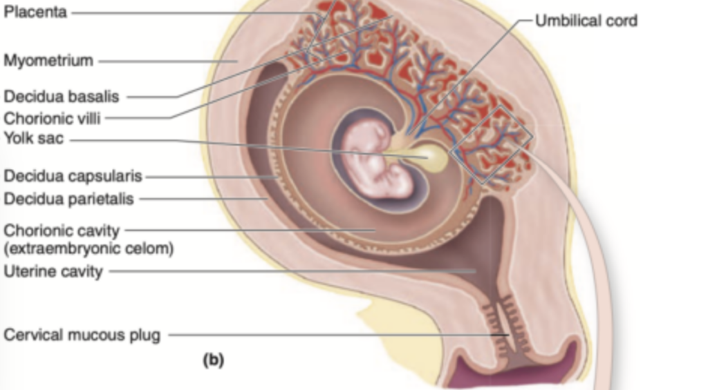
Question 6: Placenta Structure (胎盤の構造)
Q: Fill in the blanks with the correct terms.
The placenta consists of two main components: the _________ from the fetus and the _________ from the maternal tissue. The fetal portion is formed by the _________.
Answer: chorion / decidua basalis / chorionic villi
Explanation:
胎盤 (placenta) は、胎児の部分 (fetal part) と母体の部分 (maternal part) の2つの主な構成要素から成り立っています。胎児の部分は、絨毛膜 (chorion) に由来し、母体の部分は基底脱落膜 (decidua basalis) から派生します。胎児の部分には絨毛 (chorionic villi) が含まれ、代謝物の交換が行われます。
Question 7: Development of Chorionic Villi (絨毛の発達)
Q: Fill in the blanks with the correct terms.
Chorionic villi undergo three stages of development. The _________ villi appear first as solid cords. These are later invaded by _________ to form secondary villi. Finally, capillary loops develop, forming _________ villi.
Answer: primary / mesenchyme / tertiary
Explanation:
絨毛 (chorionic villi) は、3つの段階を経て発達します。最初の段階は、固形のコードとして現れる一次絨毛 (primary villi) です。これらの構造は間充織 (mesenchyme) によって侵入され、二次絨毛 (secondary villi) になります。最終的に、毛細血管のループ (capillary loops) が発達し、三次絨毛 (tertiary villi) が形成されます。
Question 8: Function of the Placenta (胎盤の機能)
Q: Fill in the blanks with the correct terms.
The placenta acts as a site of _________ exchange, allowing the transfer of oxygen, nutrients, and wastes. It also functions as an _________ organ, producing hormones such as _________ to support pregnancy.
Answer: metabolic / endocrine / progesterone
Explanation:
胎盤 (placenta) は、酸素 (oxygen)、栄養 (nutrients)、および老廃物 (wastes) の代謝交換 (metabolic exchange) を行う場として機能します。また、内分泌器官 (endocrine organ) としても機能し、妊娠を維持するためのプロゲステロン (progesterone) などのホルモンを生成します。
Question 1: Cervical Structure (子宮頸部の構造)
Q: Fill in the blanks with the correct terms.
The endocervical mucosa is lined by _________ epithelium and contains _________ cervical glands. Unlike the endometrium, it does not shed during _________.
Answer: simple columnar / mucus-secreting / menstruation
Explanation:
子宮頸部の内腔 (Endocervical Mucosa) は単層円柱上皮 (simple columnar epithelium) で裏打ちされており、粘液分泌性の子宮頸腺 (mucus-secreting cervical glands) が多数存在します。子宮内膜 (endometrium) とは異なり、月経 (menstruation) 中に剥がれ落ちることはありません。
Question 2: Transformation Zone (変換帯)
Q: Fill in the blanks with the correct terms.
The transformation zone is the junction where _________ epithelium meets _________ epithelium. It is located just outside the _________ of the cervix.
Answer: squamous / columnar / external os
Explanation:
変換帯 (transformation zone) は、扁平上皮 (squamous epithelium) と円柱上皮 (columnar epithelium) の接合部であり、外子宮口 (external os) のすぐ外側に位置します。変換帯は、子宮頸がん (cervical cancer) のリスクが最も高い領域です。
Question 3: Cervical Mucus (子宮頸部の粘液)
Q: Fill in the blanks with the correct terms.
During ovulation, cervical mucus becomes _________, facilitating sperm entry. During the luteal phase, it becomes _________, blocking sperm passage. During pregnancy, it forms a _________ in the cervical canal.
Answer: watery / viscous / mucus plug
Explanation:
排卵時 (ovulation) には、子宮頸部の粘液 (cervical mucus) は水様性 (watery) になり、精子 (sperm) の子宮内への進入が容易になります。黄体期 (luteal phase) では、粘液は粘性 (viscous) になり、精子の通過を妨げます。妊娠中 (pregnancy) は、粘液が子宮頸管 (cervical canal) に粘液栓 (mucus plug) を形成し、感染を防ぎます。
Question 4: Cervical Wall (子宮頸部の壁)
Q: Fill in the blanks with the correct terms.
The cervical wall is composed mainly of _________ connective tissue with a small amount of _________ muscle. Before birth, the cervix undergoes _________, a process that softens and dilates the cervical canal.
Answer: dense / smooth / effacement
Explanation:
子宮頸部の壁 (wall of the cervix) は、主に高密度の結合組織 (dense connective tissue) で構成されており、平滑筋 (smooth muscle) の割合は子宮の他の部分よりも少ないです。出産前 (before birth)、子宮頸部は熟化 (effacement) によって軟化 (softens) し、子宮頸管 (cervical canal) が拡張 (dilates) します。
Question 5: Vaginal Mucosa (膣の粘膜)
Q: Fill in the blanks with the correct terms.
The vaginal mucosa is lined by _________ epithelium. Under the influence of estrogen, epithelial cells accumulate _________, which is metabolized into _________ by bacteria to lower vaginal pH.
Answer: stratified squamous / glycogen / lactic acid
Explanation:
膣の粘膜 (vaginal mucosa) は重層扁平上皮 (stratified squamous epithelium) で裏打ちされています。エストロゲン (estrogen) の影響で、上皮細胞 (epithelial cells) はグリコーゲン (glycogen) を蓄積します。このグリコーゲンは、細菌 (bacteria) によって乳酸 (lactic acid) に変換され、膣内のpHを低下させ、病原菌の成長を抑制します。
Question 6: Vaginal Wall (膣の壁)
Q: Fill in the blanks with the correct terms.
The vaginal wall consists of three main layers: the _________, the _________, and the _________.
Answer: mucosa / muscular layer / adventitia
Explanation:
膣の壁 (vaginal wall) は、粘膜 (mucosa)、筋層 (muscular layer)、および外膜 (adventitia) の3つの層で構成されています。それぞれが、膣の柔軟性と強度を提供し、性交や出産時の伸展を可能にします。
Question 7: Changes During Pregnancy (妊娠中の変化)
Q: Fill in the blanks with the correct terms.
During pregnancy, the cervical glands increase mucus production, forming a _________. The cervical wall becomes _________ due to extensive remodeling, and the cervical canal undergoes _________ before birth.
Answer: mucus plug / softer / dilation
Explanation:
妊娠中 (during pregnancy)、子宮頸腺 (cervical glands) は粘液 (mucus) の産生を増加させ、粘液栓 (mucus plug) を形成します。さらに、子宮頸部の壁 (cervical wall) は軟化 (softer) し、出産前 (before birth) に子宮頸管 (cervical canal) が拡張 (dilation) します。
Question 8: Pap Smear (パパニコロウ塗抹検査)
Q: Fill in the blanks with the correct terms.
The Pap smear is a diagnostic test developed by _________ to detect _________ in the cervix. It can identify abnormal cells, which may indicate _________ changes.
Answer: George Papanicolaou / dysplasia / precancerous
Explanation:
パパニコロウ塗抹検査 (Pap smear) は、ジョージ・パパニコラウ (George Papanicolaou) によって開発された診断テストで、子宮頸部 (cervix) の異形成 (dysplasia) を検出します。異形成 (dysplasia) は、前がん性の変化 (precancerous changes) を示す可能性があるため、早期のがん検出に役立ちます。
Question 9: Medical Application (医療応用)
Q: Fill in the blanks with the correct terms.
Cervical cancer is commonly caused by infection with _________. Precancerous changes often occur in the _________ of the cervix, and these changes can be detected using a _________ test.
Answer: HPV / transformation zone / Pap smear
Explanation:
子宮頸がん (cervical cancer) は、ヒトパピローマウイルス (HPV) の感染によって引き起こされることが多いです。前がん状態の変化 (precancerous changes) は、子宮頸部の変換帯 (transformation zone) に発生することが多く、パパニコラウ塗抹検査 (Pap smear) によって検出されます。
Question 1: Mammary Gland Structure (乳腺の構造)
Q: Fill in the blanks with the correct terms.
The mammary gland consists of _________ lobes, each of which is a _________ gland separated by dense connective tissue. Each lobe has a single _________ duct that opens at the nipple.
Answer: 15-25 / separate / lactiferous
Explanation:
乳腺 (mammary gland) は、15〜25個の小葉 (15-25 lobes) で構成されており、それぞれが独立した腺 (separate gland) です。各小葉には1本の排泄乳管 (lactiferous duct) があり、それぞれが乳頭 (nipple) に独立して開口します。
Question 2: Puberty and Mammary Gland Development (思春期と乳腺の発達)
Q: Fill in the blanks with the correct terms.
During puberty, the growth of mammary glands is stimulated by _________, leading to the accumulation of _________ cells and elongation of the _________ system.
Answer: estrogen / adipose / duct
Explanation:
思春期 (puberty) において、エストロゲン (estrogen) の作用により、脂肪細胞 (adipose cells) の蓄積が促進され、管系 (duct system) の伸長が行われます。これにより、乳房 (breast) が成長します。
Question 3: Nipple and Areola (乳頭と乳輪)
Q: Fill in the blanks with the correct terms.
The areola surrounds the _________ and contains _________ glands, while the connective tissue of the nipple has abundant _________ muscle fibers.
Answer: nipple / sebaceous / smooth
Explanation:
乳輪 (areola) は乳頭 (nipple) を囲む皮膚であり、皮脂腺 (sebaceous glands) が多く存在します。さらに、乳頭 (nipple) の結合組織 (connective tissue) には平滑筋繊維 (smooth muscle fibers) が豊富に含まれており、収縮によって乳頭の勃起が起こります。
Question 4: Hormonal Regulation During Pregnancy (妊娠中のホルモン調節)
Q: Fill in the blanks with the correct terms.
During pregnancy, the growth of mammary glands is stimulated by the synergistic action of _________, _________, and _________, which promote the development of secretory alveoli.
Answer: estrogen / progesterone / prolactin
Explanation:
妊娠中 (during pregnancy)、エストロゲン (estrogen)、プロゲステロン (progesterone)、およびプロラクチン (prolactin) の相乗効果により、分泌性肺胞 (secretory alveoli) の発達が促進されます。これにより、出産後の授乳 (lactation) に備えた準備が整います。
Question 5: Alveoli and Milk Secretion (肺胞と乳の分泌)
Q: Fill in the blanks with the correct terms.
The secretory alveoli are composed of _________ epithelium and contain _________ cells between the secretory cells and the basal lamina. These cells contract under the influence of _________ to facilitate milk ejection.
Answer: cuboidal / myoepithelial / oxytocin
Explanation:
肺胞 (alveoli) は、立方上皮 (cuboidal epithelium) から構成され、筋上皮細胞 (myoepithelial cells) が分泌細胞 (secretory cells) と基底膜 (basal lamina) の間に存在します。オキシトシン (oxytocin) によって、これらの筋上皮細胞が収縮し、乳射出反射 (milk ejection reflex) を引き起こします。
Question 6: Colostrum (初乳) and IgA Production
Q: Fill in the blanks with the correct terms.
Late in pregnancy, alveoli and ducts accumulate _________, which is rich in _________ and contains _________ produced by plasma cells.
Answer: colostrum / proteins / IgA
Explanation:
妊娠後期 (late in pregnancy) には、初乳 (colostrum) が肺胞 (alveoli) と管 (ducts) に蓄積します。初乳 (colostrum) はタンパク質 (proteins) に富み、形質細胞 (plasma cells) によって合成された免疫グロブリンA (IgA) が含まれています。このIgAは新生児の受動的免疫 (passive immunity) を提供します。
Question 7: Milk Secretion After Birth (出産後の乳の分泌)
Q: Fill in the blanks with the correct terms.
After parturition, the alveoli begin active milk production under the influence of _________. Lipids are secreted by the _________ mechanism, and proteins are secreted by the _________ mechanism.
Answer: prolactin / apocrine / merocrine
Explanation:
出産後 (after parturition)、プロラクチン (prolactin) によって乳腺 (mammary gland) の分泌が促進されます。脂質 (lipids) は頂泌分泌 (apocrine secretion) によって放出され、タンパク質 (proteins) は部分分泌 (merocrine secretion) によって放出されます。
Question 8: Milk Ejection Reflex (乳射出反射)
Q: Fill in the blanks with the correct terms.
During breastfeeding, tactile stimulation of the _________ triggers the release of _________ from the posterior pituitary, causing the contraction of _________ cells, leading to milk ejection.
Answer: nipple / oxytocin / myoepithelial
Explanation:
授乳中 (during breastfeeding)、乳頭 (nipple) にある触覚受容体 (tactile receptors) の刺激が、下垂体後葉 (posterior pituitary) からのオキシトシン (oxytocin) の放出を引き起こします。筋上皮細胞 (myoepithelial cells) の収縮により、乳射出反射 (milk ejection reflex) が起こります。
Question 9: Breast Cancer (乳がん)
Q: Fill in the blanks with the correct terms.
Most breast cancers originate from the _________ cells of the terminal duct lobular units (TDLU). The most common form of breast cancer is _________ carcinoma, and cancer cells can metastasize to distant organs through the _________.
Answer: epithelial / invasive ductal / lymphatic system
Explanation:
乳がん (breast cancer) は、主に末端乳管小葉単位 (terminal duct lobular units, TDLU) にある上皮細胞 (epithelial cells) に由来します。最も一般的な形態は浸潤性乳管がん (invasive ductal carcinoma) であり、がん細胞はリンパ系 (lymphatic system) や血流 (bloodstream) を介して遠隔転移 (metastasis) する可能性があります。

コメント