
Contents
- 1 陰嚢、精巣、精巣上体、PAW
- 1.1 陰嚢 (Scrotum)
- 1.2 精巣 (Testes)
- 1.3 精巣動脈および静脈 (Testicular Arteries and Veins)
- 1.4 精巣上体 (Epididymis)
- 1.5 前外側腹壁の表面解剖 (Surface Anatomy of Anterolateral Abdominal Wall)
- 1.6 前外側腹壁と鼠径部の内面 (Internal Surface of Anterolateral Abdominal Wall and Inguinal Region)
- 1.7 精索、陰嚢、精巣、および鼠径ヘルニア (Spermatic Cord, Scrotum, Testes, and Inguinal Hernias)
- 1.8 精巣挙筋反射 (Cremasteric Reflex)
- 1.9 女性のヌック管の嚢胞およびヘルニア (Cysts and Hernias of Canal of Nuck)
- 1.10 精索および精巣の水腫 (Hydrocele of Spermatic Cord and/or Testis)
- 1.11 精巣の血腫 (Hematocele of Testis)
- 1.12 精索の捻転 (Torsion of Spermatic Cord)
- 1.13 陰嚢の麻酔 (Anesthetizing Scrotum)
- 1.14 精液瘤と精巣上体嚢胞 (Spermatocele and Epididymal Cyst)
- 1.15 胚性生殖管の遺残 (Vestigial Remnants of Embryonic Genital Ducts)
- 1.16 静脈瘤 (Varicocele)
- 1.17 精巣と陰嚢のがん (Cancer of Testis and Scrotum)
- 2 PENIS
- 2.1 陰茎 (Penis)
- 2.2 陰茎の構造 (Structure of the Penis)
- 2.3 陰茎の靭帯 (Ligaments of the Penis)
- 2.4 男性の会陰筋および勃起・射精の生理 (Perineal Muscles of Male and the Physiology of Erection, Emission, Ejaculation, and Remission)
- 2.5 男性の会陰筋 (Perineal Muscles of Male)
- 2.6 勃起、射精前分泌、射精、弛緩 (Erection, Emission, Ejaculation, and Remission)
- 2.7 男性尿生殖三角と関連する臨床事項 (Male Urogenital Triangle and Clinical Considerations)
- 2.8 尿道カテーテル挿入 (Urethral Catheterization)
- 2.9 陰嚢の膨張 (Distension of Scrotum)
- 2.10 精巣の触診 (Palpation of Testes)
- 2.11 先天性疾患:尿道下裂 (Hypospadias)
- 2.12 包茎、嵌頓包茎、割礼 (Phimosis, Paraphimosis, and Circumcision)
- 2.13 勃起不全 (Impotence and Erectile Dysfunction)
- 3 後腹壁
- 3.1 後腹壁 (Posterior Abdominal Wall)
- 3.2 後腹壁の筋膜 (Fascia of Posterior Abdominal Wall)
- 3.3 後腹壁の筋肉 (Muscles of Posterior Abdominal Wall)
- 3.4 大腰筋 (Psoas Major)
- 3.5 腸骨筋 (Iliacus)
- 3.6 腰方形筋 (Quadratus Lumborum)
- 3.7 後腹壁の神経 (Nerves of Posterior Abdominal Wall)
- 3.8 腰交感神経幹 (Lumbar Sympathetic Trunks)
- 3.9 腰神経叢 (Lumbar Plexus)
- 3.10 後腹壁の血管 (Vessels of Posterior Abdominal Wall)
- 3.11 腹部大動脈 (Abdominal Aorta)
- 3.12 腹部大動脈の位置関係 (Relations of Abdominal Aorta)
- 3.13 腹部大動脈の分岐 (Branches of the Abdominal Aorta)
- 3.14 後腹壁の静脈 (Veins of Posterior Abdominal Wall)
- 3.15 下大静脈の構造 (Structure of the IVC)
- 3.16 下大静脈の枝 (Tributaries of the IVC)
- 3.17 後腹壁のリンパ管とリンパ節 (Lymphatic Vessels and Lymph Nodes of Posterior Abdominal Wall)
- 3.18 胸管 (Thoracic Duct)
- 3.19 横隔膜 (Diaphragm)
- 3.20 先天性横隔膜ヘルニア (Congenital Diaphragmatic Hernia)
- 3.21 後腹壁 (Posterior Abdominal Wall)
- 3.22 大動脈の拍動と腹部大動脈瘤 (Pulsations of Aorta and Abdominal Aortic Aneurysm)
- 3.23 腹部骨盤静脈血の側副経路 (Collateral Routes for Abdominopelvic Venous Blood)
- 4 骨盤臓器のリンパ
- 5 骨盤臓器
- 6 骨盤(PPT)
- 7 教科書サマリーより
- 8 重要点穴埋め問題
陰嚢、精巣、精巣上体、PAW
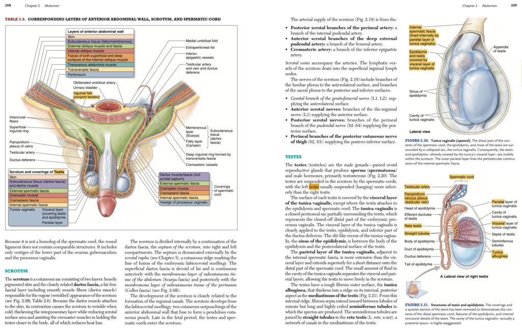
陰嚢 (Scrotum)
構造 (Structure)
陰嚢は、重色素性の皮膚 (Heavily pigmented skin) と平滑筋線維を含む脂肪のない筋膜層 (Dartos fascia) の2層からなる皮膚性嚢です。
ダートス筋 (Dartos muscle) は、陰嚢のしわ状 (Rugose) の外観を生じさせ、収縮することで以下を助けます:
- 皮膚を厚くして陰嚢表面積を減少させる。
- 精巣挙筋 (Cremaster muscle) とともに精巣を体に近づけ、熱損失を抑える。
陰嚢は内部でダートス筋膜の延長である陰嚢中隔 (Septum of the scrotum) によって右室と左室に分けられています。中隔は外部では陰嚢縫線 (Scrotal raphe) として示され、これは胎児期の唇陰嚢隆起 (Labioscrotal swellings) の融合線を表します。
血管供給とリンパ (Arterial Supply and Lymphatic Drainage)
血液供給:
陰嚢の血液供給は以下の動脈からなります:
- 会陰動脈の後陰嚢枝 (Posterior scrotal branches):内陰部動脈 (Internal pudendal artery) の枝。
- 深外陰部動脈の前陰嚢枝 (Anterior scrotal branches):大腿動脈 (Femoral artery) の枝。
- 精巣挙筋動脈 (Cremasteric artery):下腹壁動脈 (Inferior epigastric artery) の枝。
リンパ:陰嚢のリンパ管は浅鼠径リンパ節 (Superficial inguinal lymph nodes) に排出されます。
神経供給 (Nerve Supply)
陰嚢の神経支配は、腰神経叢 (Lumbar plexus) と仙骨神経叢 (Sacral plexus) の枝によります:
- 生殖大腿神経の生殖枝 (Genital branch of the genitofemoral nerve, L1, L2):前外側表面を支配。
- 腸骨鼠径神経の前陰嚢神経 (Anterior scrotal nerves):前表面を支配。
- 陰部神経の会陰枝の後陰嚢神経 (Posterior scrotal nerves):後表面を支配。
- 大腿後皮神経の会陰枝 (Perineal branches of the posterior cutaneous nerve of the thigh, S2, S3):後下表面を支配。
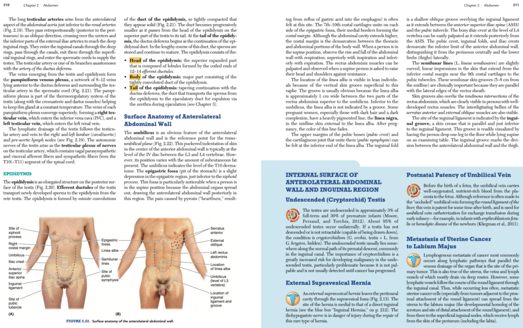
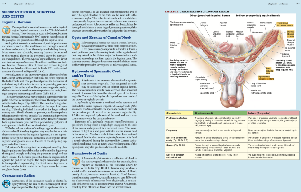
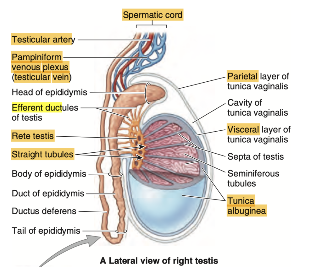
精巣 (Testes)
構造と機能 (Structure and Function)
精巣は男性の生殖腺 (Male gonads) であり、精子 (Spermatozoa) と男性ホルモン (Primarily testosterone) を産生する一対の卵状の器官です。
- 精巣は精索 (Spermatic cords) によって陰嚢内に吊り下げられています。通常、左側の精巣は右側よりも低く位置します。
精巣の表面は臓側精巣鞘膜 (Visceral layer of the tunica vaginalis) に覆われています。これは胎児期の腹膜突起 (Processus vaginalis) の閉鎖された遠位部分を表しています。
精巣内の構造 (Internal Structure)
- 精巣鞘膜 (Tunica vaginalis)
- 臓側層 (Visceral layer) と壁側層 (Parietal layer) の間には少量の液体が存在し、精巣が陰嚢内で自由に動けるようにします。
- 精巣上体洞 (Sinus of the epididymis):精巣上体 (Epididymis) と精巣後外側表面の間に存在する隙間状の構造。
- 精巣白膜 (Tunica albuginea)
- 精巣の外表面を覆う強靭な線維性外膜。内側の後方部分では肥厚して精巣縦隔 (Mediastinum of the testis) を形成。
- 精巣縦隔から線維性中隔が伸び、小さな小葉 (Lobules) を形成します。
- 精細管 (Seminiferous tubules)
- 各小葉内には長く高度にらせん状の精細管が存在し、ここで精子が産生されます。
- 精細管は直精細管 (Straight tubules) を介して精巣網 (Rete testis) に接続します。精巣網は精巣縦隔内にある管状ネットワークです。
精巣動脈および静脈 (Testicular Arteries and Veins)
精巣動脈 (Testicular Arteries)
- 起始:精巣動脈は腹大動脈 (Abdominal aorta) の前外側から腎動脈 (Renal arteries) のすぐ下で分岐します(図2.19参照)。
- 経路:後腹膜 (Retroperitoneal) を斜めに進み、尿管 (Ureters) と外腸骨動脈 (External iliac arteries) の下部を越え、深鼠径輪 (Deep inguinal rings) に到達します。
- 精索への進入:深鼠径輪を通過して鼠径管 (Inguinal canals) を抜け、浅鼠径輪 (Superficial inguinal rings) を通過して精索 (Spermatic cords) に入り、精巣に血液を供給します。
精巣動脈の枝の1つは、精管動脈 (Artery of the ductus deferens) と吻合します。
精巣静脈 (Testicular Veins)
- 精巣および精巣上体 (Epididymis) から出る静脈は、蔓状静脈叢 (Pampiniform venous plexus) を形成します。
- 蔓状静脈叢は精索内で精巣動脈を取り囲み、熱調節システム (Thermoregulatory system) の一部として機能します。
静脈叢は上部で収束し、以下の静脈を形成します:
- 右精巣静脈 (Right testicular vein):下大静脈 (Inferior vena cava, IVC) に注ぎます。
- 左精巣静脈 (Left testicular vein):左腎静脈 (Left renal vein) に注ぎます。
リンパと自律神経 (Lymphatic Drainage and Autonomic Nerves)
- リンパ排出:精巣のリンパは精巣動静脈に沿って、右および左腰リンパ節 (Lumbar lymph nodes) および前大動脈リンパ節 (Pre-aortic lymph nodes) に排出されます。
- 自律神経支配:精巣神経叢 (Testicular plexus) が精巣動脈上に形成され、迷走神経由来の副交感神経 (Vagal parasympathetic fibers)、内臓求心性線維 (Visceral afferent fibers)、および脊髄T10~T11からの交感神経線維 (Sympathetic fibers) を含みます。
精巣上体 (Epididymis)
概要 (Overview)
精巣上体は精巣の後面に位置する細長い構造で、精巣網 (Rete testis) からの輸出小管 (Efferent ductules) によって精巣から精子を輸送します(図2.20参照)。
- 精巣上体は、緊密に巻き込まれた精巣上体管 (Duct of the epididymis) で構成されています。
- 精巣上体の尾部 (Tail of the epididymis) は精管 (Ductus deferens) と連続し、精子を射精管 (Ejaculatory duct) に運びます。
精巣上体の構造 (Structure)
- 頭部 (Head of the epididymis):精巣上体の上部で拡張した部分。12~14の輸出小管の巻き込みによって形成されています。
- 体部 (Body of the epididymis):主要部分で、精巣上体管の緊密に巻き込まれた構造から成ります。
- 尾部 (Tail of the epididymis):精管に続く部分で、精子の成熟と貯蔵が行われます。
前外側腹壁の表面解剖 (Surface Anatomy of Anterolateral Abdominal Wall)
臍 (Umbilicus)
- 臍は前外側腹壁 (Anterolateral abdominal wall) の明瞭な特徴で、臍平面 (Trans-umbilical plane) の基準点となります(図2.22参照)。
- 通常、臍はL3~L4椎間円板 (IV disc between L3 and L4 vertebrae) の高さに位置しますが、皮下脂肪の量によってその位置が異なる場合があります。
- 臍はT10皮膚分節 (T10 dermatome) のレベルを示します。
剣状突起下窩 (Epigastric Fossa)
- 剣状突起 (Xiphoid process) の直下に位置するわずかなくぼみ。
- 仰臥位 (Supine position) のとき、腹部臓器が広がり、前外側腹壁が後方に引かれるため特に目立ちます。
- 胃酸逆流 (Reflux of gastric acid) による胸やけ (Pyrosis) の痛みはこの部位で感じられることが多いです。
- 第7~10肋軟骨 (7th–10th costal cartilages) がこの窩の両側で結合し、その内側縁が肋骨弓 (Costal margin) を形成します。
腹直筋と白線 (Rectus Abdominis and Linea Alba)
- 腹直筋 (Rectus abdominis muscles)
- 仰臥位で、頭部と肩を抵抗に逆らって持ち上げることで触診可能。
- 腱画 (Tendinous intersections) がよく発達している場合、皮膚の溝として視認可能。
- 白線 (Linea alba)
- 臍の上方では2つの腹直筋間に約1 cm幅の溝として明瞭に見えます。
- 臍より下方ではこの溝は目立ちません。
- 一部の妊娠女性では、白線黒線 (Linea nigra) と呼ばれる色素沈着が白線の外側に現れます。妊娠後、この線の色は薄くなります。
恥骨と鼠径部 (Pubic Bones and Inguinal Region)
- 恥骨稜 (Pubic crest) と恥骨結合 (Pubic symphysis)
- 白線の下端に位置し、触診可能。
- 鼠径溝 (Inguinal fold)
- 鼠径靭帯 (Inguinal ligament) を覆う浅い斜めの溝で、上前腸骨棘 (ASIS) と恥骨結節 (Pubic tubercle) を結びます。
- 鼠径溝は前外側腹壁と大腿 (Thigh) の境界を示します。
- 腸骨稜 (Iliac crest)
- L4椎骨の高さに位置し、ASISから後方に延びる骨で触診可能。
半月線 (Semilunar Lines)
- 腹直筋鞘 (Rectus sheath) の外側縁に沿って伸びる、皮膚上のわずかに湾曲した線状の溝。
- 第9肋軟骨付近の肋骨下縁から恥骨結節に向かって延びます。
- 半月線は、腹部手術や臨床診察における重要な解剖学的指標です。
臨床的観察 (Clinical Observations)
- 腹壁は呼吸に伴って上下動します。仰臥位では、吸気で上昇し、呼気で下降します。
- 外腹斜筋 (External oblique) と前鋸筋 (Serratus anterior) の筋腹の交互作用が、特に発達した筋肉を持つ人で明瞭に見られます。
- 鼠径靭帯の部位は、仰臥位で片脚を床に下ろすことで視認可能になり、前外側腹壁と大腿の分界を示します。
前外側腹壁と鼠径部の内面 (Internal Surface of Anterolateral Abdominal Wall and Inguinal Region)
停留精巣 (Undescended Testis)
- 頻度:停留精巣 (Cryptorchidism) は、正期産児の約3%、早産児の約30%に見られます。約95%のケースは片側性です。
- 定義:精巣が下降せず、または下降可能な状態でない場合、この状態を停留精巣と呼びます。
- 位置:停留精巣は通常、胎児期に精巣が下降する経路上、特に鼠径管 (Inguinal canal) に存在します。
- 重要性:停留精巣は悪性腫瘍のリスクが大幅に増加します。この問題は、触診ができず、がんが進行するまで検出されにくいことにあります。
外側膀胱上ヘルニア (External Supravesical Hernia)
- ヘルニアの位置:膀胱上窩 (Supravesical fossa) を通じて腹膜腔から突出します(図2.13参照)。
- 特徴:このヘルニアは、直接鼠径ヘルニア (Direct inguinal hernia) の部位より内側に位置します。
- 注意点:このまれなヘルニアの修復時には、腸骨下腹神経 (Iliohypogastric nerve) が損傷を受ける危険があります。
臍静脈の出生後開存 (Postnatal Patency of Umbilical Vein)
- 胎児期には、臍静脈 (Umbilical vein) が胎盤から酸素と栄養を豊富に含む血液を胎児に運びます。
- 一般に、閉塞された臍静脈は肝円索 (Round ligament of the liver) を形成するとされていますが、出生後もしばらくは開存しており、臍静脈カテーテル挿入 (Umbilical vein catheterization) に使用されます。
- 例:新生児の溶血性疾患 (Hemolytic disease of the newborn) や胎児赤芽球症 (Erythroblastosis fetalis) における交換輸血。
子宮がんの大陰唇への転移 (Metastasis of Uterine Cancer to Labium Majus)
- リンパ行性転移 (Lymphogenous metastasis):腫瘍の一次病巣に対応する静脈およびリンパ管を経由します。
- 子宮の場合、大部分のリンパは深部経路を通じて排出されますが、一部のリンパ管は子宮円索 (Round ligament of the uterus) を介して鼠径管を通過します。
- これにより、腫瘍細胞が大陰唇 (Labium majus) に転移し、そこから浅鼠径リンパ節 (Superficial inguinal nodes) に広がることがあります。
精索、陰嚢、精巣、および鼠径ヘルニア (Spermatic Cord, Scrotum, Testes, and Inguinal Hernias)
鼠径ヘルニア (Inguinal Hernias)
- 頻度:腹部ヘルニア全体の75%を占め、約86%が男性に発生します。これは精索 (Spermatic cord) が鼠径管を通過することが原因です。
- ヘルニアの定義:腹膜壁 (Parietal peritoneum) および内臓 (Viscera) が本来の腔所から正常または異常な開口部を通じて突出することを指します。
- 還納性 (Reducibility):多くのヘルニアは、適切な操作で元の位置に戻すことが可能です。
鼠径ヘルニアの種類と特徴
- 間接鼠径ヘルニア (Indirect Inguinal Hernia):
- 鼠径管を通過し、場合によっては陰嚢まで延びます。
- ヘルニア嚢は閉鎖されなかった腹膜鞘状突起 (Processus vaginalis) によって形成されます。
- 直接鼠径ヘルニア (Direct Inguinal Hernia):
- 鼠径三角 (Inguinal triangle) の部位から腹壁を通じて突出します。
ヘルニアの診察方法
- 浅鼠径輪 (Superficial inguinal ring):
- 恥骨結節 (Pubic tubercle) の上外側で触診可能。
- ヘルニアがある場合、患者に咳をさせると、指先に突然の衝動が感じられます。
- 深鼠径輪 (Deep inguinal ring):
- 鼠径靭帯 (Inguinal ligament) の上、恥骨結節から2~4 cm上外側に位置する皮膚のくぼみとして触診可能。
- 間接鼠径ヘルニアの場合、深鼠径輪で塊が触れることがあります。
- 直接鼠径ヘルニアの診察:
- 鼠径三角の部位に指を置き、患者に咳または腹圧をかけさせると、指の腹部に衝撃が感じられます。
精巣挙筋反射 (Cremasteric Reflex)
反射のメカニズム
- 刺激方法:大腿上部内側 (Medial aspect of the superior thigh) の皮膚を軽く擦ることで精巣挙筋 (Cremaster muscle) が収縮します。この部分の皮膚は腸骨鼠径神経 (Ilio-inguinal nerve) によって支配されています。
- 反射の結果:同側の精巣が急速に持ち上がる現象が精巣挙筋反射です。
子供における特徴
- この反射は特に小児で活発であり、過敏な精巣挙筋反射 (Hyperactive cremasteric reflex) が停留精巣 (Undescended testis) と誤診されることがあります。
- 診断の補助方法:子供を足を組んでしゃがむ姿勢にさせることで反射を抑制でき、精巣が陰嚢内に下降している場合は触診可能になります。
女性のヌック管の嚢胞およびヘルニア (Cysts and Hernias of Canal of Nuck)
ヌック管の遺残
- 女性において腹膜鞘状突起 (Processus vaginalis) が遺残すると、ヌック管 (Canal of Nuck) と呼ばれる小さな腹膜嚢が形成される場合があります。
- 位置:この嚢は鼠径管内にあり、大陰唇 (Labium majus) にまで延びることがあります。
- 病態:ヌック管の遺残が膨らみを形成すると鼠径管内に嚢胞が生じ、間接鼠径ヘルニア (Indirect inguinal hernia) に進行する可能性があります。
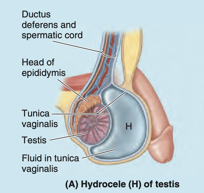
精索および精巣の水腫 (Hydrocele of Spermatic Cord and/or Testis)
水腫の原因
- 水腫 (Hydrocele) は、腹膜鞘状突起の遺残部分に過剰な漿液性液体が蓄積することで発生します。この異常は間接鼠径ヘルニアと関連する場合があります。
- 発生機序:漿液性液体は精巣鞘膜の臓側層 (Visceral layer of the tunica vaginalis) から異常に分泌されます。
- 種類:
- 精巣の水腫:陰嚢内に限局し、精巣鞘膜を膨らませます(図B2.4A)。
- 精索の水腫:精索に限局し、腹膜鞘状突起の遺残部分を膨らませます(図B2.4B)。
- 先天性水腫:腹腔と連絡している場合があります。
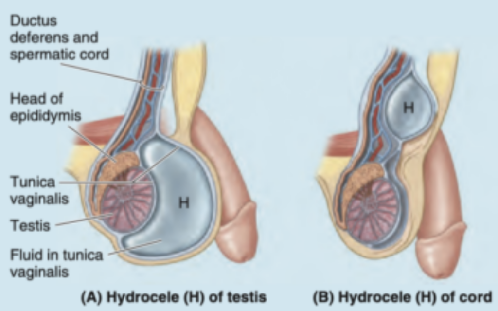
検査方法
- 透光法 (Transillumination):暗室で陰嚢に明るい光を当て、光が赤い輝きとして透過する場合、漿液性液体が過剰に蓄積していることを示します。
- 新生児の場合:新生児の男性では、精巣鞘膜内に腹膜液が残存することがありますが、通常1年以内に吸収されます。
- 成人の場合:外傷や精巣上体炎 (Epididymitis) などの病態が水腫を引き起こすことがあります。
精巣の血腫 (Hematocele of Testis)
血腫の特徴
- 原因:精巣動脈 (Testicular artery) の分枝が破裂し、精巣鞘膜内に血液が蓄積します。
- 病態:陰嚢および/または精巣内に血液が溜まり、血腫 (Hematoma) を形成します。
区別のための透光法
- 血液は光を透過しないため、透光法を用いることで水腫と血腫を区別できます。
- 合併症:精巣血腫は、陰嚢組織への血液の滲出による陰嚢血腫 (Scrotal hematocele) を伴う場合があります。
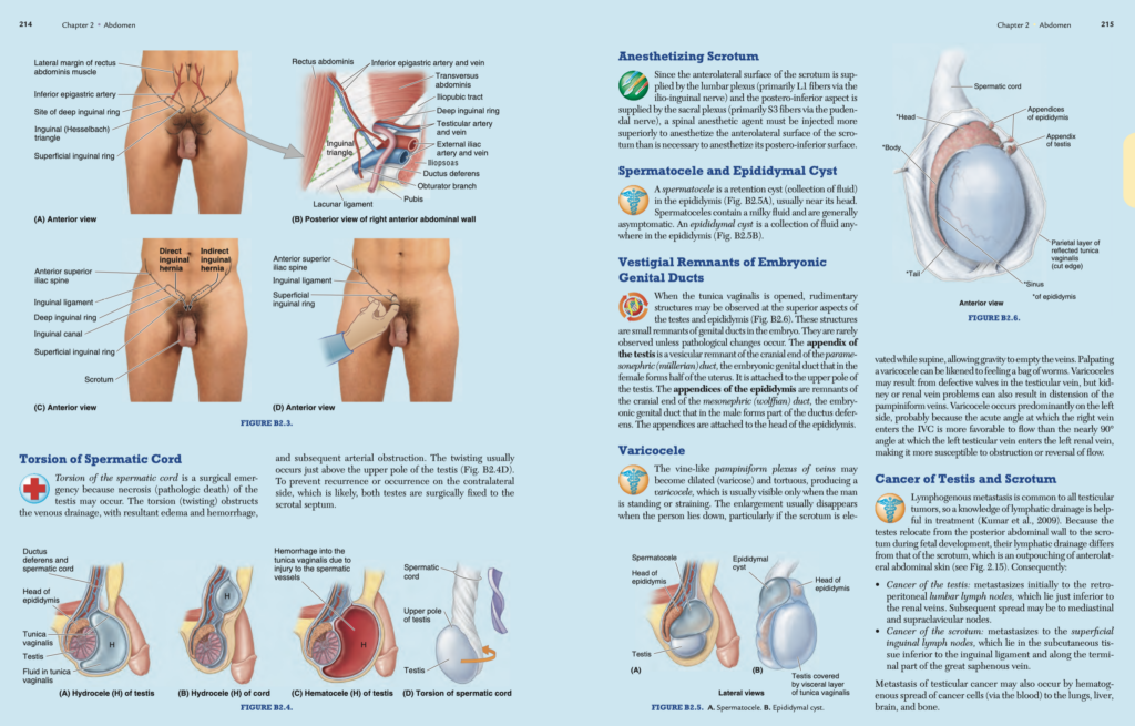
精索の捻転 (Torsion of Spermatic Cord)
概要 (Overview)
- 精索の捻転は外科的緊急事態であり、精巣壊死 (Necrosis of the testis) を引き起こす可能性があります。
- 影響:捻転により静脈排出が妨げられ、浮腫 (Edema)、出血 (Hemorrhage)、さらには動脈閉塞 (Arterial obstruction) を引き起こします。
- 発生部位:通常、精巣の上極直上で発生します(図B2.4D参照)。
治療 (Treatment)
- 再発や対側精巣での発症を防ぐため、両側の精巣を陰嚢中隔 (Scrotal septum) に固定する外科手術が行われます。
陰嚢の麻酔 (Anesthetizing Scrotum)
神経支配 (Nerve Supply)
- 前外側表面:腰神経叢 (Lumbar plexus)、主にL1線維(腸骨鼠径神経 [Ilio-inguinal nerve] 経由)。
- 後下表面:仙骨神経叢 (Sacral plexus)、主にS3線維(陰部神経 [Pudendal nerve] 経由)。
麻酔の注意点 (Anesthetic Considerations)
- 前外側表面を麻酔するためには、後下表面よりも高位に脊髄麻酔剤を注射する必要があります。
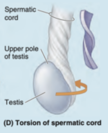
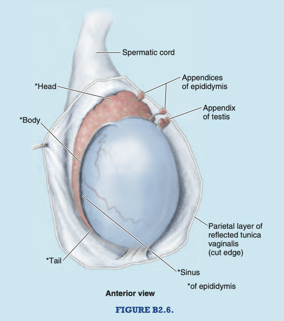
精液瘤と精巣上体嚢胞 (Spermatocele and Epididymal Cyst)
精液瘤 (Spermatocele)
- 定義:精巣上体 (Epididymis) の保持嚢胞 (Retention cyst)。
- 位置:通常、精巣上体の頭部付近に発生します(図B2.5A参照)。
- 内容物:乳白色の液体を含むが、一般的に無症状です。
精巣上体嚢胞 (Epididymal Cyst)
- 定義:精巣上体のいずれかの部位に発生する液体の収集(図B2.5B参照)。
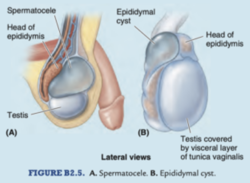
胚性生殖管の遺残 (Vestigial Remnants of Embryonic Genital Ducts)
主要な構造 (Key Structures)
- 精巣付属体 (Appendix of the Testis)
- 中腎傍管 (パラメソネフリック管) の頭側端の小型の遺残。
- 精巣の上極に付着しています。
- 精巣上体付属体 (Appendices of the Epididymis)
- 中腎管 (メソネフリック管) の頭側端の遺残。
- 精巣上体の頭部に付着しています。
静脈瘤 (Varicocele)
概要 (Overview)
- 原因:蔓状静脈叢 (Pampiniform plexus) の静脈が拡張(静脈瘤化)し、蛇行する状態。
- 特徴:
- 立位または腹圧時に目視でき、触診では「虫袋 (Bag of worms)」のように感じられます。
- 仰臥位で陰嚢を持ち上げると、重力により静脈が空になり、消失する場合があります。
発生要因 (Causes)
- 主因:精巣静脈の弁機能不全。
- 左側に多い理由:左精巣静脈が左腎静脈に直角で流入するため、逆流や閉塞が起こりやすい。
精巣と陰嚢のがん (Cancer of Testis and Scrotum)
精巣がん (Cancer of the Testis)
- リンパ転移 (Lymphogenous Metastasis):初期転移は後腹膜腰リンパ節 (Retroperitoneal lumbar lymph nodes) に起こります(図2.15参照)。
- その後、縦隔 (Mediastinal) や鎖骨上窩リンパ節 (Supraclavicular nodes) へ転移します。
- 血行性転移 (Hematogenous Metastasis):肺、肝臓、脳、骨に転移する場合があります。
陰嚢がん (Cancer of the Scrotum)
- リンパ転移:浅鼠径リンパ節 (Superficial inguinal lymph nodes) に転移します。これらのリンパ節は鼠径靭帯下の皮下組織および大伏在静脈の終末部に沿って位置します。
PENIS
陰茎 (Penis)
概要 (Overview)
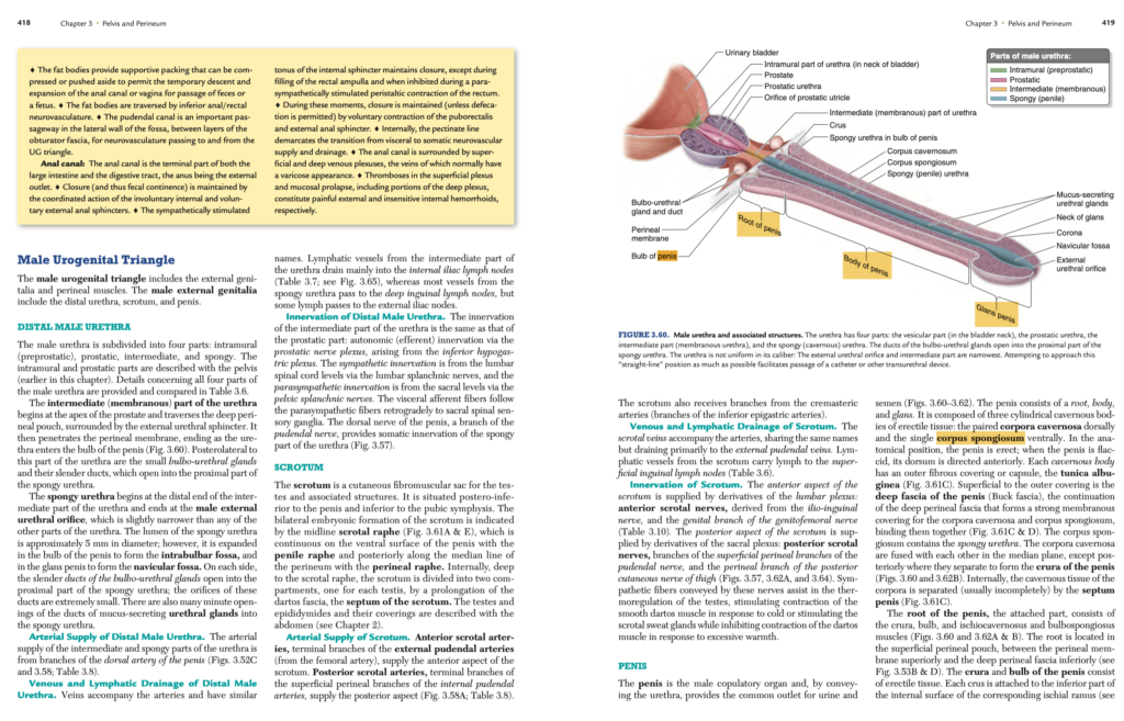
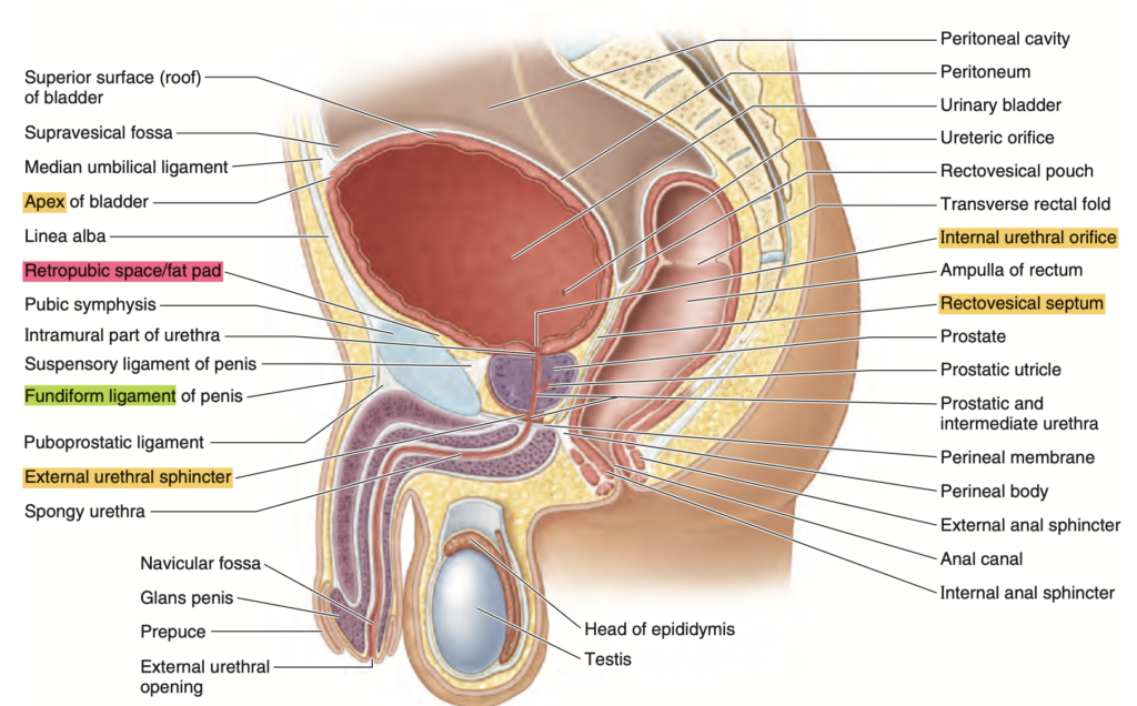
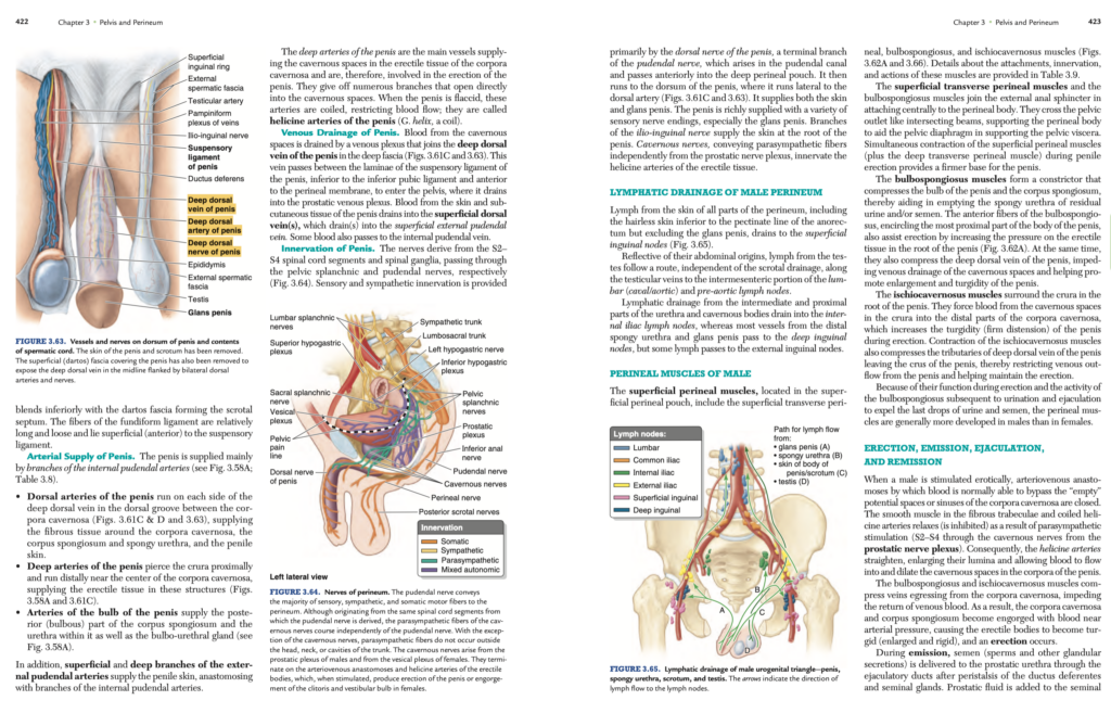
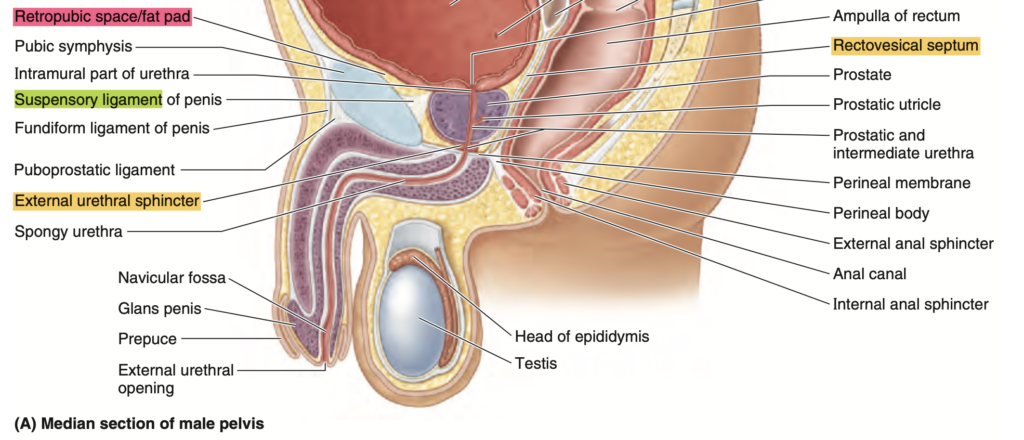
陰茎は男性の交接器官であり、尿 (Urine) と精液 (Semen) の共通の出口として機能します(図3.60~3.62参照)。
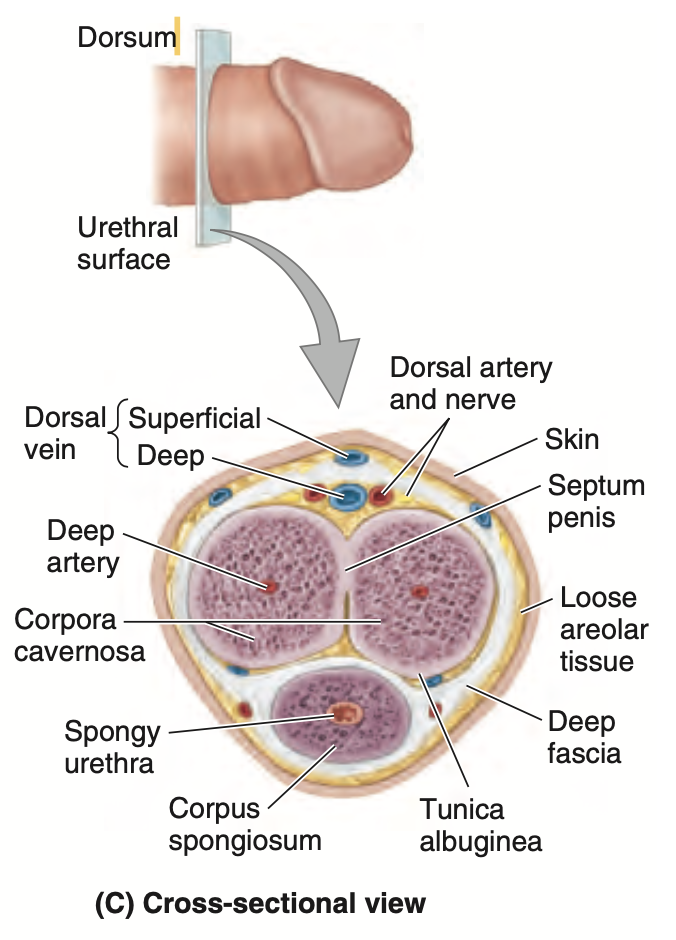
陰茎は根部 (Root)、体部 (Body)、亀頭 (Glans) の3つの部分で構成されています。内部には以下の3つの海綿体組織 (Cylindrical cavernous bodies of erectile tissue) があります:
- 背側にある左右一対の陰茎海綿体 (Corpora cavernosa)
- 腹側にある単一の尿道海綿体 (Corpus spongiosum)
解剖学的位置 (Anatomical Position)
- 陰茎が直立 (Erect) しているときが解剖学的位置とされます。
- 弛緩状態 (Flaccid) のときは、背側が前方を向きます。
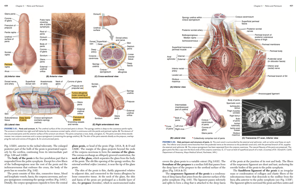
陰茎の構造 (Structure of the Penis)
海綿体の特徴 (Cavernous Bodies)
- 線維性被膜 (Tunica albuginea)
- 各海綿体を覆う外膜。
- 陰茎深筋膜 (Buck fascia)
- 深会陰筋膜 (Deep perineal fascia) の延長で、海綿体をまとめて強固な膜を形成します。
- 尿道海綿体 (Corpus spongiosum)
- 内部に海綿部尿道 (Spongy urethra) を含みます。
陰茎の根部 (Root of the Penis)
- 構成要素:陰茎脚 (Crura)、球部 (Bulb)、坐骨海綿体筋 (Ischiocavernosus muscle)、球海綿体筋 (Bulbospongiosus muscle)
- 位置:浅会陰嚢 (Superficial perineal pouch) 内に位置し、上部は会陰膜 (Perineal membrane)、下部は深会陰筋膜に挟まれています。
陰茎の体部 (Body of the Penis)
- 特徴:自由に動く下垂部で、恥骨結合 (Pubic symphysis) から吊り下げられています。筋肉はほとんど含まれません。
陰茎の亀頭 (Glans of the Penis)
- 構造:
- 尿道海綿体(Corpus spongiosum)の遠位端が拡張して形成される円錐状の構造。
- 冠状部 (Corona of the glans):亀頭の縁が陰茎海綿体の端を越えて突出。
- 亀頭頸部 (Neck of the glans):亀頭と陰茎体部を分ける溝状の構造。
- 尿道口 (External urethral orifice):亀頭の先端近くにあるスリット状の開口部。
陰茎の皮膚と包皮 (Skin and Prepuce)
- 陰茎の皮膚は薄く、周囲の皮膚よりも濃色で、線維性被膜 (Tunica albuginea) に疎性結合組織で接続されています。
- 包皮 (Prepuce):亀頭を覆う二重層の皮膚(包茎の場合、覆われる程度は個人差があります)。
- 包皮小帯 (Frenulum of the prepuce):包皮の深層から亀頭の尿道面に伸びる正中のひだ。
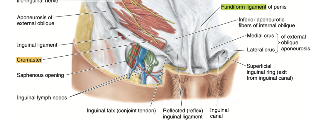
陰茎の靭帯 (Ligaments of the Penis)
陰茎懸垂靭帯 (Suspensory Ligament of the Penis)
- 起始:恥骨結合の前面から起こる深筋膜の凝集体。
- 機能:陰茎の根部と体部の接合部に付着し、陰茎の海綿体を恥骨結合に固定します。
陰茎支帯靭帯 (Fundiform Ligament of the Penis)
- 起始:白線 (Linea alba) の前面から降り、陰茎を囲みます。
- 構造:弾性繊維とコラーゲンの不規則な凝集体で、陰茎深筋膜の前方に位置します。
- 機能:陰嚢中隔 (Scrotal septum) に融合し、陰茎と陰嚢を補助的に支えます。
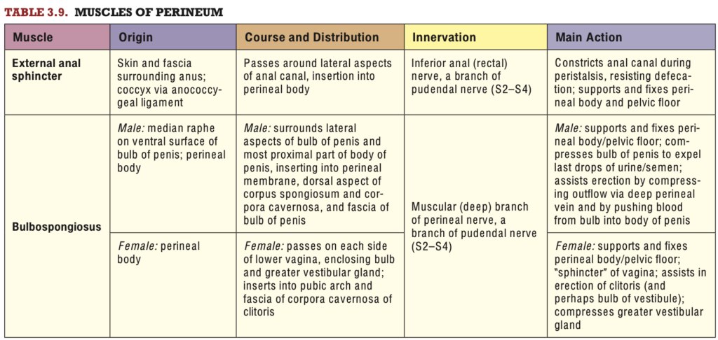
男性の会陰筋および勃起・射精の生理 (Perineal Muscles of Male and the Physiology of Erection, Emission, Ejaculation, and Remission)
男性の会陰筋 (Perineal Muscles of Male)
概要 (Overview)
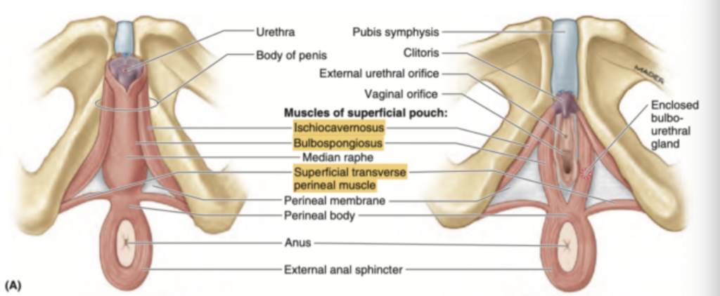
男性の浅会陰筋 (Superficial perineal muscles) は、浅会陰嚢 (Superficial perineal pouch) に位置し、以下を含みます(図3.62Aおよび図3.66参照):
- 浅横会陰筋 (Superficial transverse perineal muscle)
- 球海綿体筋 (Bulbospongiosus muscle)
- 坐骨海綿体筋 (Ischiocavernosus muscle)
これらの筋肉の詳細(付着、神経支配、作用)は表3.9に記載されています。
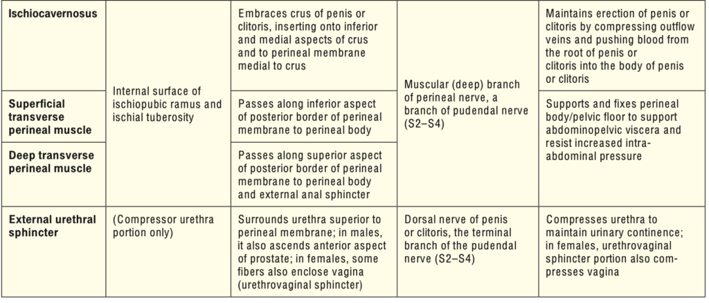
浅会陰筋の機能 (Functions of the Superficial Perineal Muscles)
- 支持機能:
- 浅横会陰筋と球海綿体筋は、肛門括約筋 (External anal sphincter) とともに会陰体 (Perineal body) に付着し、骨盤隔膜 (Pelvic diaphragm) と協調して骨盤臓器を支えます。
- 勃起時には浅会陰筋および深横会陰筋が収縮し、陰茎の基部を強化します。
- 球海綿体筋 (Bulbospongiosus Muscle):
- 役割:
- 陰茎球部と尿道海綿体を圧迫して、尿道海綿部 (Spongy urethra) の残尿や精液を排出します。
- 陰茎体部の近位部を取り囲む筋繊維は、陰茎根部の海綿体組織の圧力を高め、勃起を補助します。
- 静脈圧迫:陰茎深背静脈を圧迫し、静脈還流を妨げることで陰茎の充血と硬直を助けます。
- 役割:
- 坐骨海綿体筋 (Ischiocavernosus Muscle):
- 役割:
- 陰茎脚 (Crura) の海綿体空隙から血液を遠位部の陰茎海綿体に送り込み、陰茎の硬直 (Turgidity) を増大させます。
- 陰茎脚からの深背静脈分枝を圧迫して静脈還流を制限し、勃起を維持します。
- 役割:
男性では、これらの筋肉は尿や射精後の残尿や精液を排出する機能を持つため、女性よりも発達しています。
勃起、射精前分泌、射精、弛緩 (Erection, Emission, Ejaculation, and Remission)
勃起 (Erection)
- 血流制御:勃起時、通常は動静脈吻合部を通じて「空虚」な陰茎海綿体の空隙をバイパスしている血流が、動静脈吻合部の閉鎖により制御されます。
- 神経支配:副交感神経(S2~S4)により、螺旋動脈 (Helicine arteries) が弛緩して直線化し、空隙に血液が流入します。
- 筋肉の役割:球海綿体筋および坐骨海綿体筋が収縮し、静脈還流を妨げ、陰茎海綿体と尿道海綿体が動脈圧近くで充血・硬直します。
射精前分泌 (Emission)
- 内容物:精液(精子および分泌物)が輸精管および精嚢の蠕動により前立腺尿道 (Prostatic urethra) に運ばれます。
- 前立腺分泌液:前立腺の平滑筋収縮により精液に加わります。
- 神経支配:交感神経(L1~L2)による制御。
射精 (Ejaculation)
- 射精時、精液が尿道口から排出されます。以下の3つのプロセスが関与します:
- 膀胱頸部内尿道括約筋 (Internal urethral sphincter) の収縮(交感神経:L1~L2)。
- 尿道筋 (Urethral muscle) の収縮(副交感神経:S2~S4)。
- 球海綿体筋の収縮(陰部神経:S2~S4)。
弛緩 (Remission)
- プロセス:射精後、陰茎は次第に弛緩状態に戻ります。
- 神経支配:交感神経が螺旋動脈の平滑筋を収縮させ、海綿体の血液排出を促進します。
- 筋肉の弛緩:球海綿体筋および坐骨海綿体筋が弛緩し、静脈還流が増加します。
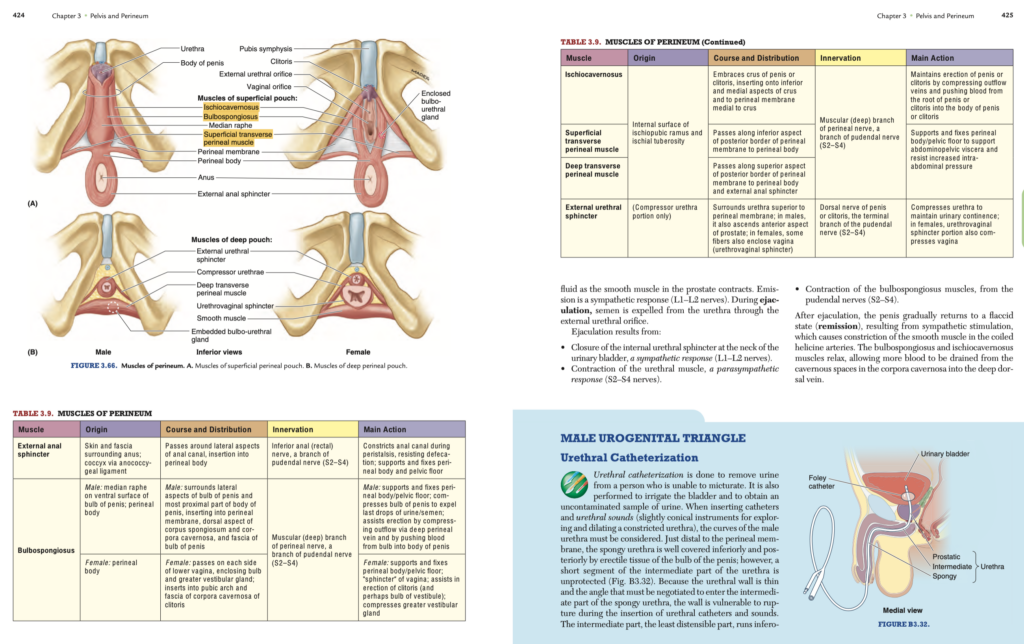
男性尿生殖三角と関連する臨床事項 (Male Urogenital Triangle and Clinical Considerations)
尿道カテーテル挿入 (Urethral Catheterization)
概要
尿道カテーテル挿入は、排尿が困難な患者の尿を排出するため、または膀胱を洗浄したり、清潔な尿サンプルを採取するために行われます。男性尿道の挿入時には、その形状とカーブに注意が必要です。
- 脆弱な部位
- 尿道海綿部 (Spongy urethra) は陰茎の球部によって保護されていますが、中間部尿道 (Intermediate part) は外尿道括約筋を通過する際に薄く柔軟性が低いため、カテーテル挿入時に損傷を受けやすい部分です。
- 構造的考慮
- 中間部は最も拡張しにくく、尿道外口(最狭部)を通過する器具が他の部分も通過可能です。
合併症
- 尿道狭窄 (Urethral stricture)
外傷や感染による場合には、尿道ブジーで拡張する必要があります。
陰嚢の膨張 (Distension of Scrotum)
主な原因
- 間接鼠径ヘルニア (Indirect inguinal hernia)
- 腸管が陰嚢内に入り、サッカーボール大になることがあります。
- 炎症性疾患
- 精巣炎 (Orchitis)(おたふく風邪に伴う場合)や、寄生虫疾患(象皮病)によるリンパ流の慢性的閉塞。
- 外傷性出血
- 皮下組織内での出血が原因で陰嚢が膨張します。
精巣の触診 (Palpation of Testes)
触診の特徴
- 陰嚢の柔らかい皮膚は、精巣や附属構造(例:精巣上体 [Epididymis]、精管 [Ductus deferens])を容易に触診可能にします。
- 通常、左精巣は右精巣より低い位置にあります。
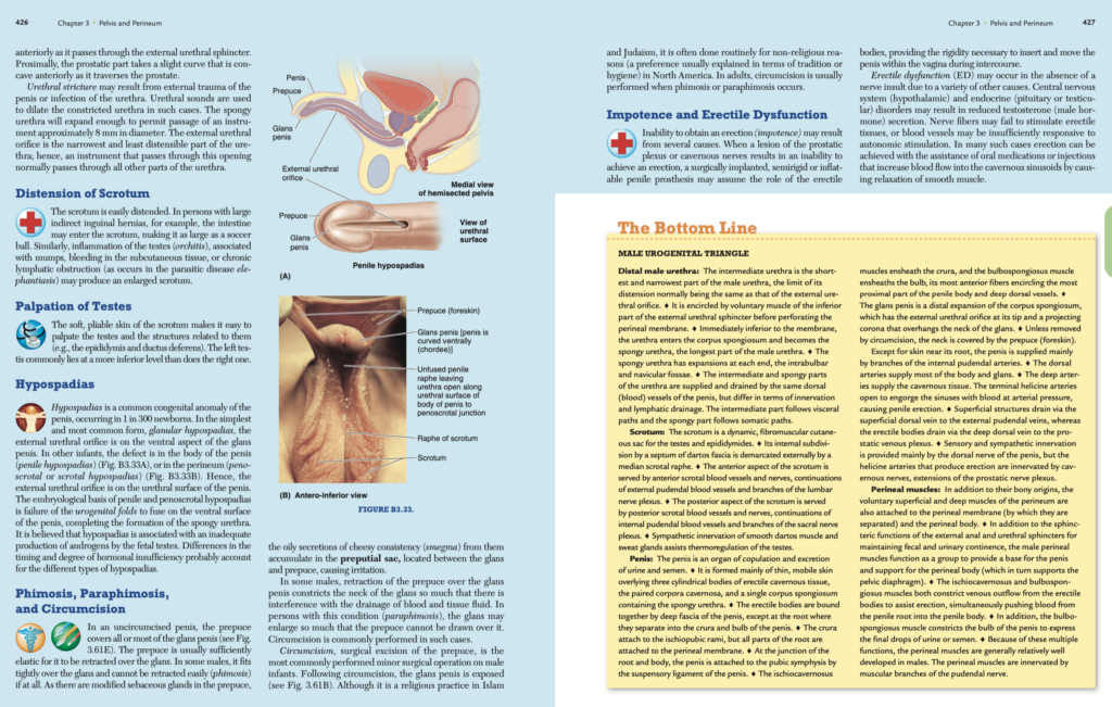
先天性疾患:尿道下裂 (Hypospadias)
概要
尿道下裂は、300人に1人の割合で見られる男性の一般的な先天性異常です。
- 種類
- 亀頭部尿道下裂 (Glanular hypospadias):最も単純で一般的な形態。外尿道口が陰茎亀頭の腹側に位置します。
- 陰茎体部尿道下裂 (Penile hypospadias) または陰茎陰嚢部尿道下裂 (Penoscrotal hypospadias):尿道口が陰茎または会陰部に位置します。
- 病因
- 胎児精巣からのアンドロゲン産生不足。ホルモン不足の程度とタイミングの違いが、多様な形態の尿道下裂を引き起こします。
包茎、嵌頓包茎、割礼 (Phimosis, Paraphimosis, and Circumcision)
包茎 (Phimosis)
- 包皮が亀頭をきつく覆い、後退困難または不可能になる状態。包皮内の皮脂腺からの分泌物(恥垢 [Smegma])が蓄積し、炎症を引き起こす場合があります。
嵌頓包茎 (Paraphimosis)
- 包皮が亀頭を締め付け、血流や組織液の排出が阻害される状態。亀頭が腫大し、包皮が元の位置に戻せなくなることがあります。
割礼 (Circumcision)
- 包皮の外科的切除。
- 理由:宗教的儀式(イスラム教、ユダヤ教)や、非宗教的理由(伝統、衛生上の理由)。
- 成人では主に包茎や嵌頓包茎の治療目的で行われます。
勃起不全 (Impotence and Erectile Dysfunction)
勃起不全 (Impotence)
- 勃起機能を失う原因には、前立腺神経叢や陰茎海綿体神経の損傷があります。
- 治療としては、半剛性または膨張式の陰茎プロテーゼの埋め込みが行われることがあります。
勃起障害 (Erectile Dysfunction, ED)
- 神経損傷がなくても、以下の要因でEDが発生します:
- 中枢神経系や内分泌疾患(例:視床下部、下垂体、精巣の異常)。
- 血管機能不全:自律神経刺激への応答性の低下。
治療
- 薬物療法:経口薬や注射剤が用いられ、陰茎海綿体の平滑筋を弛緩させて血流を増加させます。
後腹壁
後腹壁 (Posterior Abdominal Wall)
概要
後腹壁(図2.95–2.97参照)は主に以下の構造から構成されています:
- 腰椎 (Lumbar vertebrae) とそれに関連する椎間板(中心部分)。
- 後腹壁の筋肉 (Posterior abdominal wall muscles):
大腰筋 (Psoas)、
腰方形筋 (Quadratus lumborum)、
腸骨筋 (Iliacus)、
腹横筋 (Transversus abdominis)、
斜筋 (Oblique muscles)(側方)。 - 横隔膜 (Diaphragm):後壁の上部を形成。
- 筋膜 (Fascia):胸腰筋膜 (Thoracolumbar fascia) を含む。
- 腰神経叢 (Lumbar plexus):腰椎の前枝から成る。
- 脂肪、神経、血管(例:大動脈 (Aorta) と下大静脈 (IVC))、リンパ節。
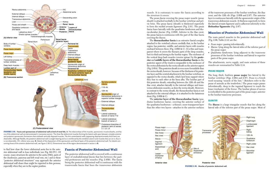
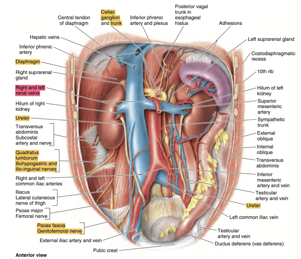
後腹壁を図2.97のような2次元の図で観察すると、それが平坦であるかのように思えるかもしれません。しかし、解剖された遺体や図2.95AおよびBのような横断断面で観察すると、腰椎が後壁の中央に顕著な隆起を形成し、左右に椎傍溝 (Paravertebral gutters) が生じていることが分かります。この溝の最も深い部分(最も後方)は腎臓とその周囲の脂肪が占めています。腹部大動脈は前方に突出した椎骨の前面に位置しています。痩せた個体では、大動脈が前腹壁に非常に近いことに驚くことがよくあります(図B2.37C参照)。もちろん、多くの構造物(上腸間膜動脈 (SMA)、十二指腸の一部、膵臓、左腎静脈など)が大動脈の前に位置しているため、これらの「後腹壁構造物」が仰臥位の痩せた人では予想以上に前腹壁に接近する場合があります。
後腹壁の筋膜 (Fascia of Posterior Abdominal Wall)
後腹壁は、腹膜壁側 (Parietal peritoneum) と筋肉の間に位置する内腹筋膜 (Endoabdominal fascia) の連続層で覆われています(図2.95B参照)。後腹壁を覆う筋膜は、腹横筋を覆う腹横筋筋膜 (Transversalis fascia) と連続しています。この筋膜は、覆う構造に応じて名前が付けられるのが一般的です。
大腰筋筋膜 (Psoas Fascia)
- 大腰筋 (Psoas major muscle) を覆う筋膜(大腰筋鞘 (Psoas sheath))は、内側で腰椎および骨盤縁に付着します。
- 上方では、この筋膜が厚くなり、内側弓状靱帯 (Medial arcuate ligament) を形成します(図2.91参照)。
- 外側では腰方形筋筋膜 (Quadratus lumborum fascia) と胸腰筋膜と融合します(図2.95B参照)。
- 腸骨稜 (Iliac crest) の下方では、腸骨筋を覆う腸骨筋筋膜 (Iliac fascia) と連続します。
胸腰筋膜 (Thoracolumbar Fascia)
胸腰筋膜は、内側で椎骨に付着し、広範囲にわたる筋膜構造です。腰部では後層、中間層、前層の3層があり、それぞれの間に筋肉が挟まれています(図2.95BおよびC参照)。
- 胸部では、この筋膜は薄く透明で、深部筋を覆っています。
- 腰部では、この筋膜は厚く強固で、脊柱起立筋群 (Erector spinae) を後層と中間層が包む構造になっています。これは前面の腹直筋鞘 (Rectus sheath) と比較されますが、胸腰筋膜の後層ははるかに厚く、腰椎に中央で付着するため、骨による支持がない白線 (Linea alba) で融合する腹直筋鞘よりも強固です。
胸腰筋膜の腰部は、第12肋骨と腸骨稜の間に広がり、外側で内腹斜筋および腹横筋に付着します。この点も腹直筋鞘に似ていますが、胸腰筋膜は外腹斜筋には付着せず、広背筋 (Latissimus dorsi) に付着しています(図2.95BおよびC参照)。
胸腰筋膜の前層 (Anterior Layer of Thoracolumbar Fascia)
- 腰方形筋 (Quadratus lumborum) の前面を覆う筋膜で、他の2層よりも薄く透明です。
- 付着部:腰椎の横突起の前面、腸骨稜、第12肋骨(図2.95Bおよび2.97参照)。
- 連続性:外側では腹横筋の腱膜起始部と連続しています。
- 厚化部:上方で外側弓状靱帯 (Lateral arcuate ligament) を形成し、下方では腸腰靱帯 (Iliolumbar ligaments) に付着しています(図2.97参照)。
後腹壁の筋肉 (Muscles of Posterior Abdominal Wall)
後腹壁の主な対となる筋肉(図2.96、表2.14参照)は以下の通りです:
- 大腰筋 (Psoas major):斜め下外側に走行。
- 腸骨筋 (Iliacus):大腰筋の下部外側に沿って位置。
- 腰方形筋 (Quadratus lumborum):腰椎の横突起に隣接し、大腰筋の上部外側に位置。
これらの筋肉の付着部、神経支配、および主な作用は表2.14にまとめられています。
| 筋肉名 | 上部付着(Superior Attachment) | 下部付着(Inferior Attachment) | 神経支配(Innervation) |
|---|---|---|---|
| 大腰筋(Psoas major) | 腰椎横突起(Transverse processes of lumbar vertebrae)、T12-L5椎体の側面(Sides of bodies of T12-L5 vertebrae)、椎間円板(Intervertebral discs) | 大腿骨小転子(Lesser trochanter of femur) | 腰神経叢の前枝L1-L3(Anterior rami of lumbar nerves L1-L3) |
| 腸骨筋(Iliacus) | 腸骨窩上部2/3(Superior two thirds of iliac fossa)、仙骨翼(Ala of sacrum)、前仙腸靭帯(Anterior sacro-iliac ligaments) | 大腿骨小転子(Lesser trochanter of femur)とその下方の骨幹(Shaft inferior to it) | 大腿神経(Femoral nerve, L2-L4) |
| 腰方形筋(Quadratus lumborum) | 第12肋骨の下縁内側半分(Medial half of inferior border of 12th rib)、腰椎横突起(Tips of lumbar transverse processes) | 腸腰靭帯(Iliolumbar ligament)、腸骨稜内側唇(Internal lip of iliac crest) | T12およびL1-L4神経の前枝(Anterior branches of T12 and L1-L4 nerves) |
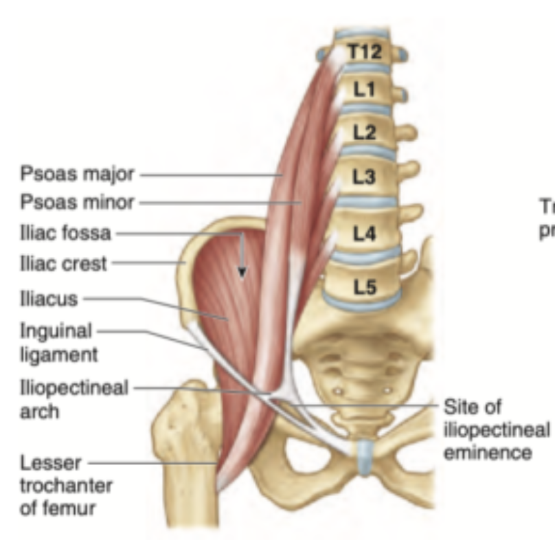
大腰筋 (Psoas Major)
長く厚い紡錘状の大腰筋は、腰椎の外側に位置します(図2.96Aおよび2.97参照)。Psoasはギリシャ語で「腰の筋肉」を意味します(食肉業者は動物の大腰筋をテンダーロイン (Tenderloin) と呼びます)。大腰筋は斜め下外側に走行し、鼠径靱帯の深部を通過して大腿骨小転子に到達します。腰神経叢 (Lumbar plexus) は大腰筋の後部、腰椎の横突起の前面に埋め込まれています。
腸骨筋 (Iliacus)
腸骨筋は、大腰筋の下部外側に沿って位置する大きな三角形の筋肉です。そのほとんどの筋線維は大腰筋の腱に合流します。大腰筋と腸骨筋は一緒に腸腰筋 (Iliopsoas) を形成し、大腿の主要な屈筋として機能します。また、股関節の安定化を助け、この関節における直立姿勢の維持に寄与します。
- 共通の作用:大腰筋と腸骨筋は股関節屈曲に関与します。
- 個別の作用:大腰筋のみが腰椎の屈曲や側屈(横方向の曲げ)を引き起こすことができます。
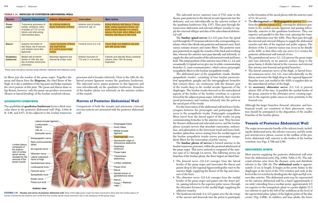
腰方形筋 (Quadratus Lumborum)
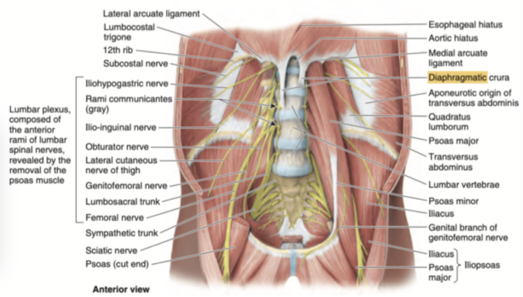
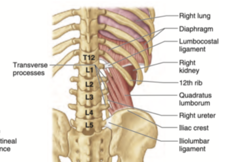
四角形の腰方形筋は、後腹壁に厚い筋板を形成しています(図2.94AおよびB、図2.96、図2.97参照)。この筋肉は腰椎の横突起に隣接し、下部が広くなっています。第12肋骨付近では、外側弓状靱帯が腰方形筋を横切っています。肋下神経(T12)はこの靱帯の後ろを通り、腰方形筋の上を斜め下外側に走行します。腰神経叢の枝は、この筋肉の前面を下方へ走ります。
後腹壁の神経 (Nerves of Posterior Abdominal Wall)
後腹壁には、体性神経系と自律神経系(内臓神経系)の両方が関与しています。
- 肋下神経 (Subcostal Nerves, T12)
肋下神経(T12の前枝)は胸部で起こり、外側弓状靱帯の後ろを通って腹部に入り、腰方形筋の前面を斜め下外側に走行します(図2.97参照)。この神経は腹横筋と内腹斜筋を貫通して、外腹斜筋および前外側腹壁の皮膚に分布します。 - 腰神経 (Lumbar Spinal Nerves, L1–L5)
腰神経は、対応する椎骨の下の椎間孔を通って脊髄から出て、後枝と前枝に分かれます。各枝には感覚神経線維と運動神経線維が含まれています。後枝は後方に進み、背部の筋肉とその上の皮膚に分布します。一方、前枝は外側および下方に進み、下部体幹および下肢の皮膚と筋肉に分布します。L1、L2、場合によってはL3の前枝の初期部分は、白交通枝(rami communicantes)を形成し、腰交感神経幹に向かう節前交感神経線維を伝達します。
腰交感神経幹 (Lumbar Sympathetic Trunks)
腹部の交感神経幹(腰交感神経幹)は、4つの腰部椎傍交感神経節およびそれらを連結する神経節間枝からなり、横隔膜の内側弓状靱帯の奥にある胸部交感神経幹と連続しています。腰神経幹は、腰椎体の前外側面を溝状に沿って下降し、その隣接部には大腰筋が存在します。下方では、仙骨岬を横切り、骨盤内で仙骨交感神経幹として続きます。
腰神経叢 (Lumbar Plexus)
腰神経叢は、大腰筋の近位付着部内で、腰椎の横突起の前面に形成される神経網です。この神経叢はL1からL4の神経前枝から構成されます。以下の神経が腰神経叢の枝です(主な3つの神経を先に挙げます):
- 大腿神経 (Femoral Nerve, L2–L4)
大腰筋の外側縁から出て腸骨筋を支配し、鼠径靱帯/腸恥帯の下を通り、大腿前面に達します。股関節の屈筋および膝の伸筋に分布します。 - 閉鎖神経 (Obturator Nerve, L2–L4)
大腰筋の内側縁から出て、小骨盤に入り、恥骨上枝の下(閉鎖孔を通る)を通って大腿内側部に達します。内転筋群を支配します。 - 腰仙幹 (Lumbosacral Trunk, L4, L5)
仙骨の翼状部(アラ)を越えて下降し、S1–S4の神経前枝とともに仙骨神経叢を形成します。 - 腸骨鼠径神経および腸骨下腹神経 (Ilio-inguinal and Iliohypogastric Nerves, L1)
L1の前枝から起こり、内側弓状靱帯の後ろを通って腹部に入り、腰方形筋の前面を斜め下外側に走行します。腸骨稜の上方および平行に走行し、ASIS付近で腹横筋を貫通します。その後、内腹斜筋と外腹斜筋を通って、腹筋と鼠径部および恥骨部の皮膚に分布します。 - 生殖大腿神経 (Genitofemoral Nerve, L1, L2)
大腰筋を貫通し、その前面を下方に走行します。この神経は外腸骨動脈の外側で分岐し、生殖枝と大腿枝に分かれます。 - 外側大腿皮神経 (Lateral Cutaneous Nerve of the Thigh, L2, L3)
腸骨筋の上を斜め下外側に走行し、鼠径靱帯/腸恥帯の下を通って大腿外側面に到達します。この神経は大腿の前外側の皮膚に分布します。 - 副閉鎖神経 (Accessory Obturator Nerve, L3, L4)
約10%の頻度で存在し、大腰筋の内側縁に沿って走行し、閉鎖神経の前を並行して走ります。この神経は恥骨上枝の上を横切り、大腿静脈に近接します。
腰神経叢の大きな枝(大腿神経、閉鎖神経、腰仙幹)は配置が一貫していますが、腰神経叢の小枝の分布には変異が予想される場合があります。
| 神経名 | 神経根(Nerve Roots) | 走行(Pathway) | 支配領域(Innervation Area) |
|---|---|---|---|
| 大腿神経(Femoral Nerve) | L2–L4 | 大腰筋の外側縁から出て腸骨筋を支配し、鼠径靱帯/腸恥帯の下を通り、大腿前面に達する。 | 股関節の屈筋(Flexors of hip)および膝の伸筋(Extensors of knee)。 |
| 閉鎖神経(Obturator Nerve) | L2–L4 | 大腰筋の内側縁から出て、小骨盤に入り、恥骨上枝の下を通り、大腿内側部に到達する。 | 大腿内転筋群(Adductor muscles of thigh)。 |
| 腰仙幹(Lumbosacral Trunk) | L4, L5 | 仙骨の翼状部(アラ)を越えて下降し、S1–S4の神経前枝とともに仙骨神経叢を形成する。 | 仙骨神経叢(Sacral plexus)を形成する一部として機能。 |
| 腸骨鼠径神経(Ilio-inguinal Nerve)および腸骨下腹神経(Iliohypogastric Nerve) | L1 | L1の前枝から起こり、腰方形筋の前面を斜め下外側に走行し、腹横筋を貫通。その後、内腹斜筋と外腹斜筋を通って皮膚に分布。 | 腹筋(Abdominal muscles)および鼠径部・恥骨部の皮膚(Skin of inguinal and pubic regions)。 |
| 生殖大腿神経(Genitofemoral Nerve) | L1, L2 | 大腰筋を貫通し、その前面を下方に走行。外腸骨動脈の外側で分岐し、生殖枝と大腿枝に分かれる。 | 生殖枝(Genital branch):外陰部の感覚。大腿枝(Femoral branch):大腿上部の皮膚(Skin over upper thigh)。 |
| 外側大腿皮神経(Lateral Cutaneous Nerve of the Thigh) | L2, L3 | 腸骨筋の上を斜め下外側に走行し、鼠径靱帯/腸恥帯の下を通って大腿外側面に到達する。 | 大腿前外側の皮膚(Skin of anterolateral thigh)。 |
| 副閉鎖神経(Accessory Obturator Nerve) | L3, L4 | 約10%の頻度で存在。大腰筋の内側縁に沿って走行し、閉鎖神経の前を並行して恥骨上枝の上を横切り、大腿静脈に近接する。 | 恥骨周辺の筋肉(Muscles near pubic region)。 |
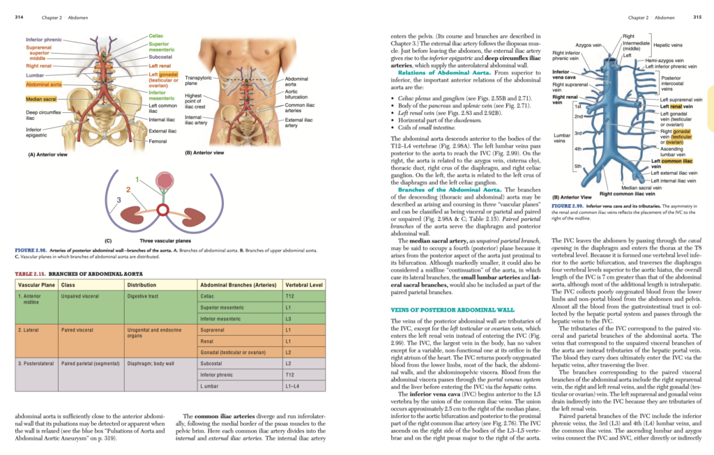
後腹壁の血管 (Vessels of Posterior Abdominal Wall)
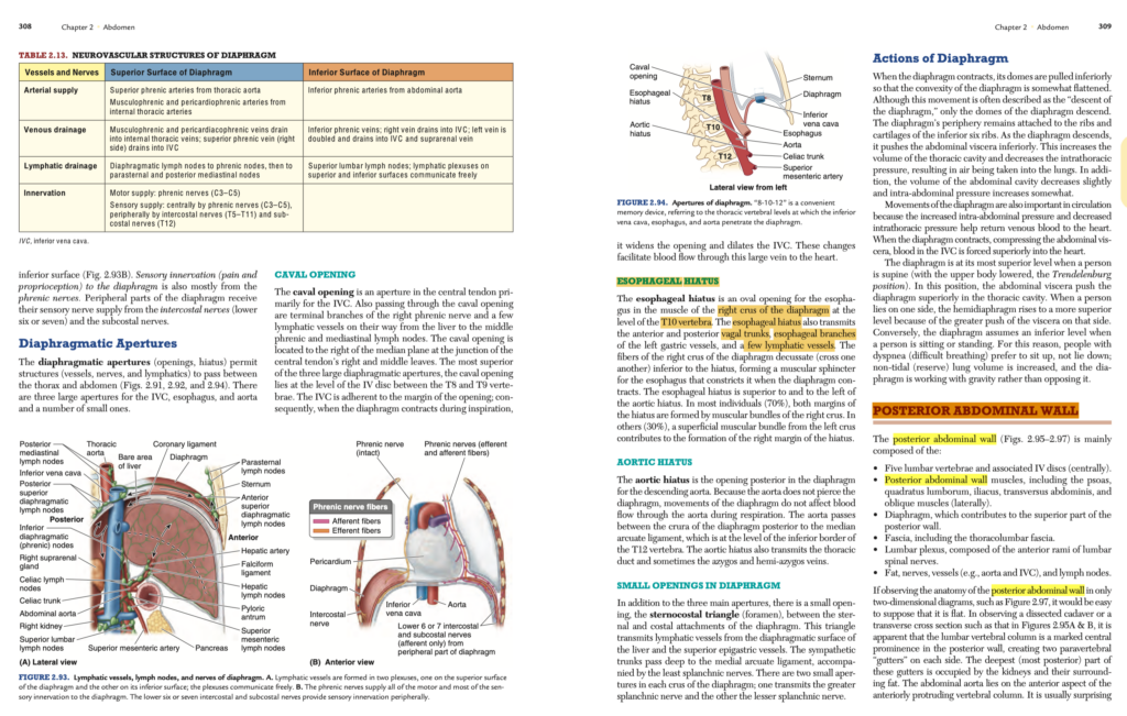
下部体幹の主要な神経血管束である腹部大動脈 (abdominal aorta)、下大静脈 (inferior vena cava)、および大動脈周囲神経叢 (aortic peri-arterial nerve plexus) は、後腹壁の中央部、腰椎体の前方を走行します(図2.70Bおよび2.89参照)。
腹部大動脈 (Abdominal Aorta)
後腹壁に血液を供給する動脈のほとんどは腹部大動脈から分岐します(図2.98A;表2.15参照)。肋下動脈 (subcostal arteries) は胸部大動脈から分岐し、第12肋骨の下方に分布します。腹部大動脈の長さは約13 cmです。この動脈は第12胸椎 (T12) の高さで横隔膜の大動脈裂孔 (aortic hiatus) から始まり、第4腰椎 (L4) の高さで左右の総腸骨動脈 (common iliac arteries) に分岐して終わります。腹部大動脈は、前腹壁上では帯状の領域として表され、この帯状領域は中央部の約2.5 cm上方に位置する幽門横断面 (transpyloric plane) の中間点から、臍のやや左下方(約2~3 cm)に位置する腸骨稜 (iliac crests) の最高点を結ぶ腸稜面 (supracristal plane) まで延びています(図2.98B参照)。
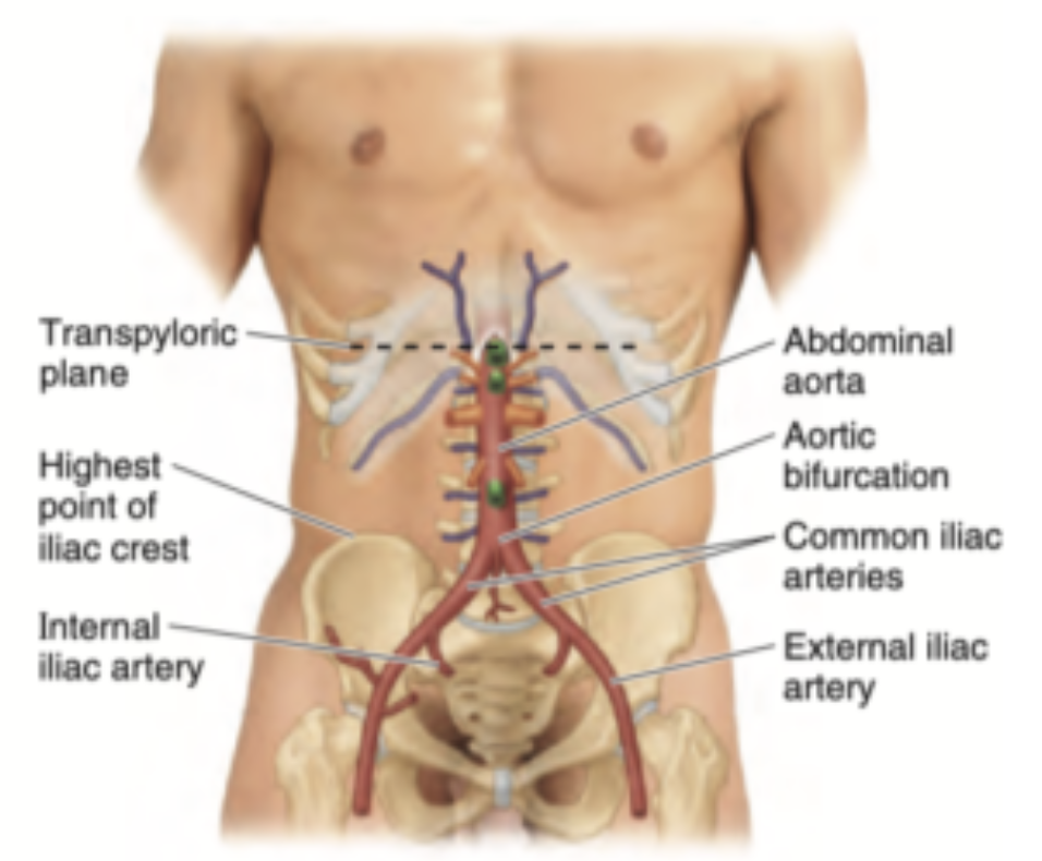
子供や痩せ型の成人では、下部腹部大動脈が前腹壁に十分近接しているため、壁が弛緩していると脈動が確認または感じられることがあります(青い枠「大動脈の脈動と腹部大動脈瘤」319ページ参照)。
総腸骨動脈は分岐して斜め下外側に走行し、大腰筋の内側縁に沿って骨盤縁に向かいます。ここで、各総腸骨動脈は内腸骨動脈 (internal iliac artery) と外腸骨動脈 (external iliac artery) に分岐します。内腸骨動脈は骨盤内に入ります(その経路と分岐は第3章で説明されています)。外腸骨動脈は腸腰筋 (iliopsoas muscle) に沿って走行します。腹腔を出る直前に、外腸骨動脈は下腹壁動脈 (inferior epigastric artery) と深腸骨回旋動脈 (deep circumflex iliac artery) を分岐させ、前外側腹壁に血液を供給します。
腹部大動脈の位置関係 (Relations of Abdominal Aorta)
腹部大動脈の重要な前方の位置関係は、上部から下部にかけて以下の通りです:
- 腹腔神経叢および腹腔神経節 (celiac plexus and ganglion)(図2.55Bおよび2.71参照)。
- 膵臓の体部および脾静脈 (splenic vein)(図2.71参照)。
- 左腎静脈 (left renal vein)(図2.83および2.92B参照)。
- 十二指腸の水平部 (horizontal part of the duodenum)。
- 小腸のコイル状部 (coils of small intestine)。
腹部大動脈は、第12胸椎から第4腰椎 (T12–L4) の椎体前方を下降します(図2.98A参照)。左腰静脈 (left lumbar veins) は大動脈の後方を通過して下大静脈に達します(図2.99参照)。右側では、大動脈は奇静脈 (azygos vein)、乳糜槽 (cisterna chyli)、胸管 (thoracic duct)、横隔膜の右脚 (right crus of the diaphragm)、および右腹腔神経節 (right celiac ganglion) に関連しています。左側では、横隔膜の左脚 (left crus of the diaphragm) および左腹腔神経節 (left celiac ganglion) に関連しています。
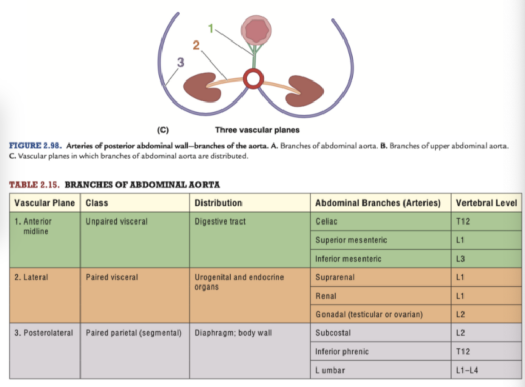
腹部大動脈の分岐 (Branches of the Abdominal Aorta)
下行大動脈(胸部および腹部)は、3つの「血管平面」に沿って分岐して走行し、内臓性または壁性、対になっているものと対になっていないものに分類されます(図2.98A&C;表2.15参照)。対になっている壁性の分岐は、横隔膜および後腹壁に供給します。
正中仙骨動脈 (median sacral artery) は、対になっていない壁性の分岐で、大動脈の分岐直前の後方から発生します。著しく小さいものの、大動脈の正中「継続部」と見なすことができ、その側枝である小腰動脈 (small lumbar arteries) および外側仙骨枝 (lateral sacral branches) も対になった壁性の分岐の一部として含まれます。
後腹壁の静脈 (Veins of Posterior Abdominal Wall)
後腹壁の静脈は、下大静脈 (inferior vena cava: IVC) の枝にあたります。ただし、左精巣静脈または卵巣静脈 (left testicular or ovarian vein) は、下大静脈ではなく左腎静脈 (left renal vein) に流入します(図2.99参照)。下大静脈は体内で最大の静脈であり、心臓の右心房での開口部に可変的で機能しない弁を除き、弁が存在しません。下大静脈は、下肢、背部の大部分、腹壁、腹腔内臓器 (abdominopelvic viscera) から酸素含有量の低い血液を還流します。腹腔内臓器からの血液は、門脈系 (portal venous system) と肝臓 (liver) を経て、肝静脈 (hepatic veins) を通じて下大静脈に入ります。
下大静脈の構造 (Structure of the IVC)
下大静脈は、第5腰椎 (L5 vertebra) の前方で総腸骨静脈 (common iliac veins) の合流によって始まります。この合流部は、正中線から約2.5 cm右側、大動脈分岐 (aortic bifurcation) の下方、および右総腸骨動脈 (right common iliac artery) の近位部の後方に位置します(図2.76参照)。下大静脈は第3~第5腰椎 (L3–L5) の椎体の右側、および大腰筋 (psoas major) の右側を上行し、大動脈の右側に位置します。
下大静脈は、横隔膜の大静脈孔 (caval opening) を通過して腹部を出て、第8胸椎 (T8 vertebral level) の高さで胸部に入ります。下大静脈は大動脈分岐部の1椎体分下部で形成され、横隔膜の大動脈裂孔 (aortic hiatus) を4椎体分上部で通過するため、その全長は腹部大動脈 (abdominal aorta) よりも7 cm長くなります。ただし、この追加の長さの大部分は肝内 (intrahepatic) 部分にあたります。
下大静脈は、下肢および腹部・骨盤 (abdomen and pelvis) の非門脈性血液 (non-portal blood) から酸素含有量の低い血液を集めます。消化管 (gastrointestinal tract) からの血液のほぼ全ては門脈系 (hepatic portal system) によって集められ、肝静脈を通じて下大静脈に入ります。
下大静脈の枝 (Tributaries of the IVC)
下大静脈の枝は、腹部大動脈の対になった内臓枝 (paired visceral branches) および壁枝 (paired parietal branches) に対応します。一方、大動脈の対になっていない内臓枝 (unpaired visceral branches) に対応する静脈は、肝門脈 (hepatic portal vein) の枝となります。これらの静脈が運ぶ血液は最終的に肝臓を通過した後、肝静脈を介して下大静脈に流入します。
腹部大動脈の対になった内臓枝に対応する静脈には以下が含まれます:
- 右副腎静脈 (right suprarenal vein)。
- 右および左腎静脈 (right and left renal veins)。
- 右性腺静脈 (右精巣静脈または卵巣静脈) (right gonadal vein: testicular or ovarian vein)。 左副腎静脈 (left suprarenal vein) および左性腺静脈 (left gonadal vein) は、左腎静脈の枝であるため、間接的に下大静脈に流入します。
対になった壁枝 (paired parietal branches) には以下が含まれます:
- 下横隔静脈 (inferior phrenic veins)。
- 第3および第4腰静脈 (3rd and 4th lumbar veins)。
- 総腸骨静脈 (common iliac veins)。
上行腰静脈 (ascending lumbar vein) と奇静脈 (azygos vein) は、直接または間接的に下大静脈と上大静脈 (superior vena cava: SVC) を接続し、側副路 (collateral pathways) を提供します(青い枠「腹腔骨盤静脈血の側副路」319ページ参照)。
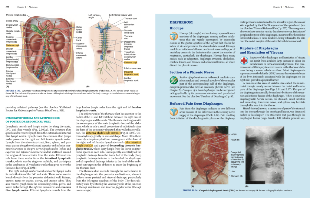
後腹壁のリンパ管とリンパ節 (Lymphatic Vessels and Lymph Nodes of Posterior Abdominal Wall)
リンパ管およびリンパ節は、大動脈 (aorta)、下大静脈 (IVC: inferior vena cava)、および腸骨動静脈 (iliac vessels) に沿って配置されています(図2.100A参照)。総腸骨リンパ節 (common iliac lymph nodes) は、外腸骨リンパ節 (external iliac lymph nodes) と内腸骨リンパ節 (internal iliac lymph nodes) からリンパを受け取ります。総腸骨リンパ節からのリンパは、右および左の腰リンパ節 (lumbar lymph nodes) に送られます。消化管 (alimentary tract)、肝臓 (liver)、脾臓 (spleen)、膵臓 (pancreas) からのリンパは、腹腔動脈 (celiac artery) および上腸間膜動脈 (superior mesenteric artery)、下腸間膜動脈 (inferior mesenteric artery) に沿って腹腔前リンパ節 (pre-aortic lymph nodes)(腹腔リンパ節 (celiac nodes)、上腸間膜リンパ節 (superior mesenteric nodes)、下腸間膜リンパ節 (inferior mesenteric nodes))に流れます。これらのリンパ節からの輸出管 (efferent vessels) は腸リンパ幹 (intestinal lymphatic trunks) を形成し、単独または複数でリンパ幹の合流 (confluence of lymphatic trunks) に参加して胸管 (thoracic duct) を形成します(図2.100B参照)。
右および左の腰リンパ節 (lumbar lymph nodes)(別名:下大静脈リンパ節 (caval lymph nodes) および大動脈リンパ節 (aortic lymph nodes))は、下大静脈と大動脈の両側に位置しています。これらのリンパ節は、後腹壁 (posterior abdominal wall)、腎臓 (kidneys)、尿管 (ureters)、精巣または卵巣 (testes or ovaries)、子宮 (uterus)、および卵管 (uterine tubes) から直接リンパを受け取ります。また、下行結腸 (descending colon)、骨盤 (pelvis)、下肢 (lower limbs) からも、下腸間膜リンパ節 (inferior mesenteric lymph nodes) と総腸骨リンパ節を介してリンパを受け取ります。これらの大きな腰リンパ節からの輸出リンパ管 (efferent lymphatic vessels) は、右および左の腰リンパ幹 (lumbar lymphatic trunks) を形成します。
| 部位 | 位置・配置 | 受け取るリンパ | 形成・接続先 |
|---|---|---|---|
| 総腸骨リンパ節(Common Iliac Lymph Nodes) | 腸骨動静脈(Iliac vessels)に沿う | 外腸骨リンパ節(External Iliac Lymph Nodes)、内腸骨リンパ節(Internal Iliac Lymph Nodes)からリンパを受け取る。 | 右および左の腰リンパ節(Lumbar Lymph Nodes)に送る。 |
| 腰リンパ節(Lumbar Lymph Nodes) | 下大静脈(IVC: Inferior Vena Cava)および大動脈(Aorta)の両側に配置。別名:下大静脈リンパ節(Caval Nodes)、大動脈リンパ節(Aortic Nodes)。 | 後腹壁(Posterior Abdominal Wall)、腎臓(Kidneys)、尿管(Ureters)、生殖器(精巣/卵巣、子宮、卵管)から直接受け取る。 | 右および左の腰リンパ幹(Lumbar Lymphatic Trunks)を形成する。 |
| 腹腔前リンパ節(Pre-aortic Lymph Nodes) | 腹腔動脈(Celiac Artery)、上腸間膜動脈(Superior Mesenteric Artery)、下腸間膜動脈(Inferior Mesenteric Artery)に沿う。 | 消化管(Alimentary Tract)、肝臓(Liver)、脾臓(Spleen)、膵臓(Pancreas)からリンパを受け取る。 | 腸リンパ幹(Intestinal Lymphatic Trunks)を形成し、胸管(Thoracic Duct)に接続。 |
| 腸リンパ幹(Intestinal Lymphatic Trunks) | 腹腔前リンパ節(Pre-aortic Lymph Nodes)の輸出管が集合して形成される。 | 腹腔前リンパ節(Pre-aortic Lymph Nodes)からのリンパを集める。 | 単独または複数でリンパ幹の合流(Confluence of Lymphatic Trunks)に参加し、胸管(Thoracic Duct)を形成。 |
| 胸管(Thoracic Duct) | リンパ幹の合流(Confluence of Lymphatic Trunks)で形成され、右胸腔を通過し、左静脈角(Left Venous Angle)に注ぐ。 | 腸リンパ幹(Intestinal Lymphatic Trunks)、腰リンパ幹(Lumbar Lymphatic Trunks)などからリンパを受け取る。 | 全身のリンパを最終的に静脈系へ還流させる。 |
胸管 (Thoracic Duct)
胸管の下端 (inferior end) は、第1および第2腰椎 (L1 and L2 vertebrae) の椎体の前方にあり、横隔膜の右脚 (right crus of the diaphragm) と大動脈の間に位置します。胸管は、腹部の主要なリンパ管の合流から始まり、この合流が一般的に描かれる薄壁の嚢状構造、すなわち乳糜槽 (cisterna chyli) を形成するのは少数の個体のみです(図2.100B参照)。乳糜槽の大きさや形状は大きく異なります。多くの場合、このレベルでは単純な合流、または複雑な網状の合流がみられます。この合流には、右および左の腰リンパ幹 (lumbar lymphatic trunks)、腸リンパ幹 (intestinal lymphatic trunk(s))、および左右6つの肋間隙 (intercostal spaces) からのリンパを運ぶ下降胸リンパ幹 (descending thoracic lymphatic trunks) が含まれます。したがって、横隔膜の下方にある体の下半分(深部リンパ排出および臍 (umbilicus) の下方にある全表在性リンパ排出)のすべてのリンパ排出が腹部で胸管の起点に集まります。
胸管は、横隔膜の大動脈裂孔 (aortic hiatus) を通過して後縦隔 (posterior mediastinum) を上行し、さらに体の左上四分の一 (left upper quadrant of the body) からの壁側および臓側のリンパ排出を収集します。最終的に胸管は左鎖骨下静脈 (left subclavian vein) と内頸静脈 (internal jugular vein) の合流部(左静脈角 (left venous angle))で静脈系に入ります。
横隔膜 (Diaphragm)
しゃっくり (Hiccups)
しゃっくり (hiccoughs) は横隔膜 (diaphragm) の不随意の痙攣性収縮であり、吸気が突然起こり、その後すぐに声門 (glottis: 喉頭の開口部) の痙攣性閉鎖により空気の流入が遮断され、特徴的な音が発生します。しゃっくりは、求心性または遠心性の神経終末の刺激、または呼吸筋(特に横隔膜)を制御する延髄 (medullary centers in the brainstem) の刺激によって生じます。原因としては、消化不良 (indigestion)、横隔膜の刺激 (irritation)、アルコール中毒 (alcoholism)、脳病変 (cerebral lesions)、胸部および腹部病変などがあります。これらの原因は横隔神経 (phrenic nerves) を刺激します。
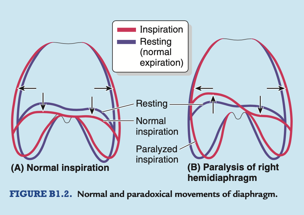
横隔神経の切断 (Section of a Phrenic Nerve)
首部で横隔神経 (phrenic nerve) を切断すると、対応する半横隔膜の筋肉部分が完全に麻痺 (paralysis) し、最終的には萎縮 (atrophy) します。ただし、副横隔神経 (accessory phrenic nerve) を持つ人では例外です(第8章参照)。半横隔膜の麻痺 (paralysis of a hemidiaphragm) は、放射線写真で永続的な挙上 (permanent elevation) と逆説的な動き (paradoxical movement) によって認識できます(青い枠「横隔膜の麻痺」85ページ参照)。
横隔膜からの関連痛 (Referred Pain from Diaphragm)
横隔膜からの痛みは、その感覚神経供給の違いにより、異なる領域に放散します(表2.12参照)。横隔膜胸膜 (diaphragmatic pleura) または横隔膜腹膜 (diaphragmatic peritoneum) の刺激による痛みは、肩部 (shoulder region) に放散します。この部位の皮膚は脊髄のC3–C5セグメントから供給されます(青い枠「内臓関連痛」257ページ参照)。これらのセグメントは横隔神経の前枝 (anterior rami) にも寄与しています。横隔膜の末梢領域の刺激(下部肋間神経 (inferior intercostal nerves) によって支配される)はより局所的であり、前外側腹壁の肋骨縁上の皮膚に関連付けられます。
横隔膜の破裂と内臓のヘルニア (Rupture of Diaphragm and Herniation of Viscera)
横隔膜の破裂 (rupture) と内臓のヘルニア (herniation of viscera) は、胸腔内または腹腔内圧の急激な増加により生じます。この損傷の一般的な原因は、自動車事故中の胸部または腹部への重大な外傷です。横隔膜破裂の大部分(95%)は左側に発生します。これは、横隔膜と密接に関連する肝臓 (liver) の実質的な質量が右側に物理的障壁を提供するためです。
横隔膜の肋骨部 (costal part) と腰部 (lumbar part) の間には、変動するサイズの非筋性領域 (non-muscular area)、すなわち腰肋三角 (lumbocostal triangle) が通常存在します(図2.91および2.97参照)。この部分は通常、横隔膜の上部および下部の筋膜の融合のみによって形成されます。外傷性横隔膜ヘルニア (traumatic diaphragmatic hernia) が発生すると、胃 (stomach)、小腸および腸間膜 (mesentery)、横行結腸 (transverse colon)、脾臓 (spleen) がこの領域を通じて胸腔に脱出する可能性があります。
先天性横隔膜ヘルニア (Congenital Diaphragmatic Hernia)
先天性横隔膜ヘルニア (CDH: congenital diaphragmatic hernia) では、胃 (stomach) と腸 (intestine) の一部が、横隔膜 (diaphragm) の腰肋三角 (lumbocostal trigone) の領域にある大きな後外側欠損 (foramen of Bochdalek) を通じてヘルニア (herniate) を形成します(図B2.34参照)。このヘルニアはほぼ常に左側で発生し、右側には肝臓 (liver) が存在するためです。このタイプのヘルニアは、横隔膜の複雑な発生過程に起因します。横隔膜の後外側欠損は比較的一般的な横隔膜の先天性異常であり、新生児2200人に1人の割合で発生します (Moore, Persaud, and Torchia, 2012)。腹部内臓 (abdominal viscera) が出生前の肺腔 (pulmonary cavity) に入り込むことで、片方の肺 (通常は左肺) が正常に発育せず、出生後に膨張することができなくなります。この結果、肺低形成 (pulmonary hypoplasia) が生じ、これらの乳児の死亡率は高く(約76%)、致命的です。
後腹壁 (Posterior Abdominal Wall)
腰部膿瘍 (Psoas Abscess)
結核 (TB: tuberculosis) の発生率は大幅に減少しましたが、現在、特にアフリカやアジアでエイズ (AIDS) や薬剤耐性の影響により結核が再流行しており、時にはパンデミック規模に達しています。脊椎結核 (TB of the vertebral column) は非常に一般的です。感染は血液を通じて脊椎に広がる (hematogenous spread) ことがあり、特に小児期に多く見られます。腰部で結核による膿瘍が形成されると、脊椎から腸腰筋筋膜 (psoas fascia: sheath) に広がり、腰部膿瘍 (psoas abscess) を引き起こします(図B2.35参照)。その結果、腸腰筋筋膜は厚くなり、強いストッキングのようなチューブを形成します。膿 (pus) はこの筋膜内で腸腰筋に沿って下方に進み、骨盤縁 (pelvic brim) を越え、鼠径靭帯 (inguinal ligament) の下を通って太ももの上部に表出します。
膿はまた、胸椎 (thoracic vertebrae) が疾患に侵されている場合、後縦隔 (posterior mediastinum) から腸腰筋筋膜に到達することもあります。腸骨筋筋膜 (iliac fascia) の下部は通常緊張しており、腸骨稜 (iliac crest) の内面に向かって折り目を形成します。一方、上部は緩く、上記の折り目の後方に腸骨下筋膜窩 (iliacosubfascial fossa) と呼ばれるポケットを形成することがあります。この窩には盲腸 (cecum) や虫垂 (appendix: appendix)(右側)またはS状結腸 (sigmoid colon)(左側)の一部が閉じ込められ、激しい痛みを引き起こすことがあります。
後腹部の痛み (Posterior Abdominal Pain)
腸腰筋 (iliopsoas muscle) は、腎臓 (kidneys)、尿管 (ureters)、盲腸 (cecum)、虫垂 (appendix)、S状結腸 (sigmoid colon)、膵臓 (pancreas)、腰部リンパ節 (lumbar lymph nodes)、および後腹壁の神経と臨床的に重要な関係があります。これらの構造のいずれかが疾患に侵されている場合、腸腰筋の運動が痛みを引き起こすことがあります。腹腔内炎症が疑われる場合、腸腰筋テスト (iliopsoas test) を行います。このテストでは、患者に健側を下にして横になってもらい、患側の太ももを検査者の手の抵抗に対して伸展させます (Bickley, 2009)。この操作によって痛みが生じる場合、腸腰筋徴候 (psoas sign) は陽性です。例えば、急性炎症を伴う虫垂 (acutely inflamed appendix) は右腸腰筋徴候 (right psoas sign) を引き起こします(図B2.36参照)。
腸腰筋が脊柱 (vertebral column) に沿って走行し、腸骨筋 (iliacus) が仙腸関節 (sacro-iliac joint) を横切るため、椎間関節 (intervertebral joints) や仙腸関節の疾患は腸腰筋の痙攣 (spasm) を引き起こすことがあります。これは防御反射 (protective reflex) として作用します。膵臓癌 (adenocarcinoma of the pancreas) の進行した段階では、後腹壁の筋肉や神経に侵入し、膵臓が後腹壁に密接しているために耐え難い痛みを引き起こします。
部分的腰部交感神経切除術 (Partial Lumbar Sympathectomy)
下肢の動脈疾患の治療には、部分的腰部交感神経切除術 (partial lumbar sympathectomy) が含まれる場合があります。この手術では、交感神経幹 (sympathetic trunks) のいくつかの交感神経節 (sympathetic ganglia) を除去します。手術は通常、後腹壁の外側からの腹膜外アプローチ (lateral extraperitoneal approach) を用いて行われます。このアプローチでは、交感神経幹が腹膜外の脂肪組織 (extraperitoneal fatty tissue) に位置しているためです(図2.97参照)。手術中、外腹壁筋を切開し、腹膜 (peritoneum) を内側および前方に動かして腸腰筋 (psoas major) の内側縁を露出させます。この腸腰筋に沿って交感神経幹が存在します。交感神経幹 (sympathetic trunks) の位置は脂肪組織やリンパ組織によって覆われていることが多く、視認が難しい場合があります。そのため、交感神経幹を誤って除去するリスクを避けるために、慎重な操作が必要です。例えば、誤って大腿生殖神経 (genitofemoral nerve)、腰部リンパ管 (lumbar lymphatics)、または尿管 (ureter) の一部を損傷する可能性があります。特に、左側の交感神経幹は大動脈 (aorta) にやや覆われ、右側の交感神経幹は下大静脈 (IVC: inferior vena cava) に覆われているため、これらの大血管が損傷を受けないよう細心の注意を払う必要があります。
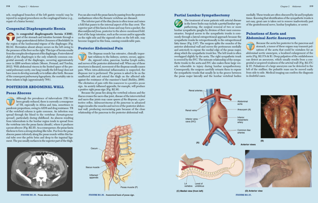
大動脈の拍動と腹部大動脈瘤 (Pulsations of Aorta and Abdominal Aortic Aneurysm)
大動脈 (aorta) は膵臓 (pancreas) および胃 (stomach) の後方に位置するため、これらの臓器に腫瘍があると、大動脈の拍動が伝わり、それが腹部大動脈瘤 (abdominal aortic aneurysm: AAA) と誤診されることがあります(図B2.37A&B参照)。腹部の深部触診により、瘤の存在を検出できる場合があります。この瘤は、先天的または後天的な動脈壁の弱化 (weakness of the arterial wall) に起因します(図B2.37C&D参照)。腹部大動脈瘤が左側の正中線に沿って拍動を示す場合、大きな瘤が疑われます。この場合、瘤は側方に簡単に動かすことができ、医療画像診断 (medical imaging) によって診断を確認します。
腹部大動脈瘤の急性破裂 (Acute Rupture of AAA)
腹部大動脈瘤が急性破裂すると、腹部または背部に激しい痛みを引き起こします。この状態が見逃されると、重度の出血によって死亡率が90%近くに達します (Swartz, 2009)。外科医は、瘤を切開し、人工血管 (prosthetic graft) を挿入して動脈壁を縫合することで修復を行います。以前は開腹手術 (open repair) が主流でしたが、現在ではカテーテル (catheter) を用いた血管内修復 (endovascular repair) による治療が多くの症例で行われています。
腹部大動脈圧迫による出血制御 (Compression of Aorta for Hemorrhage Control)
腹壁が弛緩している場合(特に子供ややせ型の成人では)、L4椎体 (L4 vertebra) の体部に対して腹部大動脈を圧迫することが可能です。この操作は、骨盤 (pelvis) または下肢 (lower limbs) の出血を制御するために行われます。圧迫は、臍 (umbilicus) の上部で腹壁にしっかりと力を加えることで行います(図B2.37C&D参照)。
腹部骨盤静脈血の側副経路 (Collateral Routes for Abdominopelvic Venous Blood)
下大静脈 (IVC: inferior vena cava) が閉塞または結紮される場合、静脈血が心臓に戻るための3つの側副経路 (collateral routes) が利用可能です。
- 上・下腹壁静脈 (Superior and Inferior Epigastric Veins): 前腹壁 (anterior abdominal wall) の静脈血流経路として機能します。
- 胸腹静脈 (Thoraco-epigastric Vein): 胸部と腹部の静脈をつなぐ経路。
- 椎骨静脈叢 (Epidural Venous Plexus): 脊柱内 (vertebral column) の静脈叢で、腰椎静脈 (lumbar veins) を介して下大静脈系 (inferior caval system) と奇静脈系 (azygos system) を結び付けます。
下大静脈の異常 (Anomalies of IVC)
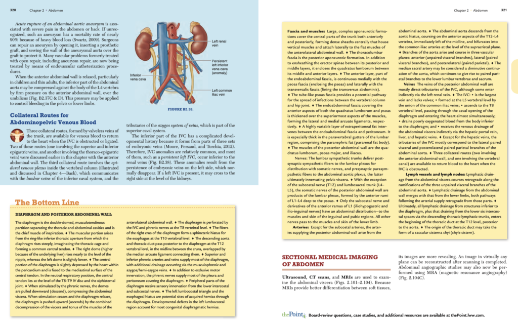
下大静脈 (IVC) は胎生期に3つの静脈セットから形成されるため、複雑な発達歴を持っています (Moore, Persaud, and Torchia, 2012)。その結果として、IVCの異常 (IVC anomalies) は比較的頻繁に見られます。例えば、持続性左側下大静脈 (persistent left IVC) が存在する場合、通常は腎静脈 (renal veins) のレベルで右側に交差します(図B2.38参照)。
骨盤臓器のリンパ
| リンパ節グループ(Lymph Node Group) | 主に排出される構造(Structures Typically Draining to Lymph Node Group) |
|---|---|
| 腰部(Lumbar) | 女性(Female): 卵巣血管(ovarian vessels)沿い – 生殖腺(Gonads)および関連構造、総腸骨リンパ節(common iliac nodes)。 排出構造(Draining Structures): 卵巣(ovary);子宮卵管(uterine tube)(峡部および子宮内部分を除く);子宮底部(fundus of uterus)。 男性(Male): 精巣血管(testicular vessels)沿い – 精巣(testis);精巣上体(epididymis)。 |
| 下腸間膜(Inferior Mesenteric) | 直腸の最上部(superiormost rectum);S状結腸(sigmoid colon);下行結腸(descending colon);直腸傍リンパ節(pararectal nodes)。 |
| 総腸骨(Common Iliac) | 外腸骨リンパ節(external iliac nodes)および内腸骨リンパ節(internal iliac nodes)。 |
| 内腸骨(Internal Iliac) | 骨盤の下部構造(inferior pelvic structures);深部会陰構造(deep perineal structures);仙骨リンパ節(sacral nodes)。 排出構造(Draining Structures): 女性(Female): 膀胱底部(base of bladder);下部骨盤尿管(inferior pelvic ureter);肛門管(anal canal)(歯状線〈pectinate line〉の上部);下部直腸(inferior rectum);膣の中・上部(middle and upper vagina);子宮頸部(cervix);子宮体部(body of uterus)。 男性(Male): 前立腺尿道(prostatic urethra);前立腺(prostate);膀胱底部;下部骨盤尿管;下部精嚢腺(inferior seminal glands);海綿体(cavernous bodies);肛門管(歯状線上部);下部直腸。 |
| 外腸骨(External Iliac) | 骨盤の前上部構造(anterosuperior pelvic structures);深部鼠径リンパ節(deep inguinal nodes)。 排出構造(Draining Structures): 女性(Female): 上部膀胱(superior bladder);上部骨盤尿管(superior pelvic ureter);膣の上部;子宮頸部;子宮体部の下部(lower body of uterus)。 男性(Male): 上部膀胱;上部骨盤尿管;上部精嚢腺(upper seminal gland);精管の骨盤部(pelvic part of ductus deferens);中部・海綿状尿道(intermediate and spongy urethra)(二次的排出経路)。 |
| 浅鼠径(Superficial Inguinal) | 下肢(lower limb);体幹の外側下部(inferolateral quadrant of trunk)の表在排出、臍部(umbilicus)より下部の腹壁前部(anterior abdominal wall)、臀部(gluteal region)、および表在会陰構造(superficial perineal structures)。 排出構造(Draining Structures): 女性(Female): 子宮の上外側部(superolateral uterus)(円靭帯〈round ligament〉の付着部付近);会陰部皮膚(skin of perineum)(外陰部〈vulva〉を含む);膣口(ostium of vagina)(処女膜〈hymen〉下部);陰核包皮(prepuce of clitoris);肛門周囲皮膚(peri-anal skin);肛門管(anal canal)(歯状線以下)。 男性(Male): 会陰部皮膚(陰茎の皮膚と包皮を含む);陰嚢(scrotum);肛門周囲皮膚;肛門管(歯状線以下)。 |
| 深鼠径(Deep Inguinal) | 女性(Female): 陰核亀頭(glans clitoris)。 男性(Male): 陰茎亀頭(glans penis);海綿状尿道の遠位部(distal spongy urethra)。 |
| 仙骨(Sacral) | 骨盤後下部構造(postero-inferior pelvic structures):下部直腸(inferior rectum);下部膣(inferior vagina)。 |
| 直腸傍(Pararectal) | 上部直腸(superior rectum)。 |
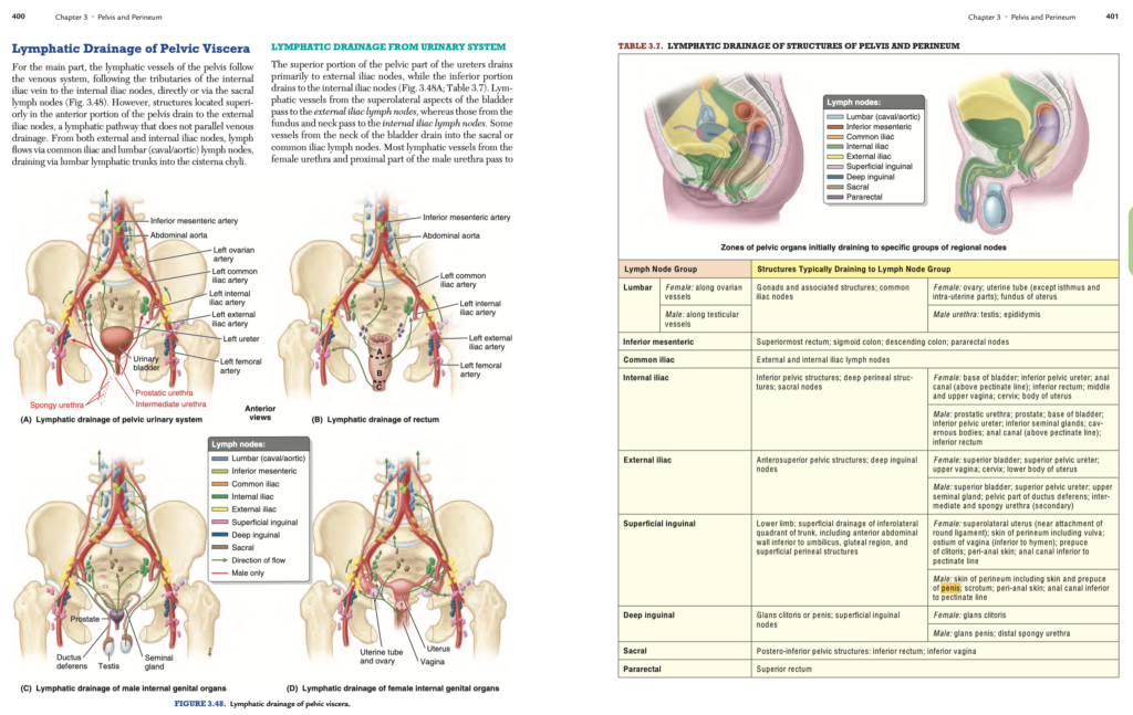
骨盤内臓(Pelvic Viscera)のリンパ排出(Lymphatic Drainage)
主な骨盤リンパ管の経路
骨盤のリンパ管(lymphatic vessels)は、主に静脈系(venous system)に沿って走行し、内腸骨静脈(internal iliac vein)の支流に沿って内腸骨リンパ節(internal iliac nodes)へ直接、または仙骨リンパ節(sacral lymph nodes)を介して排出されます(図3.48参照)。ただし、骨盤の前方上部に位置する構造は静脈排出経路と一致しない経路を取り、外腸骨リンパ節(external iliac nodes)へ排出されます。外腸骨リンパ節および内腸骨リンパ節の両方からのリンパは、総腸骨リンパ節(common iliac nodes)および腰部リンパ節(lumbar nodes)(大静脈側〈caval〉および大動脈側〈aortic〉)を経由し、腰部リンパ管幹(lumbar lymphatic trunks)を介して乳び槽(cisterna chyli)に流れ込みます。
尿路(Urinary System)のリンパ排出
- 骨盤部尿管(pelvic part of the ureters)の上部は主に**外腸骨リンパ節(external iliac nodes)へ排出され、下部は内腸骨リンパ節(internal iliac nodes)**へ排出されます(図3.48Aおよび表3.7参照)。
- 膀胱(bladder)の外側上部(superolateral aspects)は外腸骨リンパ節へ排出される一方で、膀胱底部(fundus)および膀胱頸部(neck)は内腸骨リンパ節へ排出されます。膀胱頸部からの一部のリンパ管は仙骨リンパ節(sacral nodes)または総腸骨リンパ節(common iliac nodes)へも排出されます。
- 女性の尿道(female urethra)および男性の尿道近位部(proximal male urethra)のリンパは主に内腸骨リンパ節へ排出されます。ただし、女性尿道の一部のリンパ管は仙骨リンパ節へ、遠位部は浅鼠径リンパ節(superficial inguinal lymph nodes)へも排出される場合があります。
直腸(Rectum)のリンパ排出
- 上部直腸(superior rectum)からのリンパ管は主に**下腸間膜リンパ節(inferior mesenteric lymph nodes)**へ流れます。その多くは直腸傍リンパ節(pararectal lymph nodes)(直腸筋層に直接付着)および仙骨リンパ節を通過します(図3.48Bおよび表3.7参照)。
- 下腸間膜リンパ節からのリンパは腰部リンパ節(lumbar lymph nodes)(大静脈側および大動脈側)に排出されます。
- 下部直腸(inferior half of the rectum)からのリンパ管は直接仙骨リンパ節へ、または特に遠位アンプラ(distal ampulla)からは中直腸血管(middle rectal vessels)に沿って内腸骨リンパ節に排出されます。
男性骨盤内臓(Male Pelvic Viscera)のリンパ排出
- 精管(ductus deferens)、射精管(ejaculatory ducts)、および精嚢下部(inferior parts of the seminal glands)からのリンパ管は**外腸骨リンパ節(external iliac nodes)**へ排出されます(図3.48Cおよび表3.7参照)。
- 精嚢上部(superior parts of the seminal glands)および前立腺(prostate)からのリンパ管は主に**内腸骨リンパ節(internal iliac nodes)**へ排出されますが、一部は仙骨リンパ節にも排出されます。
女性骨盤内臓(Female Pelvic Viscera)のリンパ排出
- 卵巣(ovaries)からのリンパ管は卵巣静脈(ovarian veins)に沿って、子宮卵管(uterine tubes)および子宮底部(fundus of the uterus)のリンパ管と合流し、右および左腰部リンパ節(lumbar nodes)(大静脈側および大動脈側)に排出されます(図3.48Dおよび表3.7参照)。
- 子宮(uterus)からのリンパ管は複数の方向に排出され、供給血管や子宮に付着する靭帯に沿って進みます:
- 子宮底部(fundus)および上部子宮体(superior uterine body): リンパ管の大部分は卵巣血管に沿って腰部リンパ節(lumbar nodes)に排出されますが、一部は円靭帯(round ligament)の付着部近くから出て浅鼠径リンパ節(superficial inguinal nodes)に排出されます。
- 子宮体(uterine body)の大部分および子宮頸部(cervix)の一部: 広靭帯(broad ligament)内を通り、外腸骨リンパ節(external iliac nodes)へ排出されます。
- 子宮頸部: 子宮血管(uterine vessels)に沿い、横頸靭帯(transverse cervical ligaments)内を通り内腸骨リンパ節へ、また仙骨子宮靭帯(uterosacral ligaments)に沿って仙骨リンパ節(sacral nodes)へ排出されます。
膣(Vagina)のリンパ排出
- 上部(Superior part): 内腸骨リンパ節および外腸骨リンパ節(internal and external iliac lymph nodes)へ排出。
- 中部(Middle part): 内腸骨リンパ節へ排出。
- 下部(Inferior part): 仙骨リンパ節(sacral nodes)および総腸骨リンパ節(common iliac nodes)へ排出。
- 外部開口部(External orifice): 浅鼠径リンパ節(superficial inguinal lymph nodes)へ排出。
骨盤臓器
骨盤内臓(Pelvic Viscera)
骨盤内臓には、**尿路系(urinary system)および消化管(gastrointestinal tract)**の遠位部(distal parts)と、**生殖器系(reproductive system)**が含まれます。**S状結腸(sigmoid colon)や小腸(small bowel)の一部は骨盤腔(pelvic cavity)に伸びていますが、これらは腹部内臓(abdominal viscera)**であり、骨盤内臓ではありません。
**膀胱(bladder)と直腸(rectum)**は、真の骨盤内臓(true pelvic viscera)であり、腹部で見られるシステムの下方への連続部(inferior continuations)です。男性の尿道(male urethra)が尿路系(urinary tract)と生殖器系(reproductive tract)で共有されていること、そしてそれぞれの生殖器(reproductive organs)との物理的な関係に関連する特徴を除けば、男女間で骨盤内の尿路系および消化管に大きな違いはありません。
骨盤(PPT)

教科書サマリーより
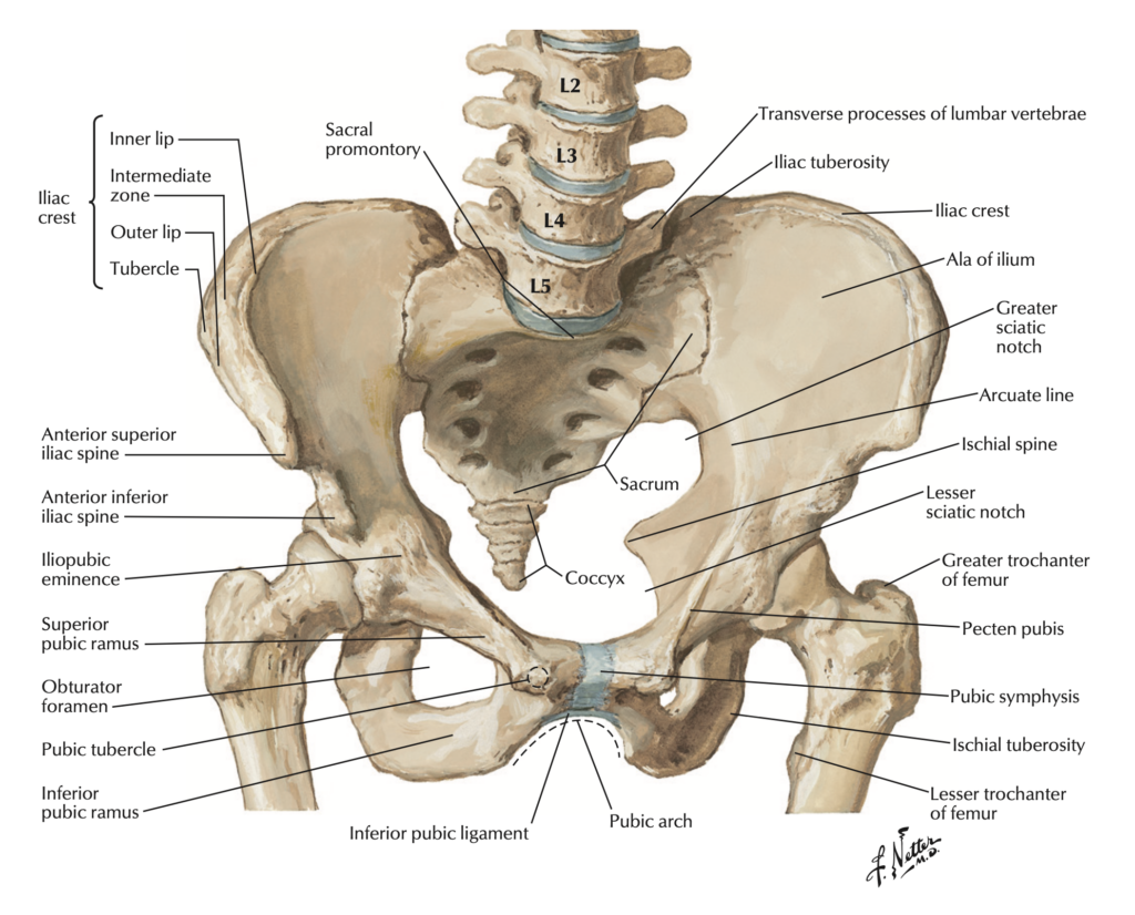
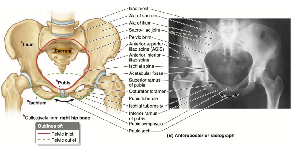
骨盤 (Pelvis) と骨盤帯 (Pelvic Girdle)
骨盤 (Pelvis)
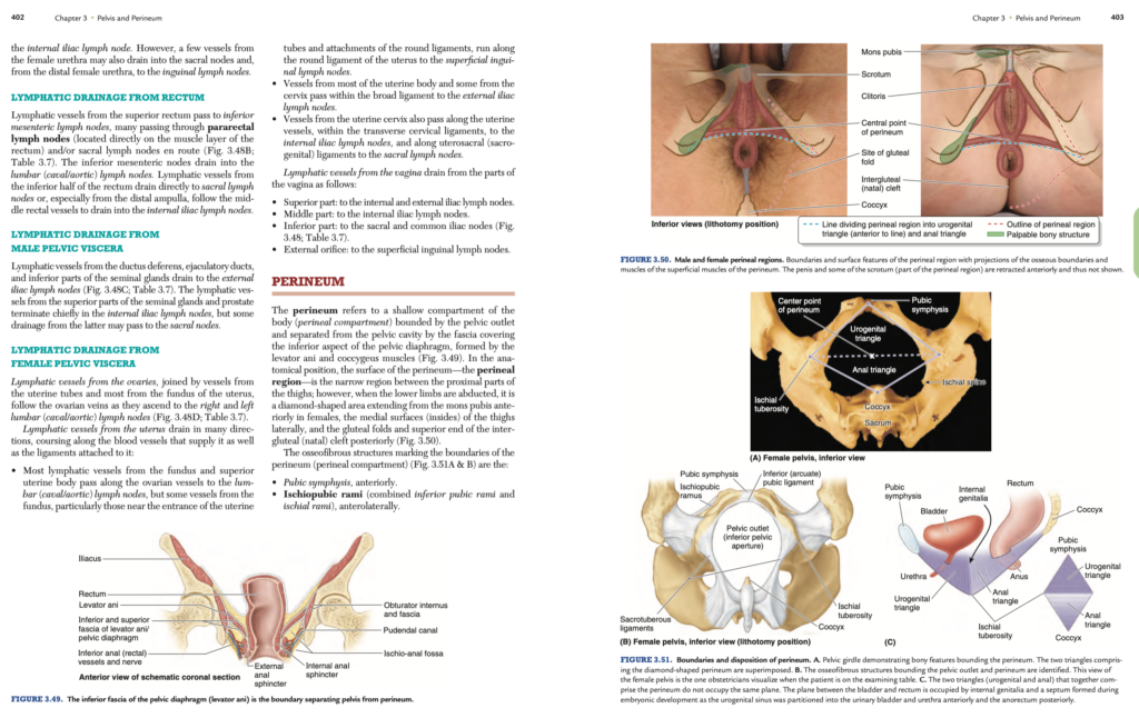
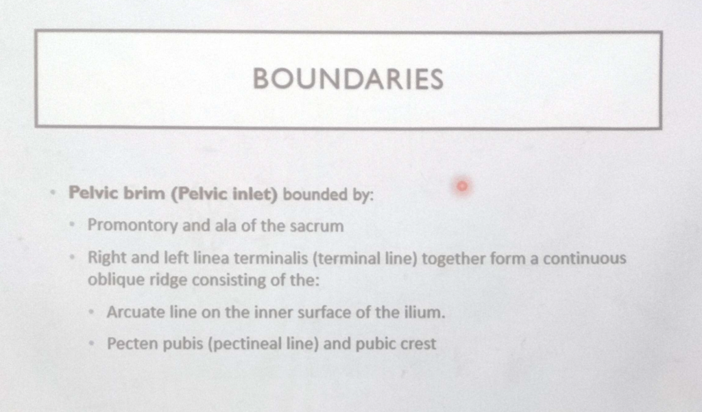
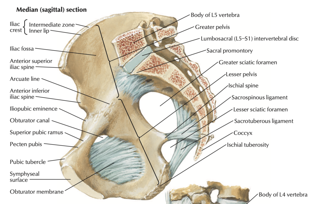
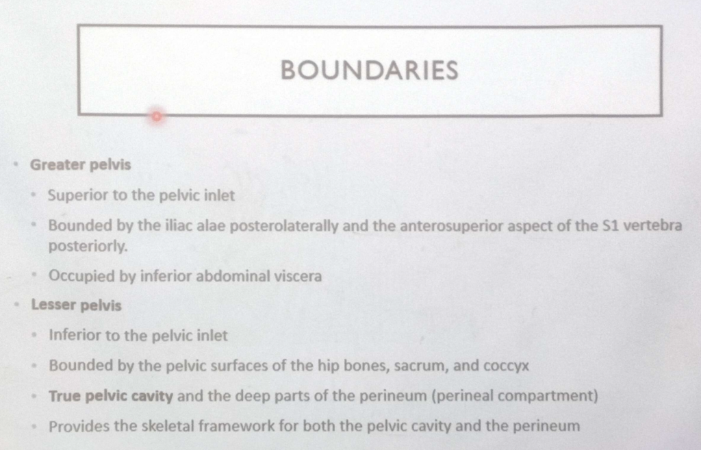
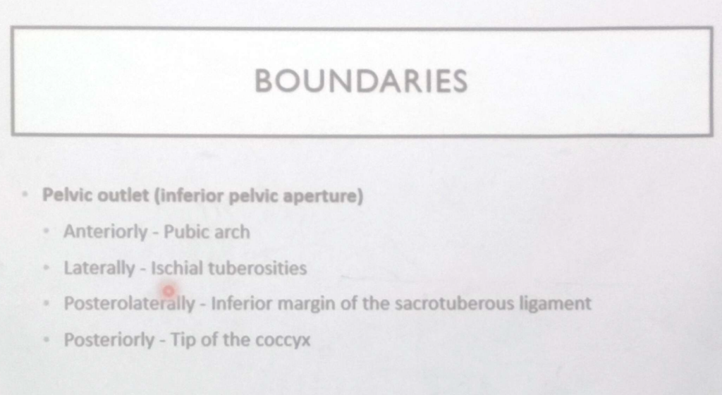
- 骨盤 (pelvis) とは、骨盤帯 (pelvic girdle) によって囲まれた空間のことを指します。骨盤は、大骨盤 (greater pelvis)(腹腔の下部で、腸骨翼 (alae of the ilia) によって保護される部分)と小骨盤 (lesser pelvis)(骨盤縁 (pelvic brim) より下方にある骨盤の骨の輪の内側の空間)に分けられます。
- 小骨盤 (lesser pelvis) は、骨盤腔 (pelvic cavity) および会陰 (perineum) の両方の骨格的な枠組みを提供します。これらは筋膜性の骨盤隔膜 (musculofascial pelvic diaphragm) によって分けられます。
- 会陰 (perineum) という用語は、肛門 (anus) と外陰部 (external genitalia) を含む領域およびその下にある浅い区画 (shallow compartment) の両方を指します。
- 下前外側腹壁 (inferior anterolateral abdominal wall)、臀部領域 (gluteal region)、および会陰 (perineum) は、骨盤と重複する領域です。
骨盤帯 (Pelvic Girdle)
- 骨盤帯 (pelvic girdle) とは、仙骨 (sacrum) と2つの寛骨 (hip bones) から構成される関節性の骨の輪 (articulated bony ring) です。
- 骨盤帯 (pelvic girdle) は、下肢の付属骨格 (appendicular skeleton of the lower limb) の一部を構成していますが、仙骨 (sacrum) は中軸骨格 (axial skeleton) の一部でもあり、上方では腰椎 (lumbar vertebrae)、下方では尾骨 (coccyx) と連続しています。
- 寛骨 (hip bones) は、腸骨 (ilium)、坐骨 (ischium)、および恥骨 (pubis) が癒合して形成されます。
- 骨盤帯の主な機能 (primary functions) は、体重の支持 (bearing of weight) および体重の移動 (transfer of weight) です。副次的な機能 (secondary functions) には、腹骨盤内臓 (abdominopelvic viscera) の保護と支持、および生殖器 (genital system) および泌尿器 (urinary system) の構造の収納と付着が含まれます。
- 解剖学的な位置 (anatomical position) では、3つの最も前方のポイント (three anteriormost points)(右および左の上前腸骨棘 (ASISs: anterior superior iliac spines) と恥骨結合の前面 (anterior aspect of the pubic symphysis))が同一の垂直平面 (same vertical plane) にあるとき、骨盤帯は解剖学的位置にあります。
- 男性と女性の骨盤 (male and female pelves) は明確に異なります。正常な (gynecoid) 女性の骨盤の特徴は、分娩 (childbirth) の際に胎児 (fetus) が骨盤管 (pelvic canal) を通過する必要があるという事実を反映しています。
- 異常な女性の骨盤 (atypical female pelves) は腟分娩 (vaginal birth) に適さない場合があるため、骨盤径 (pelvic diameters) を測定することが臨床的に重要です。
骨盤の関節 (Joints of Pelvis)
- 仙腸関節 (sacro-iliac joints) は、特殊な複合関節 (specialized compound joints) であり、滑膜関節 (synovial joint) と靭帯性結合 (syndesmotic joint) で構成されています。これらの関節の構造は、一次機能 (primary functions)(体重の支持 (weight-bearing)、体重の移動 (weight transfer) および安定性 (stability))と二次機能 (secondary function)(分娩 (parturition))の両方を反映しています。
- 強靭な靭帯 (strong ligaments)、特に骨間仙腸靭帯 (interosseous sacro-iliac ligaments) および後仙腸靭帯 (posterior sacro-iliac ligaments) によって仙骨 (sacrum) は腸骨 (ilia) の間に吊り下げられ、体重を支え (weight transfer)、骨盤の骨の輪 (bony ring of the pelvis) を安定させます。
- 滑膜関節 (synovial joints) は、分娩時 (childbirth) にわずかながらも重要な動きを可能にします。これは、恥骨結合 (pubic symphysis) および靭帯 (ligaments) がホルモン (hormones) によって柔らかくなるためです。
- 上半身の重量 (weight of the upper body) と、ジャンプや重量物の持ち上げ (jumping and load bearing) などの活動によって発生する追加の力 (additional forces) を仙腸関節 (sacro-iliac joints) に伝達する際、仙骨 (sacrum) の回転軸 (rotatory axis) の前方にある仙骨の上部 (superior sacrum) に重みがかかります。これを相殺するために、仙骨の下端 (inferior end of the sacrum) は、坐骨 (ischium) に仙結節靭帯 (sacrotuberous ligament) および仙棘靭帯 (sacrospinous ligament) によって固定されています。
重要点穴埋め問題
陰嚢 (Scrotum)
Q1: Structure of the Scrotum
The scrotum consists of heavily ________ skin and a layer of ________ fascia containing smooth muscle fibers known as the ________ muscle.
Answer: pigmented / fat-free / dartos
Explanation:
陰嚢 (Scrotum) は、色素が多く含まれる (heavily pigmented) 皮膚と、平滑筋線維を含む脂肪のない (fat-free) 筋膜層 (fascia) で構成されています。この筋膜に存在するのがダートス筋 (dartos muscle) で、これが陰嚢のしわ状の外観を生じさせます。
Q2: Function of the Dartos Muscle
The contraction of the dartos muscle thickens the scrotal ________, reduces the scrotal ________ area, and works together with the ________ muscle to regulate temperature.
Answer: skin / surface / cremaster
Explanation:
ダートス筋 (Dartos muscle) の収縮は、陰嚢の皮膚 (skin) を厚くし、表面積 (surface area) を減少させます。また、精巣挙筋 (cremaster muscle) と協力して、精巣を体に近づけ、温度を調節します。
Q3: Blood Supply of the Scrotum
The scrotum receives arterial blood supply from the posterior scrotal branches of the ________ artery, the anterior scrotal branches of the ________ artery, and the cremasteric artery from the ________ epigastric artery.
Answer: internal pudendal / femoral / inferior
Explanation:
陰嚢の血液供給は、内陰部動脈 (internal pudendal artery) の後陰嚢枝 (posterior scrotal branches)、大腿動脈 (femoral artery) の前陰嚢枝 (anterior scrotal branches)、および下腹壁動脈 (inferior epigastric artery) からの精巣挙筋動脈 (cremasteric artery) から供給されます。
Q4: Lymphatic Drainage of the Scrotum
The lymphatic drainage of the scrotum is directed to the ________ inguinal lymph nodes, while the lymphatic drainage of the testes flows into the ________ and ________ aortic lymph nodes.
Answer: superficial / lumbar / pre-
Explanation:
陰嚢のリンパ排出は、浅鼠径リンパ節 (superficial inguinal lymph nodes) に向かいますが、精巣のリンパ排出は、腰リンパ節 (lumbar lymph nodes) と前大動脈リンパ節 (pre-aortic lymph nodes) に排出されます。
Q5: Nerve Supply of the Scrotum
The anterior surface of the scrotum is supplied by the anterior scrotal nerves from the ________ nerve, while the posterior surface is supplied by the posterior scrotal nerves from the ________ nerve and the perineal branches of the ________ nerve.
Answer: ilioinguinal / pudendal / posterior cutaneous
Explanation:
陰嚢の前面は、腸骨鼠径神経 (ilioinguinal nerve) からの前陰嚢神経 (anterior scrotal nerves) に支配され、後面は陰部神経 (pudendal nerve) の後陰嚢神経 (posterior scrotal nerves) および大腿後皮神経 (posterior cutaneous nerve of the thigh) の会陰枝 (perineal branches) によって支配されます。
精巣 (Testes)
Q6: Structure of the Testes
The testes are paired male ________ that produce spermatozoa and ________. Each testis is suspended in the scrotum by the ________ cords.
Answer: gonads / testosterone / spermatic
Explanation:
精巣 (testes) は、男性の生殖腺 (male gonads) であり、精子 (spermatozoa) とテストステロン (testosterone) を産生します。精巣は精索 (spermatic cords) によって陰嚢内に吊り下げられています。
Q7: Layers Surrounding the Testes
The outer surface of the testes is covered by the ________ layer of the tunica vaginalis, while the fibrous ________ albuginea lies beneath it and forms the ________ of the testis.
Answer: visceral / tunica / mediastinum
Explanation:
精巣の表面は、精巣鞘膜 (tunica vaginalis) の臓側層 (visceral layer) に覆われており、その下には線維性の精巣白膜 (tunica albuginea) が存在します。精巣白膜の内側後部が肥厚して精巣縦隔 (mediastinum of the testis) を形成します。
Q8: Structure of Seminiferous Tubules
Each lobule of the testis contains several highly coiled ________ tubules, where ________ production occurs. These tubules connect to the rete testis via the ________ tubules.
Answer: seminiferous / sperm / straight
Explanation:
精巣の小葉 (lobules) 内には、高度にらせん状の精細管 (seminiferous tubules) があり、ここで精子 (sperm) が産生されます。精細管は直精細管 (straight tubules) を介して精巣網 (rete testis) に接続します。
Q9: Blood Supply of the Testes
The testes receive arterial blood from the testicular arteries, which branch from the ________ aorta. These arteries pass through the ________ canals and enter the ________ cords.
Answer: abdominal / inguinal / spermatic
Explanation:
精巣の血液供給は、腹大動脈 (abdominal aorta) から分岐する精巣動脈 (testicular arteries) によって行われます。これらの動脈は鼠径管 (inguinal canals) を通過し、精索 (spermatic cords) に入り、精巣に血液を供給します。
Q10: Venous Drainage of the Testes
The pampiniform plexus surrounds the testicular ________ and drains into the right ________ vena cava on the right side and the left ________ vein on the left side.
Answer: artery / inferior / renal
Explanation:
精巣の静脈排出は、精索内にある蔓状静脈叢 (pampiniform venous plexus) によって行われます。右側の蔓状静脈叢は下大静脈 (inferior vena cava) に、左側は左腎静脈 (left renal vein) に注ぎます。
Q11: Lymphatic Drainage of the Testes
The lymph from the testes drains into the lumbar lymph nodes and ________ aortic lymph nodes, following the path of the ________ artery.
Answer: pre- / testicular
Explanation:
精巣のリンパは、腰リンパ節 (lumbar lymph nodes) および前大動脈リンパ節 (pre-aortic lymph nodes) に排出され、精巣動脈 (testicular artery) に沿って進みます。
Q12: Innervation of the Testes
The testicular nerve plexus contains parasympathetic fibers from the ________ nerve, sympathetic fibers from the ________ spinal cord, and visceral afferent fibers.
Answer: vagus / T10-T11
Explanation:
精巣神経叢 (testicular plexus) には、副交感神経 (parasympathetic fibers) が迷走神経 (vagus nerve) 由来であり、交感神経 (sympathetic fibers) は脊髄のT10~T11から来ています。また、内臓求心性線維 (visceral afferent fibers) も含まれています。
精巣上体 (Epididymis)
Q1: Structure of the Epididymis
The epididymis is located on the ________ surface of the testis and is composed of the ________, body, and ________, which continues into the ductus deferens.
Answer: posterior / head / tail
Explanation:
精巣上体 (epididymis) は精巣の後面 (posterior surface) に位置しており、頭部 (head)、体部 (body)、および尾部 (tail) の3つの部分で構成されています。尾部は精管 (ductus deferens) に連続しています。
Q2: Function of the Epididymis
Sperm are transported from the rete testis via the ________ ductules to the head of the epididymis, where they mature and are stored in the ________ of the epididymis before being transported to the ________ duct.
Answer: efferent / tail / ejaculatory
Explanation:
精子は精巣網 (rete testis) から輸出小管 (efferent ductules) を介して精巣上体の頭部 (head) に輸送され、成熟し、尾部 (tail) に貯蔵されます。その後、射精管 (ejaculatory duct) へと運ばれます。
e epididymis contains a single, highly coiled ________ of the epididymis, which is responsible for sperm maturation and ________. The coiled structure provides a large surface area for the reabsorption of ________.
Answer: duct / storage / fluid
Explanation:
精巣上体管 (duct of the epididymis) は単一の高度に巻き込まれた管で、精子の成熟 (maturation) および貯蔵 (storage) に関与します。この巻き込まれた構造は、管腔の表面積を増大させ、液体 (fluid) の再吸収を可能にします。
前外側腹壁 (Anterolateral Abdominal Wall)
Q4: Position of the Umbilicus
The umbilicus is located at the level of the ________ disc between the L3 and L4 vertebrae and lies at the dermatome level of ________. Its position may vary depending on the amount of ________ tissue present.
Answer: intervertebral / T10 / subcutaneous
Explanation:
臍 (umbilicus) は、第3腰椎 (L3) と第4腰椎 (L4) の間の椎間円板 (intervertebral disc) の高さに位置し、T10皮膚分節 (dermatome) に対応します。皮下脂肪 (subcutaneous tissue) の量によって位置が変化することがあります。
Q5: Components of the Linea Alba
The linea alba is a midline structure that extends from the ________ process to the pubic ________ and separates the two ________ muscles.
Answer: xiphoid / symphysis / rectus abdominis
Explanation:
白線 (linea alba) は、剣状突起 (xiphoid process) から恥骨結合 (pubic symphysis) まで伸びる中線構造で、2つの腹直筋 (rectus abdominis) を分ける線維性結合組織の帯です。
Q6: Abdominal Muscles and Surface Anatomy
The external oblique muscle is located on the ________ surface of the abdominal wall, while the rectus abdominis is positioned along the ________ line. The tendinous intersections divide the rectus abdominis into several ________ segments.
Answer: outer / midline / muscular
Explanation:
外腹斜筋 (external oblique) は前外側腹壁 (anterolateral abdominal wall) の外側 (outer surface) に位置し、腹直筋 (rectus abdominis) は白線 (midline) に沿って位置しています。腱画 (tendinous intersections) によって、腹直筋は複数の筋肉のセグメント (muscular segments) に分割されます。
Q7: Inguinal Ligament and Surface Landmarks
The inguinal ligament extends from the anterior superior iliac spine (ASIS) to the pubic ________. It forms the base of the ________ canal and is marked externally by the inguinal ________.
Answer: tubercle / inguinal / fold
Explanation:
鼠径靭帯 (inguinal ligament) は、上前腸骨棘 (ASIS) から恥骨結節 (pubic tubercle) まで延び、鼠径管 (inguinal canal) の基底部を形成します。外部では、浅い斜めの線として鼠径溝 (inguinal fold) によって示されます。
臨床観察 (Clinical Observations)
Q8: Undescended Testis (Cryptorchidism)
An undescended testis occurs when the testis fails to ________ during fetal development. It is found in approximately ________% of full-term newborns and often remains in the ________ canal.
Answer: descend / 3 / inguinal
Explanation:
停留精巣 (undescended testis) では、胎児発生中に精巣が正常に下降 (descend) しません。正期産児 (full-term newborns) の約3% (3%) に見られ、通常は鼠径管 (inguinal canal) に留まります。
Q9: Cremasteric Reflex
The cremasteric reflex occurs when the ________ of the superior thigh is stroked, activating the ilioinguinal nerve. This causes contraction of the ________ muscle, leading to elevation of the ________ on the same side.
Answer: medial aspect / cremaster / testis
Explanation:
精巣挙筋反射 (cremasteric reflex) は、大腿の内側 (medial aspect of the superior thigh) を刺激することで引き起こされ、腸骨鼠径神経 (ilioinguinal nerve) が活性化されます。その結果、精巣挙筋 (cremaster muscle) が収縮し、同側の精巣 (testis) が上昇します。
Q10: Hydrocele of the Testis
A hydrocele occurs when ________ fluid accumulates between the layers of the tunica vaginalis. This condition is caused by excessive fluid secretion or failed closure of the ________ vaginalis. Hydroceles may be present in the scrotum or along the ________.
Answer: serous / processus / spermatic cord
Explanation:
精巣の水腫 (hydrocele) は、精巣鞘膜 (tunica vaginalis) の2つの層の間に漿液性液体 (serous fluid) が蓄積することで発生します。この状態は、過剰な液体の分泌や腹膜鞘状突起 (processus vaginalis) の閉鎖不全が原因です。水腫は、陰嚢 (scrotum) または精索 (spermatic cord) に存在する場合があります。
Q11: Testicular Artery and Vein
The testicular artery originates from the ________ aorta and passes through the inguinal canal. The right testicular vein drains into the ________ vena cava, while the left testicular vein drains into the left ________ vein.
Answer: abdominal / inferior / renal
Explanation:
精巣動脈 (testicular artery) は、腹大動脈 (abdominal aorta) から分岐し、鼠径管 (inguinal canal) を通過します。右精巣静脈 (right testicular vein) は下大静脈 (inferior vena cava) に、左精巣静脈 (left testicular vein) は左腎静脈 (left renal vein) に排出されます。
精液瘤と精巣上体嚢胞 (Spermatocele and Epididymal Cyst)
Q1: Location and Contents of Spermatocele
A spermatocele is a ________ cyst that usually occurs near the ________ of the epididymis and contains a ________-colored fluid.
Answer: retention / head / milky
Explanation:
精液瘤 (spermatocele) は、精巣上体の頭部 (head) 付近に発生する保持嚢胞 (retention cyst) であり、乳白色 (milky) の液体が含まれています。通常、無症状のままです。
胚性生殖管の遺残 (Vestigial Remnants of Embryonic Genital Ducts)
Q2: Key Structures of Embryonic Duct Remnants
The appendix of the testis is a remnant of the ________ duct, while the appendix of the epididymis is a remnant of the ________ duct. Both structures are located near the ________ of their respective organs.
Answer: paramesonephric / mesonephric / head
Explanation:
精巣付属体 (appendix of the testis) は中腎傍管 (paramesonephric duct) の遺残であり、精巣の上極に付着しています。精巣上体付属体 (appendix of the epididymis) は中腎管 (mesonephric duct) の遺残であり、精巣上体の頭部 (head) に付着しています。
静脈瘤 (Varicocele)
Q3: Varicocele Causes and Characteristics
A varicocele is caused by the dilation of the ________ plexus, most commonly affects the ________ side, and is often described as feeling like a “________” on palpation.
Answer: pampiniform / left / bag of worms
Explanation:
静脈瘤 (varicocele) は、蔓状静脈叢 (pampiniform plexus) の静脈が拡張することによって発生し、左側 (left) に多く見られます。触診では「虫袋 (bag of worms)」のように感じられます。
精巣と陰嚢のがん (Cancer of Testis and Scrotum)
Q4: Lymphatic Metastasis of Testicular Cancer
Testicular cancer initially spreads to the ________ lumbar lymph nodes, while metastasis of scrotal cancer occurs to the ________ inguinal lymph nodes, which lie near the termination of the ________ vein.
Answer: retroperitoneal / superficial / great saphenous
Explanation:
精巣がん (testicular cancer) のリンパ転移は、最初に後腹膜 (retroperitoneal) の腰リンパ節 (lumbar lymph nodes) に起こります。一方、陰嚢がん (scrotal cancer) のリンパは浅鼠径リンパ節 (superficial inguinal lymph nodes) に排出され、これらのリンパ節は大伏在静脈 (great saphenous vein) の終末部に沿って位置しています。
陰茎 (Penis)
Q5: Parts of the Penis
The penis is composed of three parts: the ________, body, and ________. It contains three cylindrical bodies of erectile tissue, including two ________ and one corpus spongiosum.
Answer: root / glans / corpora cavernosa
Explanation:
陰茎 (penis) は、根部 (root)、体部 (body)、および亀頭 (glans) の3つの部分で構成されます。さらに、左右の陰茎海綿体 (corpora cavernosa) と1つの尿道海綿体 (corpus spongiosum) の3つの円柱状の海綿体組織が含まれています。
Q6: Structure of the Penis
The corpus spongiosum contains the ________ urethra, while the two corpora cavernosa are surrounded by the fibrous ________. All three structures are enclosed within the ________ fascia.
Answer: spongy / tunica albuginea / Buck’s
Explanation:
尿道海綿体 (corpus spongiosum) には、海綿部尿道 (spongy urethra) が含まれ、陰茎海綿体 (corpora cavernosa) は線維性被膜 (tunica albuginea) に包まれています。これら3つの構造は、陰茎深筋膜 (Buck’s fascia) によってさらに包まれています。
Q7: External Anatomy of the Penis
The glans of the penis is an enlargement of the ________ spongiosum. The ridge of the glans is known as the ________ of the glans, and it is separated from the body of the penis by the ________ of the glans.
Answer: corpus / corona / neck
Explanation:
陰茎の亀頭 (glans) は、尿道海綿体 (corpus spongiosum) の拡大部分です。亀頭の縁は冠状部 (corona of the glans) として知られ、亀頭と陰茎体部は亀頭頸部 (neck of the glans) で区分されます。
Q8: Ligaments of the Penis
The suspensory ligament of the penis arises from the ________ symphysis and attaches to the junction of the ________ and body of the penis, while the fundiform ligament arises from the ________.
Answer: pubic / root / linea alba
Explanation:
陰茎懸垂靭帯 (suspensory ligament of the penis) は、恥骨結合 (pubic symphysis) から起こり、陰茎の根部 (root) と体部 (body) の接合部に付着します。一方、陰茎支帯靭帯 (fundiform ligament) は、白線 (linea alba) の前面から降りてきます。
Q9: Erectile Tissue of the Penis
The two corpora cavernosa are located on the ________ side of the penis, while the single corpus spongiosum lies on the ________ side. The anatomical position of the penis is defined when it is in the ________ state.
Answer: dorsal / ventral / erect
Explanation:
陰茎 (penis) の左右にある陰茎海綿体 (corpora cavernosa) は背側 (dorsal) に位置し、単一の尿道海綿体 (corpus spongiosum) は腹側 (ventral) に位置します。解剖学的位置は、陰茎が勃起 (erect) している状態で定義されます。
Q10: Prepuce and Frenulum
The prepuce is a double layer of skin that covers the ________ of the penis. The frenulum of the prepuce is a median fold that attaches the prepuce to the ________ aspect of the glans, near the ________ orifice.
Answer: glans / ventral / external urethral
Explanation:
包皮 (prepuce) は、亀頭 (glans) を覆う二重層の皮膚であり、包皮小帯 (frenulum of the prepuce) は包皮と亀頭の腹側 (ventral aspect) にある尿道口 (external urethral orifice) 近くを接続する正中のひだです。
男性の会陰筋 (Perineal Muscles of Male)
Q1: Superficial Perineal Muscles
The superficial perineal muscles include the ________ muscle, the bulbospongiosus muscle, and the ________ muscle. These muscles are located within the ________ pouch.
Answer: superficial transverse perineal / ischiocavernosus / superficial perineal
Explanation:
男性の浅会陰筋 (superficial perineal muscles) は、浅横会陰筋 (superficial transverse perineal muscle)、球海綿体筋 (bulbospongiosus muscle)、および坐骨海綿体筋 (ischiocavernosus muscle) で構成されており、浅会陰嚢 (superficial perineal pouch) に位置しています。
Q2: Functions of the Bulbospongiosus Muscle
The bulbospongiosus muscle compresses the ________ of the penis, increases pressure within the ________ of the penis, and assists in the expulsion of ________ and residual urine.
Answer: bulb / root / semen
Explanation:
球海綿体筋 (bulbospongiosus muscle) は、陰茎の球部 (bulb) を圧迫し、陰茎根部 (root) 内の圧力を高めます。また、精液 (semen) や残尿を排出する役割も果たします。
Q3: Functions of the Ischiocavernosus Muscle
The ischiocavernosus muscle increases blood pressure within the ________ of the penis, compresses the ________ veins, and helps maintain the rigidity of the ________ during erection.
Answer: crura / deep dorsal / penis
Explanation:
坐骨海綿体筋 (ischiocavernosus muscle) は、陰茎の陰茎脚 (crura) 内の血圧を増加させ、深背静脈 (deep dorsal veins) を圧迫し、陰茎 (penis) の硬直を維持するのを助けます。
勃起、射精前分泌、射精、弛緩 (Erection, Emission, Ejaculation, and Remission)
Q4: Erection Process
Erection is triggered by ________ nerve stimulation, causing the ________ arteries to relax, allowing blood to flow into the erectile spaces. The contraction of the ________ muscle further prevents venous return, maintaining erection.
Answer: parasympathetic / helicine / bulbospongiosus
Explanation:
勃起 (erection) は、副交感神経 (parasympathetic nerves) の刺激によって引き起こされ、螺旋動脈 (helicine arteries) が弛緩し、血液が海綿体の空隙に流入します。球海綿体筋 (bulbospongiosus muscle) が収縮し、静脈還流を防ぐことで勃起が維持されます。
Q5: Ejaculation Process
Ejaculation involves the contraction of the ________ urethral sphincter to prevent retrograde flow, the contraction of the ________ muscle to propel semen through the urethra, and the action of the ________ muscle to expel semen.
Answer: internal / urethral / bulbospongiosus
Explanation:
射精 (ejaculation) では、内尿道括約筋 (internal urethral sphincter) が収縮して逆行性射精を防ぎます。次に、尿道筋 (urethral muscle) が精液を前進させ、最後に球海綿体筋 (bulbospongiosus muscle) が収縮して精液を外尿道口から排出します。
男性尿生殖三角と関連する臨床事項 (Male Urogenital Triangle and Clinical Considerations)
Q6: Urethral Catheterization
Urethral catheterization can be challenging due to the curvature of the ________ part of the urethra, the narrowing at the ________ orifice, and the fragile nature of the ________ part of the urethra.
Answer: spongy / external urethral / intermediate
Explanation:
尿道カテーテルの挿入は、尿道の曲がった部分である尿道海綿部 (spongy part) と、最狭部である外尿道口 (external urethral orifice) で困難が生じる可能性があります。また、中間部尿道 (intermediate part) は特に柔軟性が低いため、挿入時に損傷しやすい部位です。
Q7: Hypospadias
Hypospadias is a congenital defect where the external urethral opening is located on the ________ side of the penis. The ________ form is the most common, while the ________ type occurs near the scrotum.
Answer: ventral / glanular / penoscrotal
Explanation:
尿道下裂 (hypospadias) は、外尿道口 (external urethral opening) が陰茎の腹側 (ventral side) に位置する先天性異常です。最も一般的な形態は亀頭部尿道下裂 (glanular hypospadias) で、陰茎陰嚢部尿道下裂 (penoscrotal hypospadias) は尿道口が陰嚢付近に位置するより重症の形態です。
包茎、嵌頓包茎、割礼 (Phimosis, Paraphimosis, and Circumcision)
Q8: Phimosis and Paraphimosis
Phimosis occurs when the ________ cannot be retracted over the glans, while paraphimosis occurs when the ________ becomes trapped behind the glans. Both conditions may require surgical intervention, such as ________.
Answer: prepuce / retracted prepuce / circumcision
Explanation:
包茎 (phimosis) は、包皮 (prepuce) が亀頭 (glans) を覆い、後退不可能な状態を指します。嵌頓包茎 (paraphimosis) は、後退した包皮 (retracted prepuce) が亀頭の後ろに留まり、締め付けが生じる状態です。これらの状態は、割礼 (circumcision) によって治療される場合があります。
勃起不全 (Impotence and Erectile Dysfunction)
Q9: Causes of Erectile Dysfunction
Erectile dysfunction may be caused by injury to the ________ plexus, hormonal imbalances in the ________ system, or vascular problems affecting the ________ arteries.
Answer: prostatic / endocrine / helicine
Explanation:
勃起不全 (erectile dysfunction) は、前立腺神経叢 (prostatic plexus) の損傷、内分泌系 (endocrine system) のホルモン不均衡、または螺旋動脈 (helicine arteries) に影響を及ぼす血管の問題によって引き起こされる可能性があります。
陰茎の解剖 (Anatomy of the Penis)
Q10: Anatomy of the Penis
The penis contains two ________ on the dorsal side and one ________ on the ventral side. These structures are surrounded by the fibrous ________, which maintains rigidity during erection.
Answer: corpora cavernosa / corpus spongiosum / tunica albuginea
Explanation:
陰茎は、背側 (dorsal side) にある2つの陰茎海綿体 (corpora cavernosa) と、腹側 (ventral side) にある1つの尿道海綿体 (corpus spongiosum) から構成されます。これらの構造は、勃起時に陰茎の硬直を維持する線維性の被膜である精巣白膜 (tunica albuginea) によって包まれています。
後腹壁 (Posterior Abdominal Wall)
Q1: Major Components of the Posterior Abdominal Wall
The posterior abdominal wall is composed of the ________ vertebrae, the ________ muscles, and the large vessels, including the abdominal ________.
Answer: lumbar / posterior abdominal wall / aorta
Explanation:
後腹壁 (posterior abdominal wall) は、腰椎 (lumbar vertebrae)、後腹壁の筋肉 (posterior abdominal wall muscles)、および大動脈 (aorta) や下大静脈 (IVC) などの大血管から構成されています。
Q2: Muscles of the Posterior Abdominal Wall
The main muscles of the posterior abdominal wall include the ________ major, the ________ lumborum, and the ________ muscle, which assists in hip flexion.
Answer: psoas / quadratus / iliacus
Explanation:
後腹壁の主な筋肉には、大腰筋 (psoas major)、腰方形筋 (quadratus lumborum)、および腸骨筋 (iliacus) が含まれます。腸腰筋 (iliopsoas) は、大腰筋と腸骨筋が協力して股関節の屈曲を助ける重要な筋肉群です。
Q3: Lumbar Plexus and Nerves
The lumbar plexus is formed by the anterior rami of ________ to ________ spinal nerves and lies within the ________ muscle.
Answer: L1 / L4 / psoas
Explanation:
腰神経叢 (lumbar plexus) は、L1からL4までの脊髄神経の前枝によって形成され、大腰筋 (psoas muscle) の内部で形成されます。この神経叢は、下肢や骨盤の筋肉と皮膚を支配する複数の末梢神経を生じます。
Q4: Fascia of the Posterior Abdominal Wall
The psoas fascia covers the ________ muscle, the ________ fascia covers the quadratus lumborum, and the thoracolumbar fascia has ________ layers in the lumbar region.
Answer: psoas / quadratus lumborum / three
Explanation:
後腹壁の筋膜は、各筋肉を包むように配されています。大腰筋 (psoas muscle) は大腰筋筋膜 (psoas fascia) に包まれ、腰方形筋 (quadratus lumborum) は腰方形筋筋膜 (quadratus lumborum fascia) に包まれます。胸腰筋膜 (thoracolumbar fascia) は腰部では3つの層 (three layers) に分かれています。
Q5: Thoracolumbar Fascia
The thoracolumbar fascia attaches to the ________ spine medially, extends laterally to the ________ muscle, and is divided into ________ layers.
Answer: lumbar / transversus abdominis / three
Explanation:
胸腰筋膜 (thoracolumbar fascia) は、内側では腰椎 (lumbar spine) に付着し、外側では腹横筋 (transversus abdominis) に連続します。この筋膜は、後層 (posterior layer)、中間層 (middle layer)、および前層 (anterior layer) の3つの層 (three layers) に分かれています。
Q6: Quadratus Lumborum Muscle
The quadratus lumborum muscle attaches to the ________ rib, the transverse processes of lumbar vertebrae, and the ________ crest. It is innervated by branches of the ________ nerve.
Answer: 12th / iliac / subcostal
Explanation:
腰方形筋 (quadratus lumborum) は、第12肋骨 (12th rib)、腰椎の横突起 (transverse processes of lumbar vertebrae)、および腸骨稜 (iliac crest) に付着しています。肋下神経 (subcostal nerve) や腰神経 (lumbar nerves) の枝から支配を受けます。
Q7: Psoas Major Muscle
The psoas major muscle originates from the ________ bodies of lumbar vertebrae, passes under the ________ ligament, and inserts onto the ________ of the femur.
Answer: vertebral / inguinal / lesser trochanter
Explanation:
大腰筋 (psoas major) は、腰椎の椎体 (vertebral bodies) から起こり、鼠径靱帯 (inguinal ligament) の下を通過して、大腿骨の小転子 (lesser trochanter) に付着します。この筋肉は腸腰筋の一部であり、股関節の主要な屈筋です。
Q8: Nerves of the Lumbar Plexus
The femoral nerve is derived from ________ to ________ spinal nerves, the obturator nerve from L2 to L4, and the ________ trunk joins the sacral plexus.
Answer: L2 / L4 / lumbosacral
Explanation:
大腿神経 (femoral nerve) はL2からL4の前枝から生じ、閉鎖神経 (obturator nerve) もL2からL4の前枝から派生します。腰仙幹 (lumbosacral trunk) はL4とL5からなり、仙骨神経叢 (sacral plexus) に接続します。
Q9: Lumbar Sympathetic Trunk
The lumbar sympathetic trunk consists of ________ ganglia, is continuous with the thoracic trunk above, and continues below as the ________ sympathetic trunk.
Answer: four / sacral
Explanation:
腰交感神経幹 (lumbar sympathetic trunk) は、4つの交感神経節 (four ganglia) から構成され、上部では胸部交感神経幹 (thoracic sympathetic trunk) に連続し、下部では仙骨交感神経幹 (sacral sympathetic trunk) に続きます。
Q10: Vascular Structures of the Posterior Abdominal Wall
The abdominal aorta lies anterior to the ________ vertebrae, gives rise to the ________ mesenteric artery, and bifurcates into the common ________ arteries.
Answer: lumbar / superior / iliac
Explanation:
腹部大動脈 (abdominal aorta) は、腰椎 (lumbar vertebrae) の前方に位置し、上腸間膜動脈 (superior mesenteric artery) などの主要な分枝を生じます。腹部大動脈は、左右の総腸骨動脈 (common iliac arteries) に分岐します。
後腹壁の血管 (Vessels of the Posterior Abdominal Wall)
Q1: Abdominal Aorta Anatomy
The abdominal aorta begins at the ________ vertebra, ends at the ________ vertebra, and bifurcates into the right and left ________ arteries.
Answer: T12 / L4 / common iliac
Explanation:
腹部大動脈 (abdominal aorta) は、第12胸椎 (T12) の高さで大動脈裂孔 (aortic hiatus) から始まり、第4腰椎 (L4) の高さで右および左の総腸骨動脈 (common iliac arteries) に分岐します。
Q2: Major Branches of the Abdominal Aorta
The abdominal aorta gives rise to unpaired visceral arteries, including the ________ artery, the superior mesenteric artery, and the ________ mesenteric artery. It also gives rise to paired ________ arteries that supply the kidneys.
Answer: celiac / inferior / renal
Explanation:
腹部大動脈 (abdominal aorta) からは、内臓への血流を供給する非対称の枝が分岐します。これには、腹腔動脈 (celiac artery)、上腸間膜動脈 (superior mesenteric artery)、および下腸間膜動脈 (inferior mesenteric artery) が含まれます。また、腎臓に血液を供給する対になった腎動脈 (renal arteries) も分岐します。
Q3: Relationship of the Abdominal Aorta
The abdominal aorta is positioned anterior to the ________ vertebral bodies, posterior to the ________ vein, and is accompanied by the ________ nerve plexus.
Answer: lumbar / left renal / aortic peri-arterial
Explanation:
腹部大動脈 (abdominal aorta) は、腰椎 (lumbar vertebral bodies) の前方に位置し、左腎静脈 (left renal vein) の後方を通ります。また、大動脈周囲神経叢 (aortic peri-arterial nerve plexus) に囲まれています。
Q4: Arterial Supply of the Abdominal Wall
The external iliac artery gives rise to the ________ artery, which supplies the anterior abdominal wall, and the ________ artery, which supplies the iliac region. These arteries originate just before the external iliac artery passes under the ________ ligament.
Answer: inferior epigastric / deep circumflex iliac / inguinal
Explanation:
外腸骨動脈 (external iliac artery) は、前外側腹壁に血液を供給する下腹壁動脈 (inferior epigastric artery) と、腸骨部に血液を供給する深腸骨回旋動脈 (deep circumflex iliac artery) を分岐させます。これらの動脈は、外腸骨動脈が鼠径靱帯 (inguinal ligament) の下を通過する直前に分岐します。
Q5: Venous Drainage of the Posterior Abdominal Wall
The inferior vena cava (IVC) is formed by the convergence of the right and left ________ veins, lies to the right of the ________ aorta, and passes through the ________ opening of the diaphragm.
Answer: common iliac / abdominal / caval
Explanation:
下大静脈 (inferior vena cava: IVC) は、右および左の総腸骨静脈 (common iliac veins) の合流によって形成されます。下大静脈は、腹部大動脈 (abdominal aorta) の右側に位置し、横隔膜の大静脈孔 (caval opening) を通過して胸腔に入ります。
Q6: Tributaries of the IVC
The tributaries of the inferior vena cava include the renal veins, the ________ gonadal vein, and the ________ suprarenal vein, while the ________ gonadal vein drains into the left renal vein.
Answer: right / right / left
Explanation:
下大静脈 (IVC) の主な枝には、腎静脈 (renal veins)、右性腺静脈 (right gonadal vein)、および右副腎静脈 (right suprarenal vein) が含まれます。一方、左性腺静脈 (left gonadal vein) は左腎静脈 (left renal vein) に流入し、下大静脈を介してではなく左腎静脈を介して心臓に戻ります。
Q7: Thoracic Duct and Lymphatic Drainage
The thoracic duct originates from the ________ located at the level of L1 and L2 vertebrae, receives lymph from the ________ trunks, and enters the venous system at the left ________ vein.
Answer: cisterna chyli / lumbar / subclavian
Explanation:
胸管 (thoracic duct) は、L1およびL2の椎体の前方にある乳糜槽 (cisterna chyli) から始まり、右および左の腰リンパ幹 (lumbar trunks) からのリンパを受け取ります。最終的に、左鎖骨下静脈 (left subclavian vein) で静脈系にリンパを排出します。
Q8: Lymph Nodes of the Posterior Abdominal Wall
Lymph nodes of the posterior abdominal wall are classified into ________ nodes located along the IVC, ________ nodes along the abdominal aorta, and ________ nodes near the iliac arteries.
Answer: caval / aortic / common iliac
Explanation:
後腹壁のリンパ節は、下大静脈に沿った下大静脈リンパ節 (caval nodes)、腹部大動脈に沿った大動脈リンパ節 (aortic nodes)、および腸骨動脈の近くにある総腸骨リンパ節 (common iliac nodes) に分類されます。
Q9: Formation and Pathway of the IVC
The inferior vena cava is formed at the level of the ________ vertebra, runs on the ________ side of the aorta, and enters the thoracic cavity at the level of the ________ vertebra.
Answer: L5 / right / T8
Explanation:
下大静脈 (IVC) は、第5腰椎 (L5) の前方で、右および左の総腸骨静脈の合流によって形成されます。IVCは、腹部大動脈の右側 (right side) を上行し、第8胸椎 (T8) の高さで胸腔に入ります。
Q10: Collateral Venous Pathways
Collateral venous pathways between the IVC and SVC include the ________ vein, the ________ lumbar vein, and the ________ vein.
Answer: azygos / ascending / hemiazygos
Explanation:
下大静脈 (IVC) と上大静脈 (SVC) との間の側副路 (collateral pathways) には、奇静脈 (azygos vein)、上行腰静脈 (ascending lumbar vein)、および半奇静脈 (hemiazygos vein) が含まれます。これらの側副路は、IVCが閉塞した場合の代替経路として機能します。
腰部膿瘍 (Psoas Abscess)
Q1: Causes and Spread of Psoas Abscess
A psoas abscess may result from ________ infection, spread hematogenously to the vertebral column, and extend along the ________ muscle sheath to the ________ region, where it can appear as a swelling.
Answer: tuberculosis / psoas / thigh
Explanation:
腰部膿瘍 (psoas abscess) は、結核 (tuberculosis) 感染により引き起こされることが多く、血行性に (hematogenous) 脊椎に波及します。腸腰筋筋膜 (psoas muscle sheath) に沿って下方に進み、鼠径靱帯を通過して太もも (thigh) に腫れを生じることがあります。
Q2: Clinical Sign of Psoas Abscess
The clinical test for psoas abscess involves extending the patient’s ________ against resistance. Pain during this movement indicates a positive ________ sign, suggesting irritation of the ________ muscle.
Answer: thigh / psoas / iliopsoas
Explanation:
腰部膿瘍 (psoas abscess) を検出するための検査では、腸腰筋テスト (iliopsoas test) を行います。患者の大腿 (thigh) を抵抗に対して伸展する際に痛みが生じる場合、腸腰筋徴候 (psoas sign) は陽性とされ、腸腰筋 (iliopsoas muscle) の刺激が示唆されます。
後腹部の痛み (Posterior Abdominal Pain)
Q3: Causes of Posterior Abdominal Pain
Posterior abdominal pain can result from irritation of the ________ muscle, which lies in close contact with organs such as the ________ and ________, among others.
Answer: iliopsoas / kidneys / pancreas
Explanation:
後腹部の痛み (posterior abdominal pain) は、腸腰筋 (iliopsoas muscle) の刺激によって生じる可能性があります。この筋肉は、腎臓 (kidneys)、膵臓 (pancreas)、盲腸 (cecum)、虫垂 (appendix)、S状結腸 (sigmoid colon) などの重要な臓器と接触しています。
Q4: Reflex Spasm of Iliopsoas
Reflex spasm of the iliopsoas muscle can result from disease of the ________ joints, irritation of the ________ nerve, or inflammation of the ________ vertebrae.
Answer: sacroiliac / lumbar / intervertebral
Explanation:
腸腰筋 (iliopsoas) の反射的な痙攣 (spasm) は、仙腸関節 (sacroiliac joints) の疾患、腰神経 (lumbar nerve) の刺激、または椎間関節 (intervertebral joints) の炎症に起因することがあります。
大動脈の拍動と腹部大動脈瘤 (Abdominal Aortic Aneurysm, AAA)
Q5: Anatomy of the Abdominal Aortic Aneurysm
An abdominal aortic aneurysm (AAA) commonly occurs near the level of the ________ vertebra, involves dilation of the ________ wall, and may present as a pulsating mass in the ________ abdomen.
Answer: L4 / arterial / midline
Explanation:
腹部大動脈瘤 (AAA: abdominal aortic aneurysm) は、L4椎体 (L4 vertebra) の高さ付近で発生することが多く、動脈壁 (arterial wall) の拡張を伴います。中央の腹部 (midline abdomen) に触れる拍動性の腫瘤 (pulsating mass) として臨床的に現れることがあります。
Q6: Clinical Symptoms of AAA Rupture
The rupture of an abdominal aortic aneurysm can cause sudden severe pain in the ________ or ________, and if undiagnosed, it can result in severe ________ due to internal bleeding.
Answer: abdomen / back / hemorrhage
Explanation:
腹部大動脈瘤 (AAA) が破裂すると、腹部 (abdomen) または背部 (back) に突然の激しい痛みが生じ、診断が遅れると大量の内出血 (hemorrhage) による死亡リスクが高まります。
Q7: Surgical Repair of AAA
Surgical repair of an abdominal aortic aneurysm may involve the insertion of a ________ graft, which can be performed either by ________ repair or by minimally invasive ________ repair.
Answer: prosthetic / open / endovascular
Explanation:
腹部大動脈瘤 (AAA) の外科的修復は、人工血管 (prosthetic graft) の挿入を伴います。これには、開腹修復 (open repair) と、カテーテルを使用した低侵襲な血管内修復 (endovascular repair) の2つの方法があります。
腹部骨盤静脈血の側副経路 (Collateral Routes for Abdominopelvic Venous Blood)
Q8: Venous Collateral Routes
When the inferior vena cava (IVC) is blocked, collateral venous pathways may involve the ________ veins of the anterior abdominal wall, the ________ vein connecting the thoracic and abdominal veins, and the ________ venous plexus of the vertebral column.
Answer: epigastric / thoraco-epigastric / epidural
Explanation:
下大静脈 (IVC) が閉塞した場合、上腹壁静脈 (epigastric veins)、胸腹静脈 (thoraco-epigastric vein)、および椎骨静脈叢 (epidural venous plexus) などの側副静脈路が利用され、心臓への静脈還流が可能になります。
Q9: Formation and Position of IVC
The inferior vena cava (IVC) is formed by the union of the ________ veins at the level of the ________ vertebra and lies to the ________ of the abdominal aorta.
Answer: common iliac / L5 / right
Explanation:
下大静脈 (IVC) は、L5椎体 (L5 vertebra) の高さで右および左の総腸骨静脈 (common iliac veins) の合流によって形成されます。IVCは腹部大動脈 (abdominal aorta) の右側 (right) に位置しています。
部分的腰部交感神経切除術 (Partial Lumbar Sympathectomy)
Q10: Approach to Partial Lumbar Sympathectomy
Partial lumbar sympathectomy is performed using a ________ approach, involving dissection of the ________ muscle and exposure of the ________ trunk, which is located in extraperitoneal fatty tissue.
Answer: lateral extraperitoneal / psoas / sympathetic
Explanation:
部分的腰部交感神経切除術 (partial lumbar sympathectomy) は、外側腹膜外アプローチ (lateral extraperitoneal approach) を用いて行います。手術中、腸腰筋 (psoas muscle) を解剖して、腹膜外脂肪組織 (extraperitoneal fatty tissue) に位置する交感神経幹 (sympathetic trunk) を露出させます。
Urinary System Lymphatic Drainage
Q1: Lymphatic Drainage of the Ureters
The upper part of the pelvic ureters drains into the ________ lymph nodes, while the lower part drains into the ________ lymph nodes. Some lymph from the lower ureters may also drain into the ________ lymph nodes.
Answer: external iliac / internal iliac / common iliac
Explanation:
尿管 (ureters) の上部は外腸骨リンパ節 (external iliac lymph nodes) に排出され、下部は内腸骨リンパ節 (internal iliac lymph nodes) に排出されます。一部のリンパは総腸骨リンパ節 (common iliac lymph nodes) にも流入する場合があります。
Q2: Lymphatic Drainage of the Bladder
The lymphatic drainage of the bladder is divided, with the superolateral parts draining into the ________ lymph nodes, while the fundus and neck drain into the ________ lymph nodes. Some lymph from the neck may also drain into the ________ lymph nodes.
Answer: external iliac / internal iliac / sacral
Explanation:
膀胱 (bladder) の外側上部は外腸骨リンパ節 (external iliac lymph nodes) に排出されますが、膀胱底 (fundus) と膀胱頸部 (neck) は内腸骨リンパ節 (internal iliac lymph nodes) に排出されます。膀胱頸部のリンパの一部は仙骨リンパ節 (sacral lymph nodes) にも排出されることがあります。
Q3: Lymphatic Drainage of the Urethra
The lymphatic drainage of the proximal male urethra and the female urethra primarily flows into the ________ lymph nodes, but some of the lymph from the female urethra also drains into the ________ lymph nodes and the ________ lymph nodes.
Answer: internal iliac / sacral / superficial inguinal
Explanation:
男性の近位部尿道 (proximal male urethra) および女性の尿道 (female urethra) は、主に内腸骨リンパ節 (internal iliac lymph nodes) に排出されます。女性の尿道の一部のリンパは仙骨リンパ節 (sacral lymph nodes) および浅鼠径リンパ節 (superficial inguinal lymph nodes) にも排出されることがあります。
Male Pelvic Viscera Lymphatic Drainage
Q4: Lymphatic Drainage of Male Pelvic Organs
The ductus deferens and the inferior parts of the seminal glands drain into the ________ lymph nodes, while the superior parts of the seminal glands and the prostate primarily drain into the ________ lymph nodes, with some lymph also draining into the ________ lymph nodes.
Answer: external iliac / internal iliac / sacral
Explanation:
精管 (ductus deferens) および精嚢の下部 (inferior parts of the seminal glands) からのリンパは外腸骨リンパ節 (external iliac lymph nodes) に排出されます。精嚢の上部 (superior parts of the seminal glands) および前立腺 (prostate) からのリンパは主に内腸骨リンパ節 (internal iliac lymph nodes) に排出されますが、仙骨リンパ節 (sacral lymph nodes) にも一部が排出されます。
Q5: Lymphatic Drainage of the Testes
The lymphatic drainage of the testes follows the path of the ________ vessels and drains into the ________ lymph nodes, while the lymph from the scrotum drains into the ________ lymph nodes.
Answer: testicular / lumbar / superficial inguinal
Explanation:
精巣 (testes) からのリンパは、精巣血管 (testicular vessels) に沿って流れ、腰リンパ節 (lumbar lymph nodes) に排出されます。一方、陰嚢 (scrotum) からのリンパは浅鼠径リンパ節 (superficial inguinal lymph nodes) に排出されます。
Pelvic Viscera
Q6: Classification of Pelvic Viscera
The pelvic viscera include parts of the urinary system, the ________ system, and the ________ system. The sigmoid colon and portions of the small intestine are considered ________ viscera, not pelvic viscera.
Answer: gastrointestinal / reproductive / abdominal
Explanation:
骨盤内臓 (pelvic viscera) には、尿路系 (urinary system)、消化管 (gastrointestinal system)、および生殖器系 (reproductive system) が含まれます。ただし、S状結腸 (sigmoid colon) および小腸 (small intestine) の一部は、解剖学的には骨盤内に位置していても、腹部内臓 (abdominal viscera) に分類されます。
Q7: True Pelvic Viscera
The true pelvic viscera include the ________ and the ________, which are inferior extensions of the urinary and gastrointestinal tracts, respectively. The ________ is shared between the urinary and reproductive systems in males.
Answer: bladder / rectum / urethra
Explanation:
真の骨盤内臓 (true pelvic viscera) には、尿路系の膀胱 (bladder) と、消化管の直腸 (rectum) が含まれます。男性では、尿路系と生殖器系が共有する構造として尿道 (urethra) があります。
Q8: Lymphatic Drainage of the Pelvic Viscera
Lymph from the pelvic viscera primarily drains into the ________ lymph nodes, although some drainage also occurs into the ________ and ________ lymph nodes, depending on the organ.
Answer: internal iliac / sacral / external iliac
Explanation:
骨盤内臓 (pelvic viscera) からのリンパは、主に内腸骨リンパ節 (internal iliac lymph nodes) に排出されますが、臓器の位置によっては仙骨リンパ節 (sacral lymph nodes) や外腸骨リンパ節 (external iliac lymph nodes) にも流れることがあります。
Q9: Clinical Considerations for Lymphatic Drainage
Lymphatic drainage of the pelvic organs is clinically important because cancer from the prostate can spread to the ________ lymph nodes, while cancer from the bladder may spread to the ________ or ________ lymph nodes.
Answer: internal iliac / external iliac / sacral
Explanation:
骨盤内臓のリンパ排出は、がんの転移経路として重要です。前立腺がんは内腸骨リンパ節 (internal iliac lymph nodes) に転移しやすく、膀胱がんは外腸骨リンパ節 (external iliac lymph nodes) や仙骨リンパ節 (sacral lymph nodes) にも転移する可能性があります。
Q1: Pelvis (骨盤)
The pelvis is divided into the ________, which is the inferior part of the abdominal cavity, and the ________, which is the space inside the bony ring of the pelvis inferior to the ________.
Answer: greater pelvis / lesser pelvis / pelvic brim
Explanation:
骨盤 (pelvis) は大骨盤 (greater pelvis) と小骨盤 (lesser pelvis) に分かれます。大骨盤は腹腔の下部 (inferior part of the abdominal cavity) にあり、腸骨翼 (alae of the ilia) によって保護されています。小骨盤 (lesser pelvis) は、骨盤縁 (pelvic brim) の下に位置する骨の輪の内側の空間です。
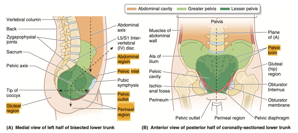
Q2: Pelvic Cavity and Perineum (骨盤腔と会陰)
The lesser pelvis forms the skeletal framework for the ________ and the ________, which are separated by the ________.
Answer: pelvic cavity / perineum / musculofascial pelvic diaphragm
Explanation:
小骨盤 (lesser pelvis) は、骨盤腔 (pelvic cavity) と会陰 (perineum) の骨格的な枠組みを提供します。これらの2つの領域は、筋膜性骨盤隔膜 (musculofascial pelvic diaphragm) によって分けられています。
Q3: Perineum (会陰)
The perineum refers to the region that includes the ________ and the ________, as well as a shallow ________ beneath it.
Answer: anus / external genitalia / compartment
Explanation:
会陰 (perineum) とは、肛門 (anus) と外陰部 (external genitalia) を含む領域を指し、その下には浅い区画 (compartment) があります。
Q4: Pelvic Girdle (骨盤帯)
The pelvic girdle is an articulated bony ring composed of the ________ and two ________. The hip bones are formed by the fusion of the ________, ischium, and pubis.
Answer: sacrum / hip bones / ilium
Explanation:
骨盤帯 (pelvic girdle) は、仙骨 (sacrum) と2つの寛骨 (hip bones) から構成される関節性の骨の輪 (articulated bony ring) です。寛骨 (hip bones) は、腸骨 (ilium)、坐骨 (ischium)、および恥骨 (pubis) が癒合して形成されます。
Q5: Functions of the Pelvic Girdle (骨盤帯の機能)
The primary functions of the pelvic girdle are ________ and ________ of weight, while its secondary functions include protection and support of the ________.
Answer: bearing / transfer / abdominopelvic viscera
Explanation:
骨盤帯 (pelvic girdle) の主な機能 (primary functions) は、体重の支持 (bearing of weight) および体重の移動 (transfer of weight) です。副次的な機能 (secondary functions) には、腹骨盤内臓 (abdominopelvic viscera) の保護と支持が含まれます。
Q6: Anatomical Position of the Pelvic Girdle (骨盤帯の解剖学的位置)
The pelvic girdle is in the anatomical position when its three anteriormost points, the right and left ________ and the anterior aspect of the ________, lie in the same ________.
Answer: ASISs / pubic symphysis / vertical plane
Explanation:
骨盤帯 (pelvic girdle) は、右および左の上前腸骨棘 (ASISs: anterior superior iliac spines) と恥骨結合の前面 (anterior aspect of the pubic symphysis) が同じ垂直平面 (vertical plane) にあるときに解剖学的な位置 (anatomical position) にあります。
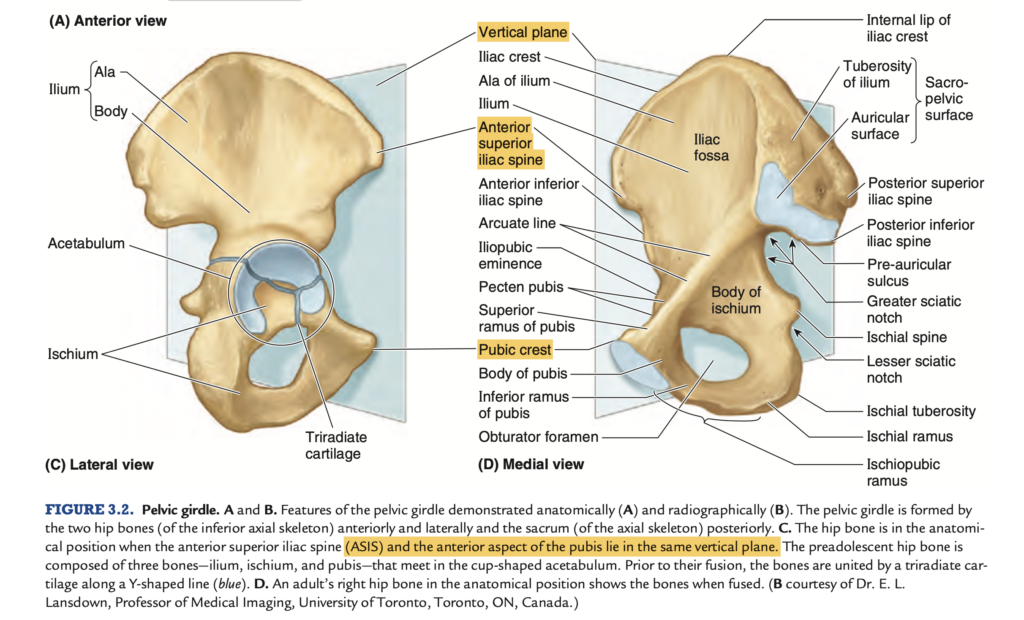
Q7: Male and Female Pelves (男性と女性の骨盤の違い)
The female pelvis has distinct features that reflect its role in allowing the ________ to traverse the ________ during ________.
Answer: fetus / pelvic canal / childbirth
Explanation:
女性の骨盤 (female pelvis) は、分娩 (childbirth) の際に胎児 (fetus) が骨盤管 (pelvic canal) を通過する必要があるため、特有の特徴を持っています。
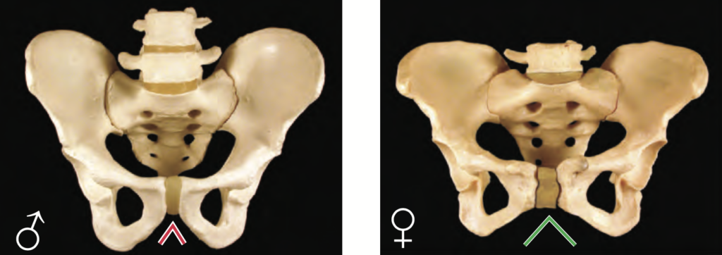
Q8: Joints of the Pelvis (骨盤の関節)
The sacro-iliac joints are specialized compound joints consisting of a ________ joint and a ________ joint, which reflect the primary functions of ________, weight transfer, and stability.
Answer: synovial / syndesmotic / weight-bearing
Explanation:
仙腸関節 (sacro-iliac joints) は、滑膜関節 (synovial joint) と靭帯性結合 (syndesmotic joint) から構成される特殊な複合関節 (specialized compound joints) です。これらの関節の主な機能 (primary functions) は、体重の支持 (weight-bearing)、体重の移動 (weight transfer) および安定性 (stability) です。
Q9: Ligaments of the Sacro-Iliac Joints (仙腸関節の靭帯)
The sacro-iliac joints are supported by strong ________ ligaments and ________ ligaments, which suspend the sacrum between the ilia, stabilizing the ________ of the pelvis.
Answer: interosseous / posterior sacro-iliac / bony ring
Explanation:
仙腸関節 (sacro-iliac joints) は、骨間仙腸靭帯 (interosseous sacro-iliac ligaments) および後仙腸靭帯 (posterior sacro-iliac ligaments) によって支えられています。これらの靭帯は、仙骨 (sacrum) を腸骨 (ilia) の間に吊り下げ、骨盤の骨の輪 (bony ring of the pelvis) を安定させます。
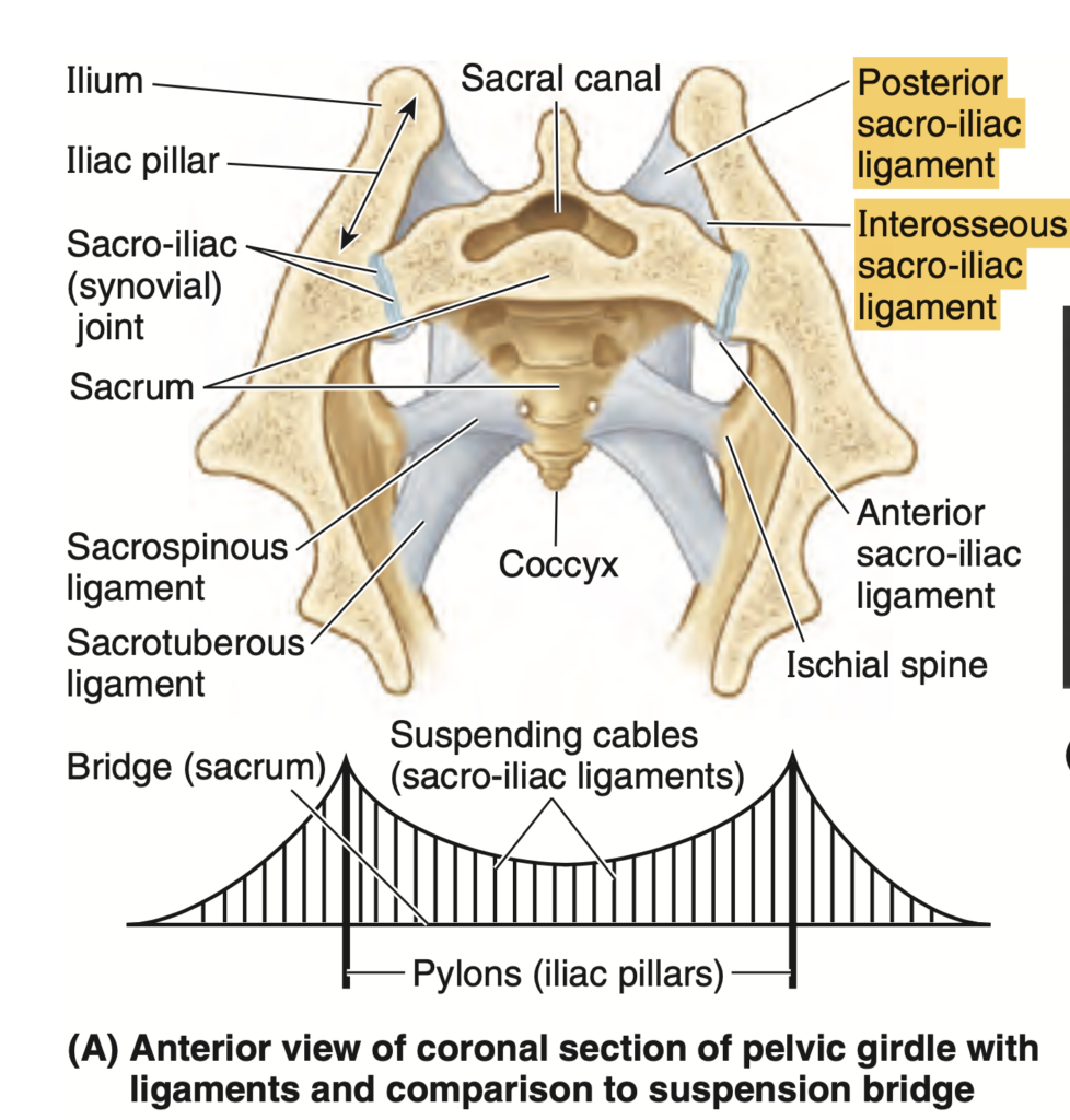
Q10: Childbirth and Movement of the Sacro-Iliac Joints (分娩時の仙腸関節の動き)
During childbirth, the ________ and ________ are softened by ________, allowing slight but significant movement in the sacro-iliac joints.
Answer: pubic symphysis / ligaments / hormones
Explanation:
分娩 (childbirth) の際、恥骨結合 (pubic symphysis) および靭帯 (ligaments) はホルモン (hormones) によって柔らかくなり、仙腸関節 (sacro-iliac joints) にわずかながらも重要な動きが生じます。
Q11: Sacral Support (仙骨の支持)
The inferior end of the sacrum is anchored to the ischium by the ________ ligament and the ________ ligament, which counterbalance the weight of the upper body.
Answer: sacrotuberous / sacrospinous
Explanation:
仙骨 (sacrum) の下端 (inferior end) は、仙結節靭帯 (sacrotuberous ligament) および仙棘靭帯 (sacrospinous ligament) によって坐骨 (ischium) に固定されています。これにより、上半身の重量 (weight of the upper body) を相殺します。
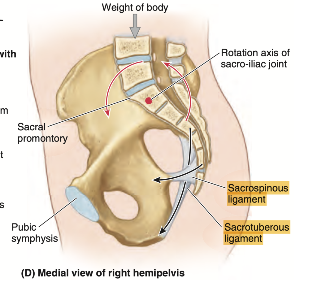

コメント