
Contents
- 1 アセス(22nd CDM)
- 1.1 Question 1
- 1.2 Question 2 ★出た
- 1.3 Question 3 ★出た
- 1.4 Question 4 ★出た
- 1.5 Question 5
- 1.6 Question 6 ★出た
- 1.7 Question 7 ★出た
- 1.8 Question 8 ★出た
- 1.9 Question 9 ★出た
- 1.10 Question 10
- 1.11 Question 11 ★出た
- 1.12 Question 12 ★出た
- 1.13 Question 13 ★出た
- 1.14 Question 14
- 1.15 Question 15
- 1.16 Question 16 ★出た
- 1.17 Question 17
- 1.18 Question 18
- 1.19 Question 19(類似9) ★出た
- 1.20 Question 20 ★出た
- 1.21 Question 21 ★出た
- 1.22 Question 22 ★出た
- 1.23 Question 23
- 1.24 Question 24 ★出た
- 1.25 Question 25(類似13) ★出た
- 1.26 Question 26 ★出た
- 1.27 Question 27 ★出た
- 1.28 Question 28
- 1.29 Question 29
- 1.30 Question 30 ★出た
- 1.31 Question 31
- 1.32 Question 32
- 1.33 Question 33
- 1.34 Question 34 ★出た
- 1.35 Question 35 ★出た
- 1.36 Question 36
- 1.37 Question 37
- 1.38 Question 38 ★出た
- 1.39 Question 39 ★出た
- 1.40 Question 40
- 1.41 Question 41(類似37)
- 1.42 Question 42
- 1.43 Question 43 ★出た
- 1.44 Question 44 ★出た
- 1.45 Question 45
- 1.46 Question 46 ★出た
- 1.47 Question 47 ★出た
- 1.48 Question 48 ★出た
- 1.49 Question 49 ★出た
- 1.50 Question 50
- 2 アセス(29th Oct Dr. Lastimoso)
- 2.1 Question 1(29th Oct)
- 2.2 Question 2(5th Nov ブロック)
- 2.3 Question 3(29th Oct)
- 2.4 Question 4(29th Oct)
- 2.5 Question 5(5th Nov ブロック)
- 2.6 Question 6(29th Oct)
- 2.7 Question 7(29th Oct)
- 2.8 Question 8(29th Oct)
- 2.9 Question 9(29th Oct)
- 2.10 Question 10(5th Nov ブロック)
- 2.11 Question 11(29th Oct)
- 2.12 Question 12(29th Oct)
- 2.13 Question 13(29th Oct)
- 2.14 Question 14(29th Oct)
- 2.15 Question 15(29th Oct)
- 2.16 Question 16(29th Oct)
- 2.17 Question 17(29th Oct)
- 2.18 Question 18(5th Nov ブロック)
- 2.19 Question 19(5th Nov ブロック)
- 2.20 Question 20(29th Oct)
- 2.21 Question 21(5th Nov ブロック)
- 2.22 Question 22(29th Oct)
- 2.23 Question 23(5th Nov ブロック)
- 2.24 Question 24(29th Oct)
- 2.25 Question 25(29th Oct)
- 2.26 Question 26(29th Oct)
- 2.27 Question 27(5th Nov ブロック)
- 2.28 Question 28(29th Oct)
- 2.29 Question 29(5th Nov ブロック)
- 2.30 Question 30(5th Nov ブロック)
- 2.31 Question 31(29th Oct)
- 2.32 Question 32(29th Oct)
- 2.33 Question 33(29th Oct)
- 2.34 Question 34(5th Nov ブロック)
- 2.35 Question 35(29th Oct)
- 2.36 Question 36(29th Oct)
- 2.37 Question 37(5th Nov ブロック)
- 2.38 Question 38(29th Oct)
- 2.39 Question 39(29th Oct)
- 2.40 Question 40(29th Oct)
- 2.41 Question 41(5th Nov ブロック)
- 2.42 Question 42(5th Nov ブロック)
- 2.43 Question 43(5th Nov ブロック)
- 2.44 Question 44(29th Oct)
- 2.45 Question 45(29th Oct)
- 2.46 Question 46(5th Nov ブロック)
- 2.47 Question 47(5th Nov ブロック)
- 2.48 Question 48(29th Oct)
- 2.49 Question 49(29th Oct)
- 2.50 Question 50(29th Oct)
- 3 ブロック(5th Nov)
- 3.1 Question 1
- 3.2 Question 2
- 3.3 Question 3
- 3.4 Question 4
- 3.5 Question 5
- 3.6 Question 6
- 3.7 Question 7
- 3.8 Question 8
- 3.9 Question 9
- 3.10 Question 10
- 3.11 Question 11
- 3.12 Question 12
- 3.13 Question 13
- 3.14 Question 14
- 3.15 Question 15
- 3.16 Question 16
- 3.17 Question 17(29th Oct)
- 3.18 Question 18
- 3.19 Question 19
- 3.20 Question 20
- 3.21 Question 21
- 3.22 Question 22
- 3.23 Question 23
- 3.24 Question 24
- 3.25 Question 25
- 3.26 Question 26
- 3.27 Question 27
- 3.28 Question 28(29th Oct)
- 3.29 Question 29
- 3.30 Question 30
- 3.31 Question 31
- 3.32 Question 32(29th Oct)
- 3.33 Question 33
- 3.34 Question 34
- 3.35 Question 35(29th Oct)
- 3.36 Question 36(29th Oct)
- 3.37 Question 37
- 3.38 Question 38
- 3.39 Question 39
- 3.40 Question 40
- 3.41 Question 41
- 3.42 Question 42
- 3.43 Question 43
- 3.44 Question 44
- 3.45 Question 45
- 3.46 Question 46
- 3.47 Question 47
- 3.48 Question 48
- 3.49 Question 49
- 3.50 Question 50
- 4 Dr Latismoso対策(ソリッド)
- 4.1 脾臓
- 4.2 Question 1
- 4.3 Question 2
- 4.4 Question 3
- 4.5 Question 4
- 4.6 Question 5
- 4.7 Question 6
- 4.8 Question 7
- 4.9 Question 8
- 4.10 膵臓
- 4.11 Question 1
- 4.12 Question 2
- 4.13 Question 3
- 4.14 Question 4
- 4.15 Question 5
- 4.16 Question 6
- 4.17 Question 7
- 4.18 Question 8
- 4.19 Question 9
- 4.20 Question 10
- 4.21 Question 11
- 4.22 Question 12
- 4.23 肝臓
- 4.24 Question 1
- 4.25 Question 2
- 4.26 Question 3
- 4.27 Question 4
- 4.28 Question 5
- 4.29 Question 6
- 4.30 Question 7
- 4.31 Question 8
- 4.32 Question 9
- 4.33 Question 10
- 4.34 Question 11
- 4.35 Question 12
- 4.36 Question 13
- 4.37 Question 14
- 4.38 Question 15
- 4.39 胆嚢
- 4.40 Question 1
- 4.41 Question 2
- 4.42 Question 3
- 4.43 Question 4
- 4.44 Question 5
- 4.45 Question 6
- 4.46 Question 7
- 4.47 Question 8
- 4.48 Question 9
- 4.49 Question 10
- 4.50 Question 11
- 4.51 Question 12
- 4.52 Question 13
- 4.53 Question 14
- 5 Question 1
- 6 自作100問(22nd Oct 2024)
アセス(22nd CDM)
Question 1
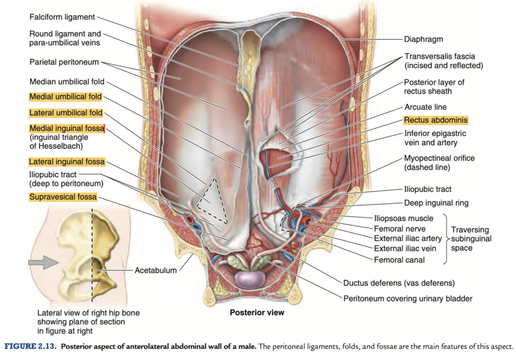
It is located between the medial and lateral umbilical fold and is also known as the Hesselbach’s triangle.
a. supravesicular fossa
b. lateral inguinal fossa
c. medial inguinal fossa
d. inguinal region
Answer: c. medial inguinal fossa
解説: Hesselbach三角(Hesselbach’s triangle)は、内側では腹直筋の外縁、下部では鼠径靭帯、外側では下腹壁動静脈によって境界づけられます。この三角は、腹壁の弱点であり、直接鼠径ヘルニアが発生しやすい場所です。他の選択肢は異なる領域を指しています。
- a. supravesicular fossa: 恥骨上方に位置し、ヘルニアが発生する領域ではありません。
- b. lateral inguinal fossa: 鼠径輪の外側にあり、間接ヘルニアが発生しやすい場所です。
- d. inguinal region: 鼠径部全体を指しますが、Hesselbach三角とは異なります。
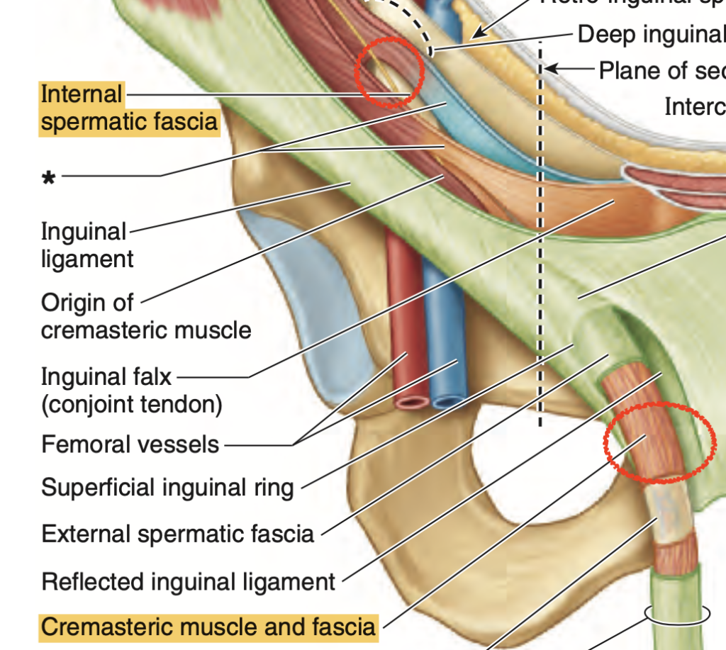
Question 2 ★出た
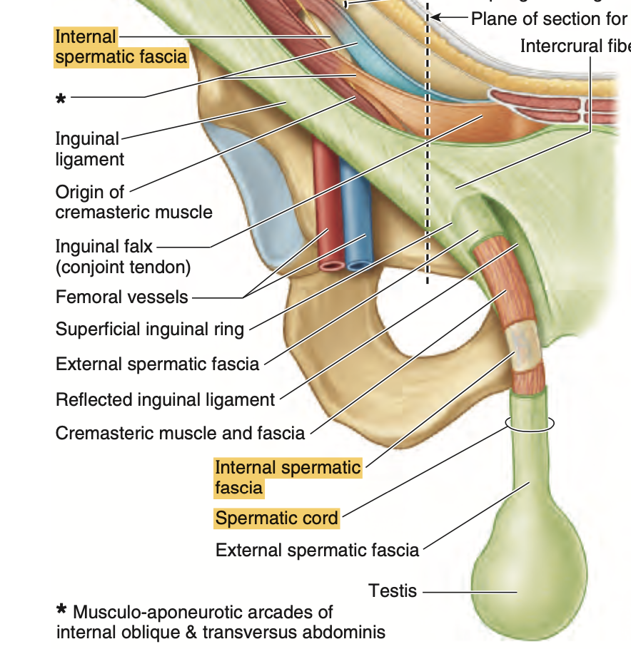
The transversalis fascia of the abdomen corresponds to what structure found in the spermatic cord?
a. External Spermatic Fascia
b. Cremasteric Fascia
c. Processus Vaginalis
d. Internal Spermatic Fascia
Answer: d. Internal Spermatic Fascia
解説: 腹部の横筋筋膜(transversalis fascia)は、精索(spermatic cord)の内精索筋膜(internal spermatic fascia)に対応しています。精索は、精巣が腹腔から陰嚢に下降する際にこれらの層が変化して形成されます。
- a. External Spermatic Fascia: これは外腹斜筋の筋膜から形成され、横筋筋膜には対応しません。
- b. Cremasteric Fascia: 内腹斜筋に由来する筋膜であり、横筋筋膜とは異なります。
- c. Processus Vaginalis: これは腹膜の延長であり、横筋筋膜とは関連しません。
Question 3 ★出た
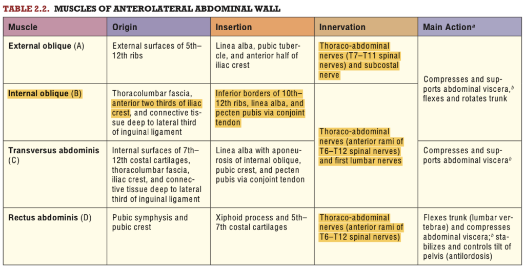
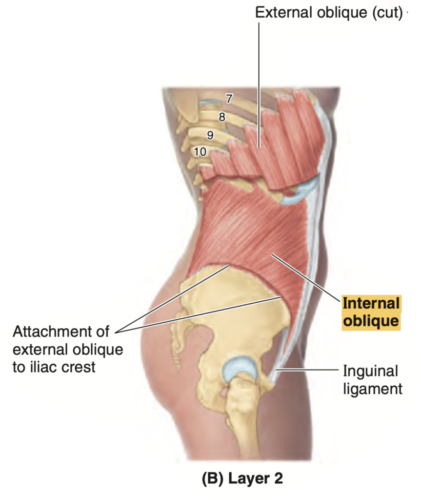
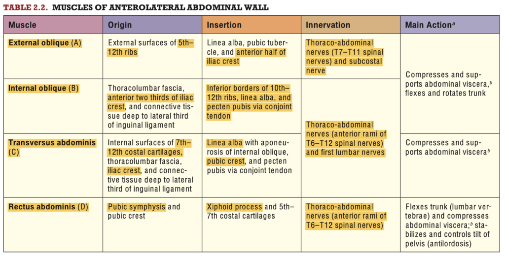
What is the largest and most superficial of the three abdominal muscles?
a. rectus abdominis M.
b. transversus abdominis M.
c. external oblique M.
d. internal oblique M.
Answer: c. external oblique M.
解説: 外腹斜筋(external oblique)は腹部筋肉の中で最も大きく、表層に位置しています。体幹の回旋や側屈に関与します。
- a. rectus abdominis M.: 腹直筋は中央に位置し、体幹の屈曲を主に行いますが、最も表層の筋肉ではありません。
- b. transversus abdominis M.: 横腹筋は最も深層に位置し、腹部の引き締めに関与します。
- d. internal oblique M.: 内腹斜筋は外腹斜筋の下にあり、体幹の回旋に関与しますが、外腹斜筋ほど表層ではありません。
Question 4 ★出た
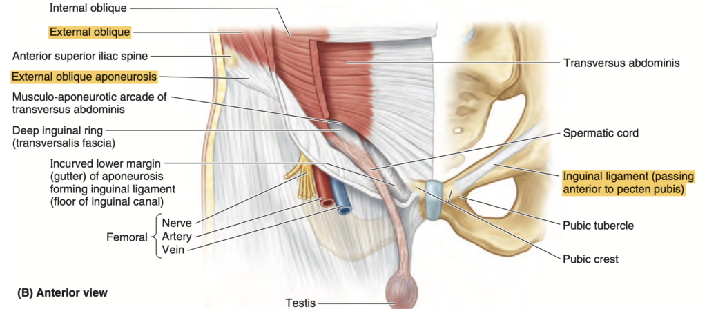
What anterior lateral abdominal wall aponeurosis(腱膜) has a thick inferior margin that forms the inguinal ligament?
a. Transversus Abdominis
b. Internal Oblique
c. External Oblique
d. Rectus Abdominis
Answer: c. External Oblique
解説: 外腹斜筋の腱膜(external oblique aponeurosis)は下部が厚く、これが鼠径靭帯(inguinal ligament)を形成します。鼠径靭帯は腹部の下部に位置し、下肢に繋がる構造の一部です。
- a. Transversus Abdominis: 横腹筋は鼠径靭帯を形成しません。
- b. Internal Oblique: 内腹斜筋も鼠径靭帯の一部を形成しますが、主に外腹斜筋が靭帯を形成します。
- d. Rectus Abdominis: 腹直筋は鼠径靭帯の形成には関与しません。
Question 5
What is the only fascia or aponeurosis that covers the rectus abdominis muscle posteriorly below the arcuate line?
a. External Oblique Aponeurosis
b. Internal Oblique Aponeurosis
c. Transversalis Fascia
d. Peritoneum
Answer: c. Transversalis Fascia
解説: 弓状線(arcuate line)より下では、腹直筋の後方は横筋筋膜(transversalis fascia)と腹膜(peritoneum)に覆われます。他の筋膜や腱膜は腹直筋の前方を覆うだけです。
- a. External Oblique Aponeurosis: 外腹斜筋の腱膜は腹直筋の前方を覆います。
- b. Internal Oblique Aponeurosis: 内腹斜筋の腱膜も前方を覆いますが、後方は覆いません。
- d. Peritoneum: 腹膜は横筋筋膜とともに後方を覆いますが、主に横筋筋膜が該当します。
Question 6 ★出た
What is the counterpart layer in the spermatic cord of the internal oblique fascia in the anterior lateral abdominal wall?
a. External Spermatic Fascia
b. Cremasteric Fascia
c. Internal Spermatic Fascia
d. Darto’s Fascia
Answer: b. Cremasteric Fascia
解説: 内腹斜筋(internal oblique fascia)の筋繊維は精索(spermatic cord)の中で挙睾筋筋膜(cremasteric fascia)に変化します。挙睾筋は精巣を持ち上げる作用があり、内腹斜筋の筋繊維によって構成されています。
- a. External Spermatic Fascia: 外腹斜筋の筋膜から形成され、挙睾筋筋膜とは異なります。
- c. Internal Spermatic Fascia: 横筋筋膜に由来し、内腹斜筋には対応しません。
- d. Darto’s Fascia: 陰嚢の皮下組織にあり、精索の層とは異なります。
Question 7 ★出た
At the Hesselbach’s Triangle of the inguinal region, which of the following structures is found medially?
a. Rectus Sheath
b. Inguinal Ligament
c. Inferior Epigastric Vein
d. Superior Epigastric Vein
Answer: a. Rectus Sheath
解説: Hesselbach三角(Hesselbach’s triangle)の内側の境界は腹直筋鞘(rectus sheath)によって形成されます。これは直接鼠径ヘルニアの発生しやすい部位です。
- b. Inguinal Ligament: 鼠径靭帯はHesselbach三角の下部境界を形成します。
- c. Inferior Epigastric Vein: 下腹壁静脈は外側境界に位置します。
- d. Superior Epigastric Vein: 上腹壁静脈は上方にあり、Hesselbach三角には関与しません。
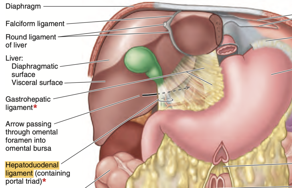
Question 8 ★出た
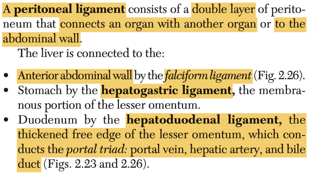
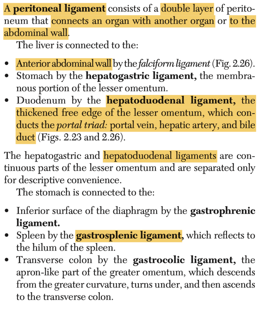
The hepatoduodenal ligament is classified under what type of peritoneum?
a. Lesser Omentum
b. Peritoneal Ligament
c. Mesentery
d. Greater Omentum
Answer: a. Lesser Omentum
解説: 肝十二指腸靭帯(hepatoduodenal ligament)は小網(lesser omentum)の一部であり、肝臓と十二指腸を結ぶ構造です。
- b. Peritoneal Ligament: 肝十二指腸靭帯自体が腹膜靭帯の一つですが、特に小網に分類されます。
- c. Mesentery: 腸間膜は小腸を支持する腹膜のひだであり、小網とは異なります。
- d. Greater Omentum: 大網は胃の下部から横行結腸にかけて垂れ下がる腹膜であり、肝十二指腸靭帯には関与しません。
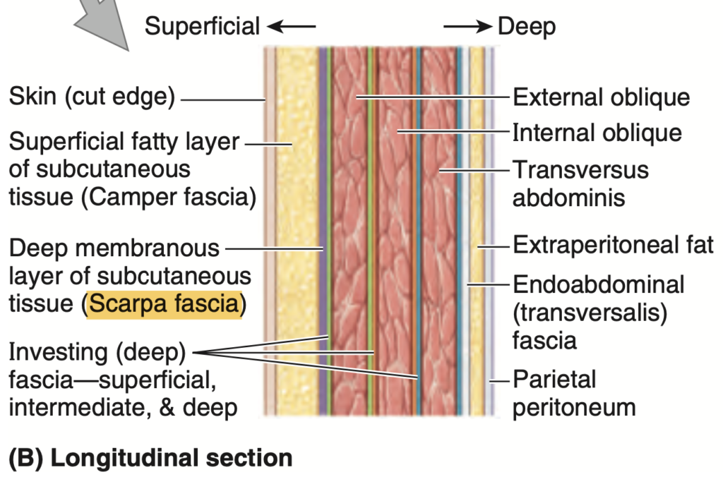
Question 9 ★出た
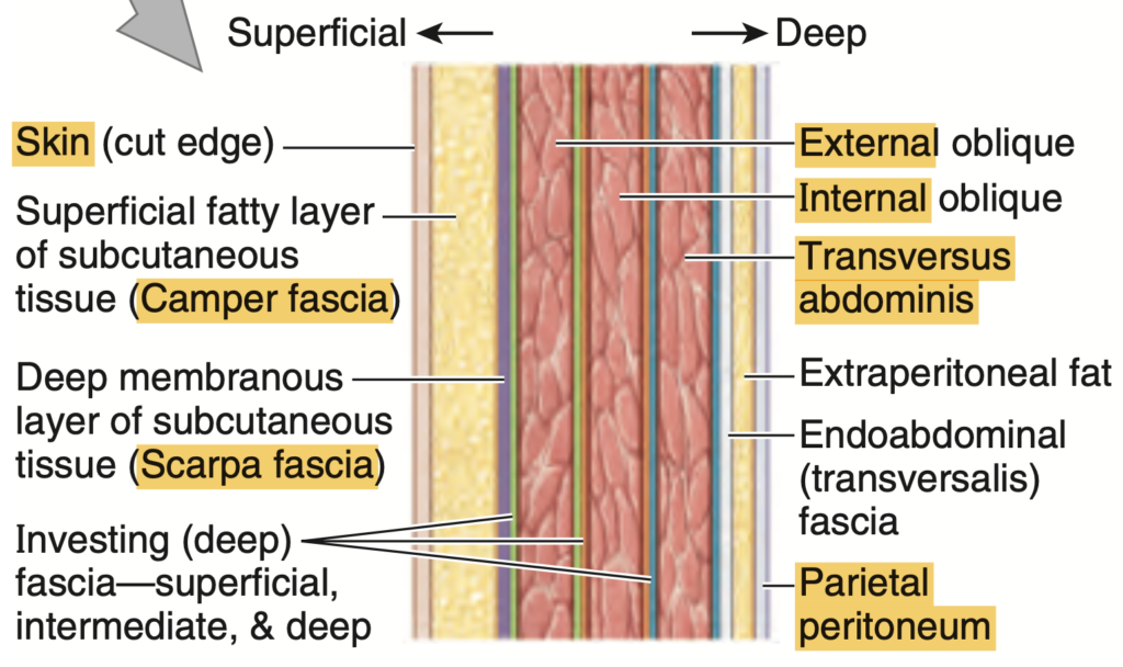
This is called the deep membranous layer of the subcutaneous tissue in the abdomen.
a. scampers fascia
b. colle’s fascia
c. scarpa’s fascia
d. campers fascia
Answer: c. Scarpa’s Fascia
解説: スカルパ筋膜(Scarpa’s fascia)は腹部の皮下組織の深層膜です。この層は皮膚の下にあり、特に下腹部で厚くなります。
- a. scampers fascia: この名称の筋膜は存在しません。
- b. colle’s fascia: 会陰部の筋膜で、腹部には位置しません。
- d. campers fascia: キャンパー筋膜は腹部の皮下組織の表層膜であり、スカルパ筋膜の上に位置します。
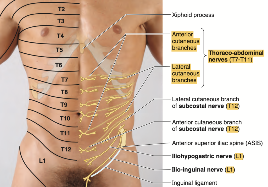
Question 10
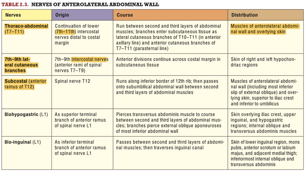
What cutaneous nerve of the anterior lateral abdominal wall comes from spinal nerves T7 to T11?
a. Thoracoabdominal
b. Lateral Thoracic
c. Subcostal
d. Iliohypogastric
Answer: a. Thoracoabdominal
解説: 胸腹神経(thoracoabdominal nerve)は脊髄神経T7〜T11から起こり、前外側腹壁の皮膚と筋肉を支配します。
- b. Lateral Thoracic: 外側胸神経は主に乳腺や側胸壁を支配し、腹壁神経とは異なります。
- c. Subcostal: 肋下神経はT12から起こり、T7〜T11からの胸腹神経とは異なります。
- d. Iliohypogastric: 腸骨下腹神経はL1から起こり、T7〜T11とは関係がありません。
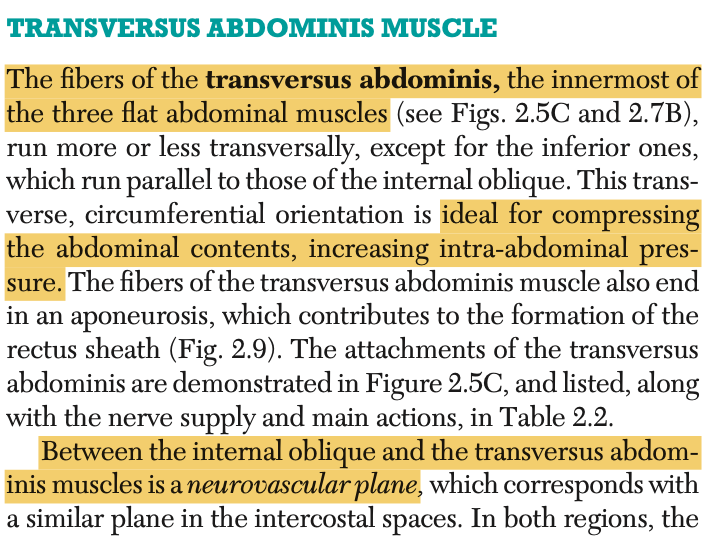
Question 11 ★出た
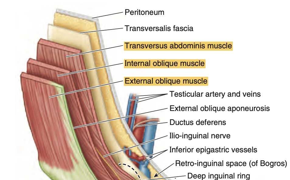
The neurovascular plane of the anterior abdomen is located between which group of muscles?
a. External Oblique and Internal Oblique
b. Internal Oblique and Transversus Abdominis
c. Transversus Abdominis and Rectus Abdominis
d. Rectus Abdominis and External Oblique
Answer: b. Internal Oblique and Transversus Abdominis
解説: 腹部の神経血管層(neurovascular plane)は、内腹斜筋(internal oblique)と横腹筋(transversus abdominis)の間に位置します。この層には肋間神経や血管が通っており、腹壁の感覚と運動を支配しています。
- a. External Oblique and Internal Oblique: 外腹斜筋と内腹斜筋の間には神経血管層は存在しません。
- c. Transversus Abdominis and Rectus Abdominis: これらの筋肉の間には神経血管層はありません。
- d. Rectus Abdominis and External Oblique: 腹直筋と外腹斜筋の間にも神経血管層はありません。
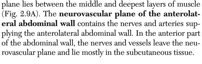
Question 12 ★出た
Which of the following muscles is attached to the anterior surface of the pubis and anterior pubic ligament that tenses the linea alba?
a. Rectus Abdominis
b. Pyramidalis
c. Transversus Abdominis
d. Internal Oblique
Answer: b. Pyramidalis
解説: 錐体筋(pyramidalis)は恥骨前面に付着し、白線(linea alba)を緊張させる小さな筋肉です。この筋肉は一部の人には欠如していますが、白線を緊張させることで役割を果たします。
- a. Rectus Abdominis: 腹直筋は白線に沿って走行しますが、直接緊張させる筋肉ではありません。
- c. Transversus Abdominis: 横腹筋は腹部を圧迫しますが、白線の緊張には関与しません。
- d. Internal Oblique: 内腹斜筋も腹壁を圧迫しますが、錐体筋とは異なります。
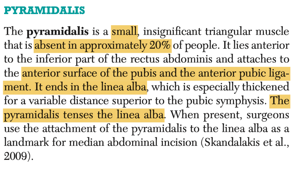
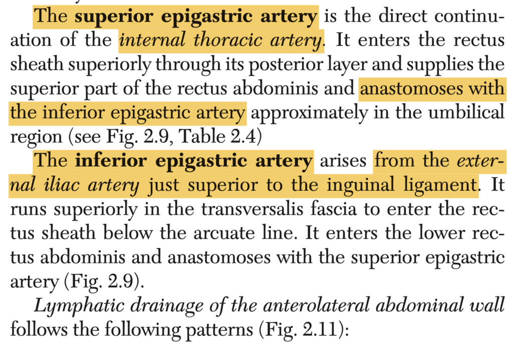
Question 13 ★出た
Where does the superior epigastric artery come from?
a. Internal Thoracic
b. Femoral
c. External Iliac
d. Internal Iliac
Answer: a. Internal Thoracic
解説: 上腹壁動脈(superior epigastric artery)は内胸動脈(internal thoracic artery)から分岐し、腹壁の上部を供給します。これは下腹壁動脈(inferior epigastric artery)と吻合し、腹壁の血液供給を助けます。
- b. Femoral: 大腿動脈は下肢に血液を供給します。
- c. External Iliac: 外腸骨動脈は下腹壁動脈の起始点ですが、上腹壁動脈には関与しません。
- d. Internal Iliac: 内腸骨動脈は骨盤内臓に血液を供給します。
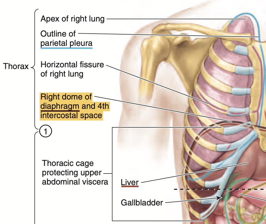
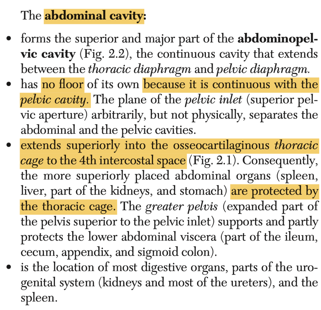
Question 14
At what level of the thoracic cage does the abdominal cavity extend superiorly?
a. 4th Intercostal Space
b. 5th Intercostal Space
c. 6th Intercostal Space
d. 7th Intercostal Space
Answer: a
Question 15
This layer of peritoneum forms a connective tissue plane in which the nerves and vessels of the descending colon continue to lie.
a. Scarpa’s fascia
b. Camper’s fascia
c. Transversalis fascia
d. Fusion fascia
Answer: d. Fusion fascia
解説: 融合筋膜(fusion fascia)は、下降結腸(descending colon)の神経や血管が存在する結合組織の層です。これは腹膜が他の層と融合して形成されます。
- a. Scarpa’s fascia: これは腹部の皮下組織の深層膜です。
- b. Camper’s fascia: これは腹部の皮下組織の表層膜です。
- c. Transversalis fascia: 横筋筋膜は腹壁の内側に位置しますが、腸の神経や血管には関連しません。

Question 16 ★出た
What is a double-layered peritoneal structure that connects an organ with another organ and is attached to the abdominal wall?
a. Lesser Omentum
b. Mesentery
c. Greater Omentum
d. Peritoneal Ligament
Answer: d. Peritoneal Ligament
解説: 腹膜靭帯(peritoneal ligament)は二重層の腹膜で、臓器同士や臓器と腹壁を結びつけます。例として、肝臓と横隔膜をつなぐ冠状靭帯があります。
- a. Lesser Omentum: 小網は肝臓と胃を結ぶ腹膜の一部ですが、臓器と腹壁を結びません。
- b. Mesentery: 腸間膜は腸を腹壁に固定しますが、他の臓器との連結には使われません。
- c. Greater Omentum: 大網は胃の下部から横行結腸にかけて垂れ下がりますが、臓器同士の主要な結合構造ではありません。
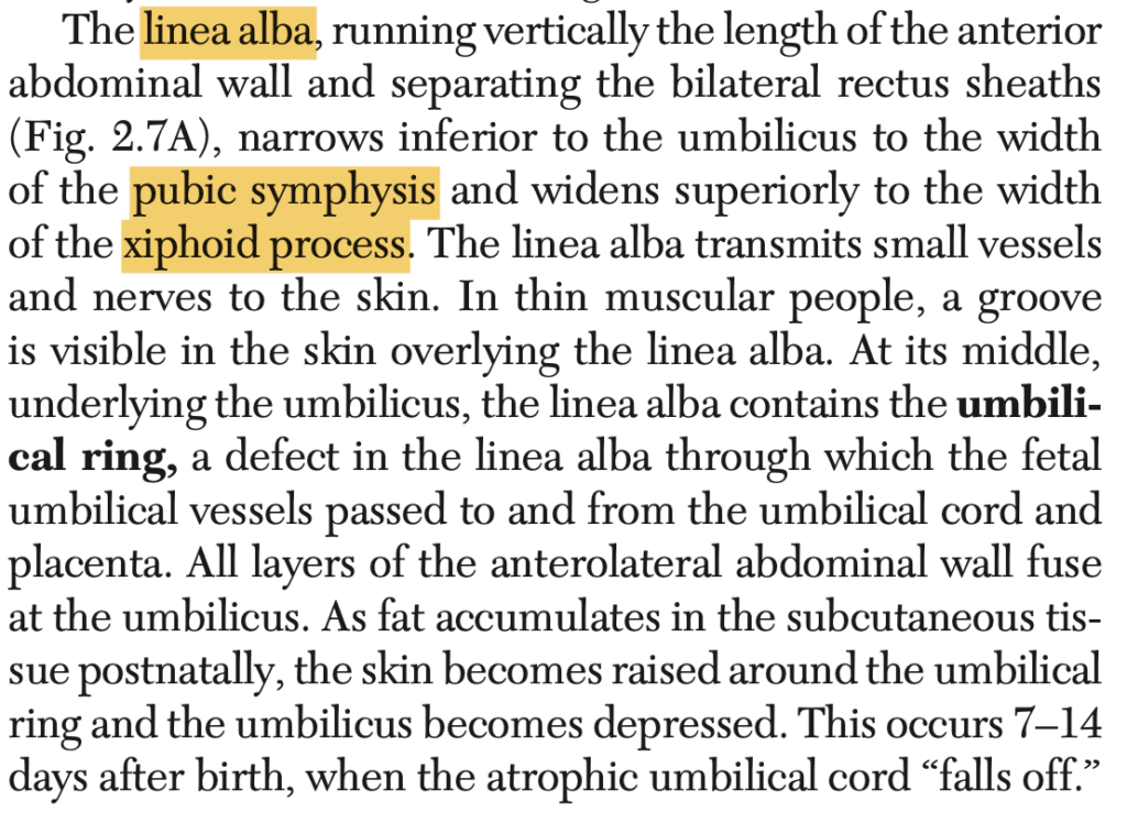
Question 17
What structure extends from the xiphoid process to the symphysis pubis?
a. Linea Semilunaris
b. Linea Alba
c. External Oblique M.
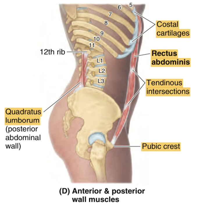
d. Tendinous Intersection
Answer: b. Linea Alba
解説: 白線(linea alba)は剣状突起(xiphoid process)から恥骨結合(symphysis pubis)まで垂直に伸びる腱膜です。この腱膜は腹直筋を左右に分けています。
- a. Linea Semilunaris: これは腹直筋の外側縁に沿って走る構造であり、白線ではありません。
- c. External Oblique M.: 外腹斜筋は腹部外側に位置し、縦方向に走行しません。
- d. Tendinous Intersection: 腱画は腹直筋の中にある水平な腱であり、白線とは異なります。
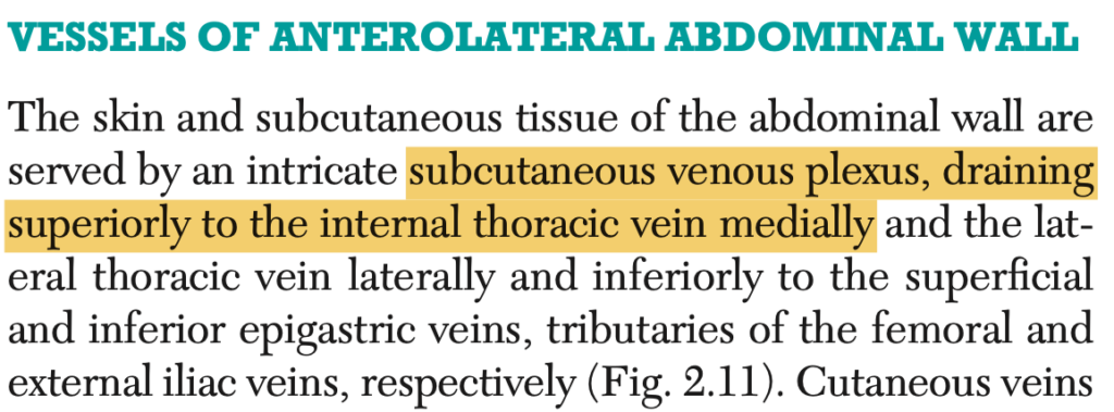
Question 18
Where does the subcutaneous venous plexus of the anterior lateral abdominal wall drain medially?
a. Lateral Thoracic
b. Inferior Epigastric
c. Internal Thoracic
d. Superficial Femoral
Answer: c. Internal Thoracic
解説: 前外側腹壁の皮下静脈叢(subcutaneous venous plexus)は、内胸静脈(internal thoracic vein)に内側に向かって流れ込みます。これは胸壁や腹壁の血液を上半身に戻す役割を担っています。
- a. Lateral Thoracic: 外側胸静脈は上肢や側胸壁に血液を送る役割がありますが、腹壁の内側ではありません。
- b. Inferior Epigastric: 下腹壁静脈は下部での血液排出に関連します。
- d. Superficial Femoral: 浅大腿静脈は下肢に関連しており、腹壁には関与しません。
Question 19(類似9) ★出た
What is considered as a deep membranous layer of the anterior lateral abdominal wall?
a. Colle’s Fascia
b. Endoabdominal Fascia
c. Scarpa’s Fascia
d. Camper’s Fascia
Answer: c. Scarpa’s Fascia
解説: スカルパ筋膜(Scarpa’s fascia)は前外側腹壁の深部に位置する膜状層です。この層は腹部の下部で最も厚く、腹壁の支持に重要です。
- a. Colle’s Fascia: これは会陰部に位置する筋膜で、腹壁には関係しません。
- b. Endoabdominal Fascia: これは腹壁の内部にある筋膜で、スカルパ筋膜ほど表層にはありません。
- d. Camper’s Fascia: キャンパー筋膜は前腹壁の浅層にある脂肪層で、スカルパ筋膜の外側に位置します。
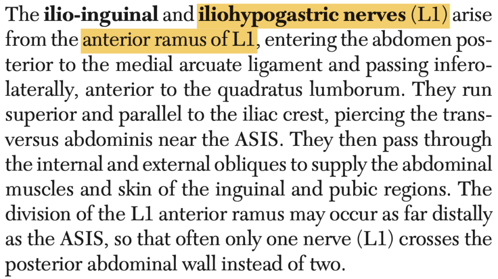
Question 20 ★出た
Which of the following nerves is derived from the terminal branches of the anterior ramus of L1?
a. Lateral Thoracic
b. Iliohypogastric
c. Subcostal
d. Thoracoabdominal
Answer: b. Iliohypogastric
解説: 腸骨下腹神経(iliohypogastric nerve)はL1の前枝の末端から派生し、下腹部と会陰部の皮膚に感覚を供給します。
- a. Lateral Thoracic: 外側胸神経は胸壁に供給する神経であり、L1には関係しません。
- c. Subcostal: 肋下神経はT12から派生し、L1とは関係がありません。
- d. Thoracoabdominal: 胸腹神経はT7〜T11から派生し、L1とは無関係です。
Question 21 ★出た
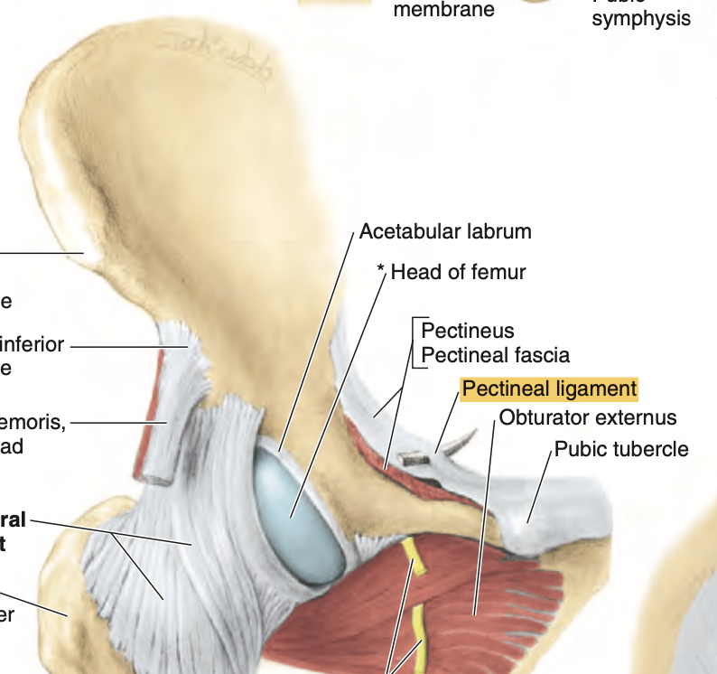
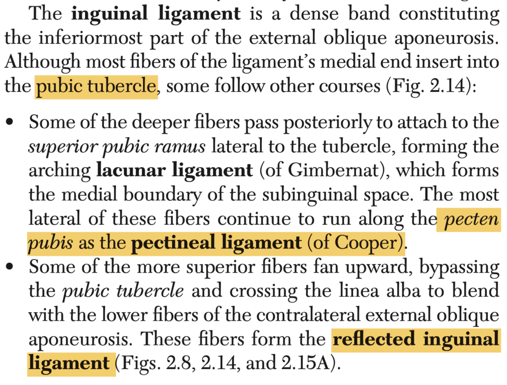
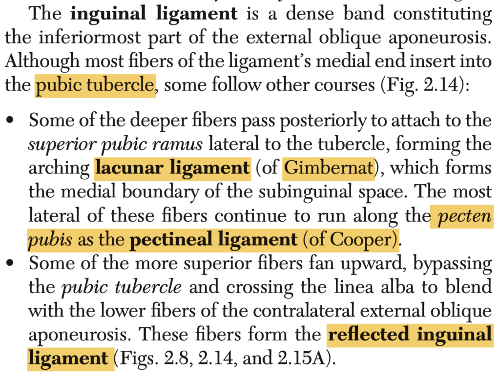
It is also known as “ligament of Cooper.”
a. Iliopubic Tract
b. Pectineal Ligament
c. Lacunar Ligament
d. Reflected Inguinal Ligament
Answer: b. Pectineal Ligament
解説: クーパー靭帯(ligament of Cooper)は恥骨櫛靭帯(pectineal ligament)とも呼ばれ、鼠径靭帯の一部が骨盤の恥骨櫛に付着する構造です。鼠径ヘルニアの修復時に重要なランドマークとなります。
- a. Iliopubic Tract: 腸恥線は鼠径靭帯の下に走行するが、クーパー靭帯とは異なる。
- c. Lacunar Ligament: 腸骨筋と恥骨筋の間に位置し、恥骨靭帯とは異なる。
- d. Reflected Inguinal Ligament: 反転鼠径靭帯は鼠径靭帯の延長部であり、クーパー靭帯とは関係がない。
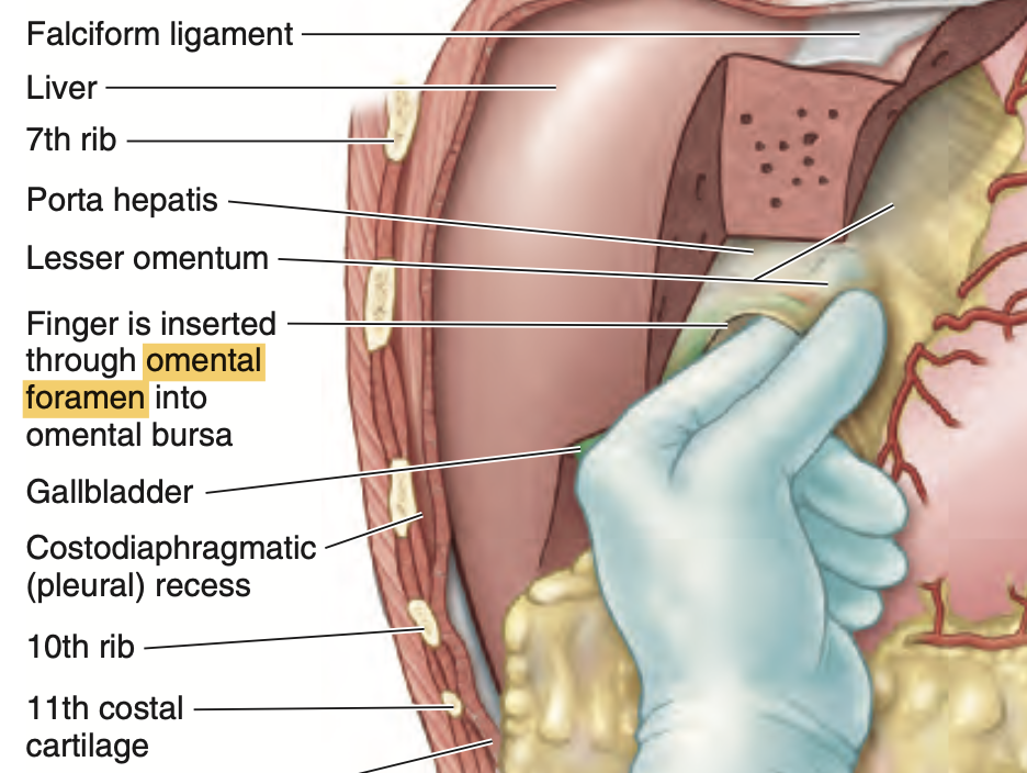
Question 22 ★出た
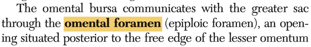
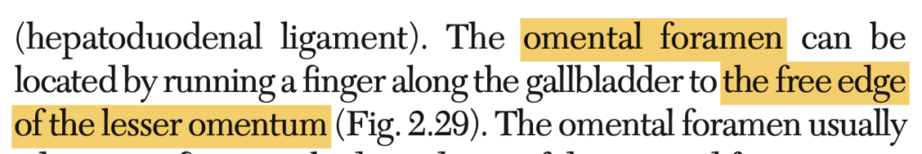
The omental foramen is found posterior to the free edge of what peritoneal structure or area?
a. Omental Bursa
b. Greater Omentum
c. Greater Peritoneal Sac
d. Lesser Omentum
Answer: d. Lesser Omentum
解説: 大網孔(omental foramen)は小網(lesser omentum)の自由縁の後ろに位置し、大網嚢(lesser sac)と大腹膜嚢(greater sac)を連絡しています。
- a. Omental Bursa: 大網嚢自体がこの構造の一部ですが、自由縁には関与しない。
- b. Greater Omentum: 大網は自由縁には関与しません。
- c. Greater Peritoneal Sac: 大腹膜嚢は大きな腹膜の腔ですが、特定の自由縁の位置ではありません。
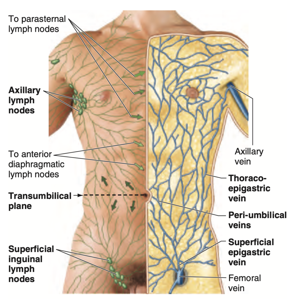
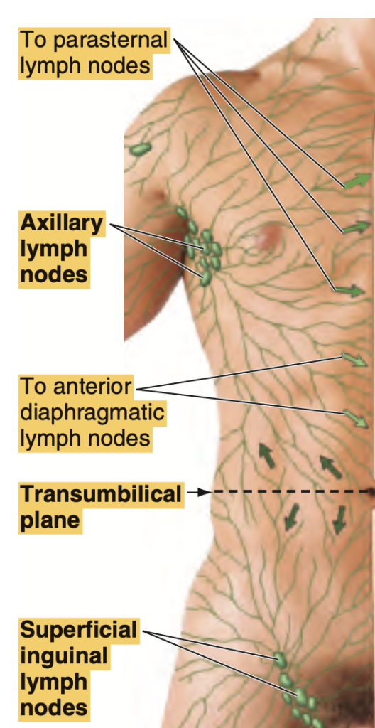
Question 23
Superficial lymphatics above the umbilical area drain mainly to what group of lymph nodes?
a. Axillary
b. External Iliac
c. Internal Thoracic
d. Jugular
Answer: a. Axillary
解説: 臍(へそ)より上部の皮下リンパ管は主に腋窩リンパ節(axillary lymph nodes)に流れ込みます。この部位のリンパ系は上肢や胸部と関連しています。
- b. External Iliac: 外腸骨リンパ節は主に下肢と関連しています。
- c. Internal Thoracic: 内胸リンパ節は胸壁の深部に関連しています。
- d. Jugular: 頸リンパ節は首や頭部に関連しており、腹部とは関係がありません。
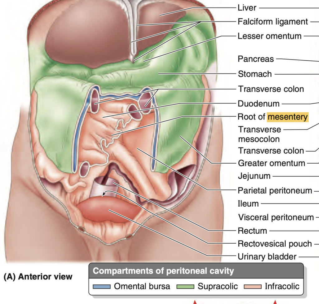
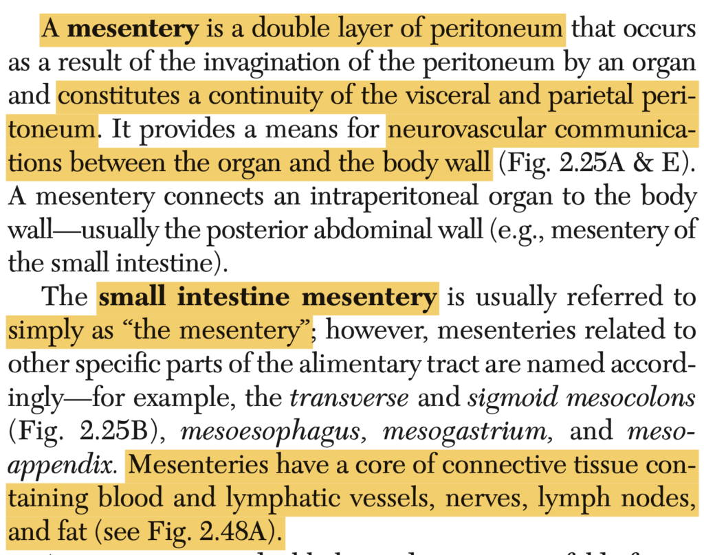
Question 24 ★出た
What do you call a double-layered peritoneum that forms a continuity between the visceral and parietal peritoneum?
a. Mesentery
b. Peritoneal Ligament
c. Lesser Omentum
d. Greater Omentum
Answer: a. Mesentery
解説: 腸間膜(mesentery)は二重層の腹膜で、臓側腹膜と壁側腹膜を連結し、腸を固定します。これにより血管や神経が腸に供給されます。
- b. Peritoneal Ligament: 腹膜靭帯は臓器同士を結びますが、腸間膜とは異なります。
- c. Lesser Omentum: 小網は肝臓と胃を結ぶ腹膜の一部です。
- d. Greater Omentum: 大網は胃の下部から横行結腸にかけて垂れ下がる腹膜です。
Question 25(類似13) ★出た
Where does the inferior epigastric artery come from?
a. Internal Thoracic
b. Femoral
c. Anterior Intercostal
d. External Iliac
Answer: d. External Iliac
解説: 下腹壁動脈(inferior epigastric artery)は外腸骨動脈(external iliac artery)から分岐し、腹壁の下部を供給します。
- a. Internal Thoracic: 内胸動脈は上腹壁動脈を供給しますが、下腹壁には関与しません。
- b. Femoral: 大腿動脈は下肢に供給されますが、腹壁とは関係がありません。
- c. Anterior Intercostal: 前肋間動脈は肋間の構造に血液を供給しますが、腹壁には関与しません。
Question 26 ★出た
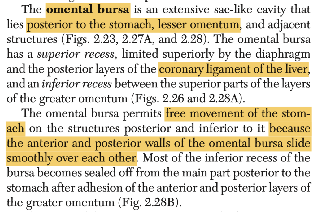
In the subdivisions of the peritoneal cavity, where do you locate the omental bursa?
a. Supracolic Compartment
b. Lesser Peritoneal Sac
c. Infracolic Compartment
d. Greater Peritoneal Sac
Answer: b. Lesser Peritoneal Sac
解説: 大網嚢(omental bursa)は小腹膜嚢(lesser peritoneal sac)とも呼ばれ、胃の後ろに位置する腹膜の空間です。
- a. Supracolic Compartment: 横隔膜の上部に位置する部分で、大網嚢とは異なります。
- c. Infracolic Compartment: 腸間膜の下部にある空間であり、大網嚢とは異なります。
- d. Greater Peritoneal Sac: 大腹膜嚢は腹腔全体の大部分を占める空間であり、小腹膜嚢とは区別されます。
Question 27 ★出た
Layers of the abdominal wall at the right lumbar region.
a. Skin, fatty layer of superficial fascia, membranous layer of superficial fascia, external oblique muscle, internal oblique muscle, extra peritoneal fat, peritoneum
b. Skin, Camper’s fascia, Scarpa’s fascia, external oblique muscle, internal oblique muscle, transversus abdominis muscle, extraperitoneal fat, parietal peritoneum
c. Skin, fatty layer of superficial fascia, membranous layer, internal oblique muscle, external oblique muscle, transversus abdominis muscle, peritoneum
d. Skin, Scarpa’s fascia, Camper’s fascia, external oblique muscle, internal oblique muscle, transversus abdominis muscle, extraperitoneal fat, peritoneum
Answer: b. Skin, Camper’s fascia, Scarpa’s fascia, external oblique muscle, internal oblique muscle, transversus abdominis muscle, extraperitoneal fat, parietal peritoneum
解説: 腹壁の層は、皮膚、キャンパー筋膜(浅層脂肪層)、スカルパ筋膜(深層膜)、外腹斜筋、内腹斜筋、横腹筋、腹膜前脂肪層、そして壁側腹膜(parietal peritoneum)の順に構成されています。これらの層は、腹壁の構造と機能を支える重要な要素です。
- a: スカルパ筋膜とキャンパー筋膜の順序が逆です。
- c: 外腹斜筋と内腹斜筋の順序が逆です。
- d: スカルパ筋膜とキャンパー筋膜の順序が逆です。
Question 28
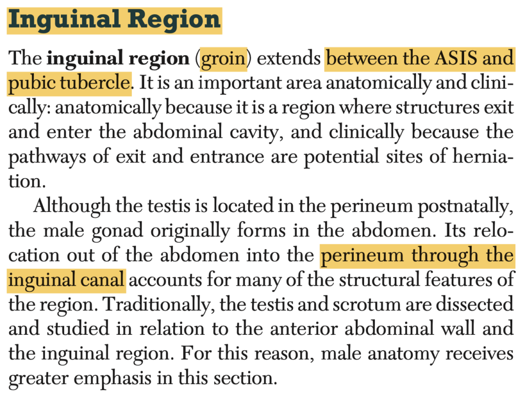
It is located between the ASIS and pubic tubercle.
a. Inguinal Region
b. Supravesicular Fossa
c. Medial Inguinal Fossa
d. Lateral Inguinal Fossa
Answer: a. Inguinal Region
解説: 鼠径部(inguinal region)は上前腸骨棘(ASIS)と恥骨結節(pubic tubercle)の間に位置し、鼠径靭帯が走行する場所です。
- b. Supravesicular Fossa: これは膀胱の上方に位置し、異なる領域です。
- c. Medial Inguinal Fossa: 内側鼠径窩は腹部の内側の部分に位置しますが、鼠径部とは異なります。
- d. Lateral Inguinal Fossa: 外側鼠径窩は鼠径輪の外側にあります。
Question 29
The internal oblique originates from the:
a. Xiphoid Process
b. Symphysis Pubis
c. 12th Rib
d. Anterior Iliac Crest
Answer: d. Anterior Iliac Crest
解説: 内腹斜筋(internal oblique muscle)は前腸骨稜(anterior iliac crest)から起始し、体幹の回旋や側屈に関与します。
- a. Xiphoid Process: これは剣状突起であり、内腹斜筋の起始点ではありません。
- b. Symphysis Pubis: 恥骨結合は起始点ではありません。
- c. 12th Rib: 第12肋骨は筋の一部が付着する部位ですが、主要な起始点ではありません。
Question 30 ★出た
How many layers are there in the anterior abdominal wall?
a. 6
b. 7
c. 8
d. 9
Answer: b. 7
解説: 前腹壁には7層の構造があります。皮膚、キャンパー筋膜、スカルパ筋膜、外腹斜筋、内腹斜筋、横腹筋、腹膜の順に並んでいます。
Question 31
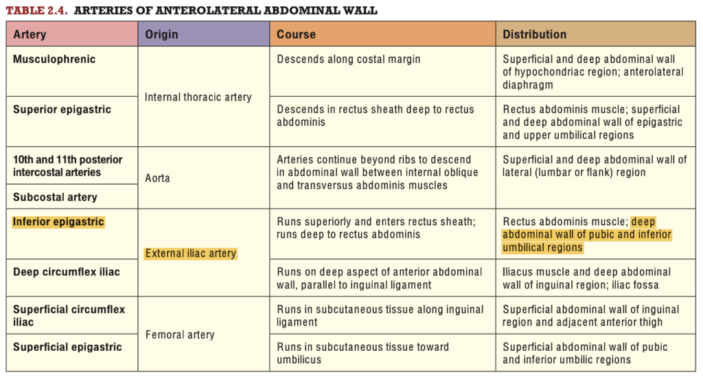
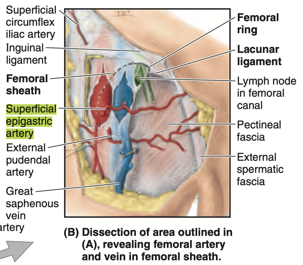
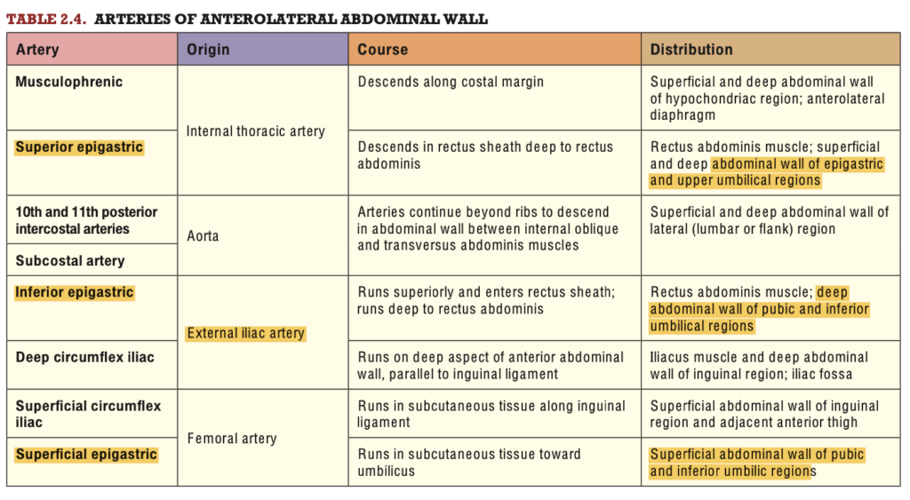
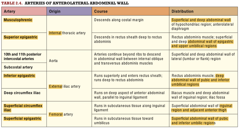
What artery supplies the superficial abdominal wall of the pubic and inferior umbilical region?
a. Musculophrenic A.
b. Internal Thoracic Artery
c. Inferior Epigastric A.
d. Superficial Epigastric A.
Answer: d. Superficial Epigastric A.
解説: 浅腹壁動脈(superficial epigastric artery)は、恥骨と下部臍領域の表層腹壁に血液を供給します。この動脈は大腿動脈から分岐し、皮下組織に血液を送ります。
- a. Musculophrenic A.: これは横隔膜と胸壁の一部に供給されますが、臍領域には供給されません。
- b. Internal Thoracic Artery: 内胸動脈は主に胸壁に供給します。
- c. Inferior Epigastric A.: 下腹壁動脈は主に腹壁の深層に血液を供給します。
Question 32
This artery that supplies the anterior abdomen originates from the external iliac artery.
a. Superficial Circumflex A.
b. Superior Epigastric A.
c. Inferior Epigastric A.
d. Musculophrenic A.
Answer: c. Inferior Epigastric A.
解説: 下腹壁動脈(inferior epigastric artery)は外腸骨動脈(external iliac artery)から分岐し、前腹壁の下部に血液を供給します。この動脈は腹直筋の後ろを走行し、上腹壁動脈と吻合します。
- a. Superficial Circumflex A.: 浅環状動脈は腹壁の外側に供給します。
- b. Superior Epigastric A.: 上腹壁動脈は内胸動脈から分岐します。
- d. Musculophrenic A.: 横隔膜と腹壁の上部に供給されますが、外腸骨動脈からは分岐しません。
Question 33
The thoraco-abdominal nerves are derived from the anterior rami of?
a. T7-11
b. T1-6
c. T1-12
d. T12
Answer: a. T7-11
解説: 胸腹神経(thoraco-abdominal nerves)は脊髄神経の前枝T7〜T11から派生し、腹壁の感覚および運動を支配します。
- b. T1-6: これらの神経は主に胸部に供給され、腹部には供給されません。
- c. T1-12: T1からT12までの神経は胸壁と腹壁の両方に関与しますが、特定の胸腹神経はT7-11です。
- d. T12: T12は肋下神経(subcostal nerve)として知られています。
Question 34 ★出た
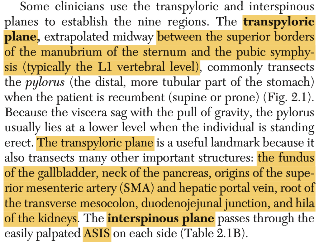
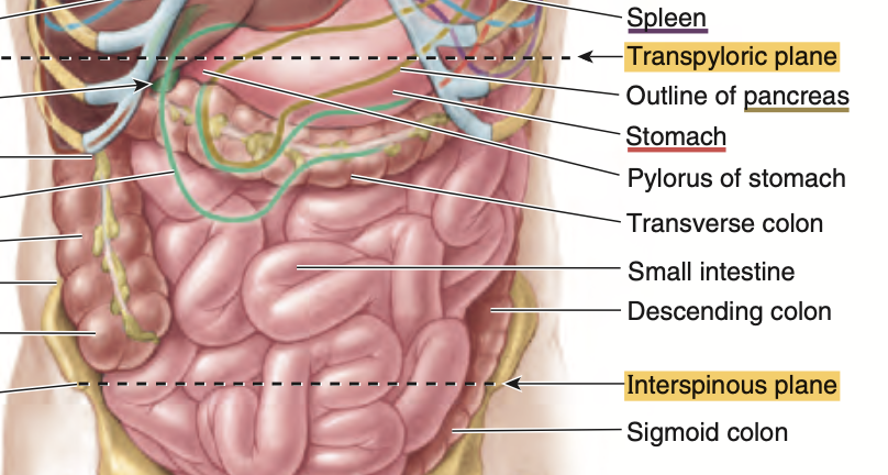
What anatomical plane is located between the superior border of the manubrium and pubic symphysis or L1 vertebral level?
a. Transumbilical
b. Transtubercular
c. Transpyloric
d. Interspinous
Answer: c. Transpyloric
解説: 幽門横断面(transpyloric plane)は、剣状突起の上端とL1椎骨の高さに位置し、胃の幽門や膵臓、腎臓の上部を含む重要な解剖学的ランドマークです。
- a. Transumbilical: 臍を通る水平面で、より下に位置します。
- b. Transtubercular: 腸骨結節を通る水平面で、L5の高さに位置します。
- d. Interspinous: 腸骨棘を通る水平面で、仙骨の高さに位置します。
Question 35 ★出た
What ligament is known as “ligament of Gimbernat” and whose fibers attach to the superior pubic ramus?
a. Round Ligament
b. Inguinal Ligament
c. Pectineal Ligament
d. Lacunar Ligament
Answer: d. Lacunar Ligament
解説: ギンベルナート靭帯(ligament of Gimbernat)は、鼠径靭帯の一部であり、恥骨の上縁に付着します。これは鼠径ヘルニア修復において重要な構造です。
- a. Round Ligament: これは子宮を固定する靭帯であり、骨盤には関与しません。
- b. Inguinal Ligament: 鼠径靭帯は腸骨と恥骨を結ぶが、ラキュナ靭帯の一部とは異なる。
- c. Pectineal Ligament: これは恥骨筋膜に沿った靭帯であり、ギンベルナート靭帯とは異なる。
Question 36
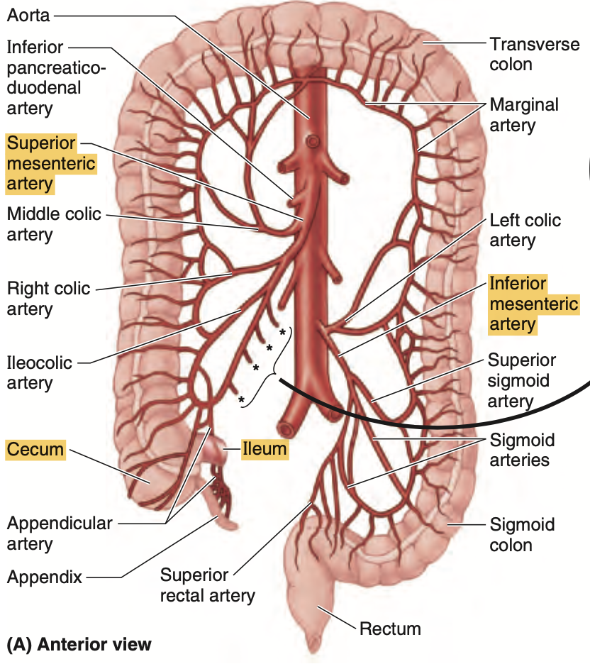
Contents of the right iliac region of the abdomen, EXCEPT:
a. External Iliac Artery
b. Inferior Mesenteric Artery
c. Ileum
d. Cecum
e. Genitofemoral Nerve
Answer: b. Inferior Mesenteric Artery
解説: 右腸骨領域には外腸骨動脈、回腸、盲腸、性大腿神経などが含まれますが、下腸間膜動脈(inferior mesenteric artery)は主に左側に位置し、大腸の供給に関与します。
- a. External Iliac Artery: 右腸骨領域に含まれます。
- c. Ileum: 回腸は右下腹部にあります。
- d. Cecum: 盲腸も右下腹部に位置します。
- e. Genitofemoral Nerve: これは腹壁や股関節領域に感覚を供給する神経で、右腸骨領域に位置します。
Question 37
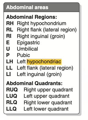
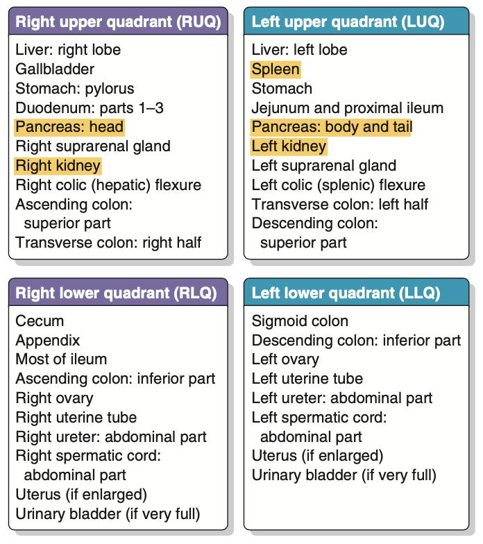
Which of the following organs are found in the left hypochondriac region, EXCEPT:
a. Spleen
b. Pancreas
c. Gallbladder
d. Kidney
e. Colon
Answer: c. Gallbladder
解説: 胆嚢(gallbladder)は右上腹部に位置し、左季肋部にはありません。左季肋部には脾臓、膵臓の一部、腎臓、そして大腸の一部があります。
- a. Spleen: 脾臓は左季肋部に位置します。
- b. Pancreas: 膵臓の尾部が左側に伸びています。
- d. Kidney: 左腎は左季肋部に位置します。
- e. Colon: 大腸の左結腸曲がこの領域にあります。
Question 38 ★出た
The gastrosplenic ligament is classified under what type of peritoneum?
a. Lesser Omentum
b. Peritoneal Ligament
c. Mesentery
d. Greater Omentum
Answer: b. Peritoneal Ligament
解説: 胃脾靭帯(gastrosplenic ligament)は腹膜靭帯(peritoneal ligament)の一つで、胃と脾臓を結びつけます。
- a. Lesser Omentum: 小網は肝臓と胃を結びますが、胃脾靭帯には含まれません。
- c. Mesentery: 腸間膜は腸を固定する構造であり、胃脾靭帯とは異なります。
- d. Greater Omentum: 大網は胃と横行結腸を結ぶが、胃脾靭帯は大網の一部ではありません。
Question 39 ★出た
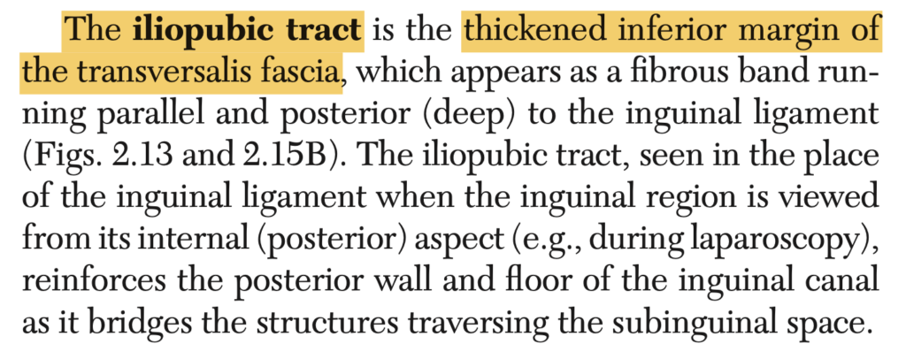
Which of the following ligaments in the inguinal region is defined as a thickened inferior margin of the transversalis fascia?
a. Lacunar
b. Inguinal
c. Iliopubic
d. Cooper’s
Answer: c. Iliopubic
解説: 腸恥靭帯(iliopubic ligament)は、横筋筋膜の下縁が厚くなった構造で、鼠径部に位置します。
- a. Lacunar: これは鼠径靭帯の延長であり、腸恥靭帯とは異なる。
- b. Inguinal: 鼠径靭帯は鼠径管を形成する構造ですが、腸恥靭帯とは異なります。
- d. Cooper’s: クーパー靭帯は恥骨に沿って走る靭帯であり、腸恥靭帯とは異なる。
Question 40
This structure is a double fold of peritoneum that extends from the stomach to the abdominal cavity.
a. Scarpa’s Fascia
b. Transversalis Fascia
c. Camper’s Fascia
d. Omentum
Answer: d. Omentum
解説: 大網(omentum)は胃から腹腔にかけて垂れ下がる二重の腹膜ひだで、主に大網と小網に分けられます。
- a. Scarpa’s Fascia: これは腹壁の深層膜であり、腹膜とは関係ありません。
- b. Transversalis Fascia: 横筋筋膜は腹壁に位置する膜であり、腹膜とは異なります。
- c. Camper’s Fascia: これは皮下脂肪層であり、腹膜とは異なります。
Question 41(類似37)
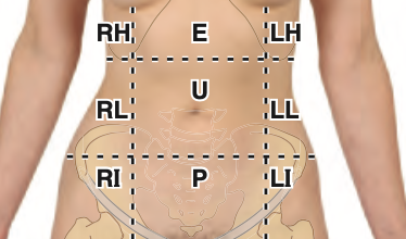
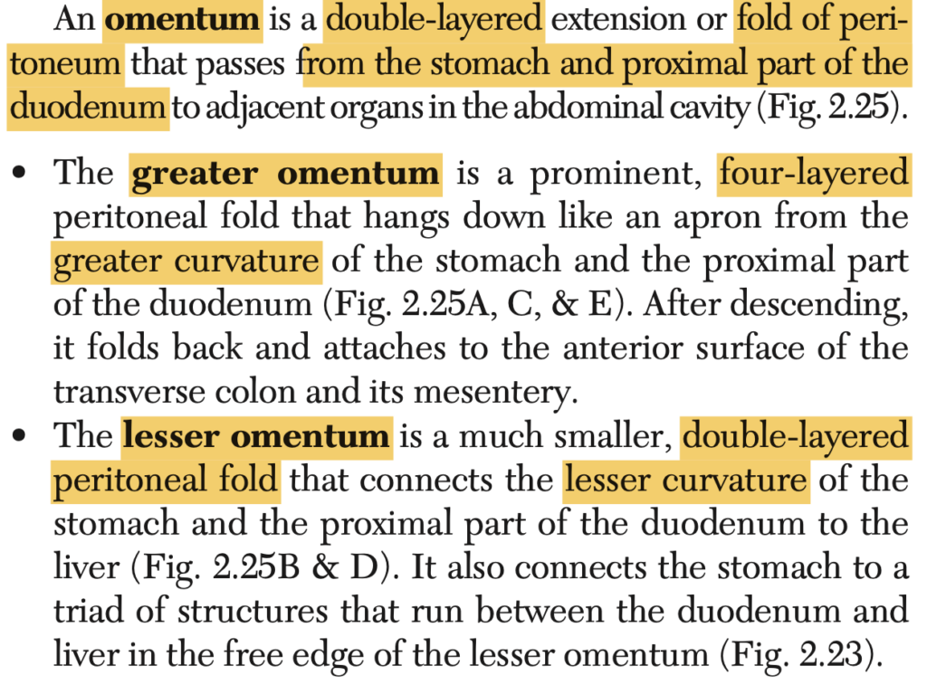
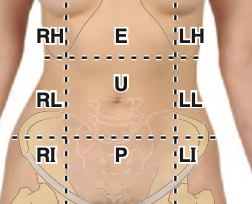
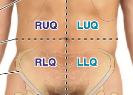
The abdomen is divided into:
a. 9 quadrants and 4 regions
b. 6 regions and 4 quadrants
c. 4 regions and 4 quadrants
d. 4 quadrants and 9 regions
Answer: d. 4 quadrants and 9 regions
解説: 腹部は4つの象限(右上象限、左上象限、右下象限、左下象限)に分かれており、また9つの領域(右季肋部、左季肋部、右腸骨領域、左腸骨領域、心窩部、臍部、恥骨部、右腰部、左腰部)に細分されています。
- a: 領域と象限の数が間違っています。
- b: 正しい数ではありません。
- c: 正しい構成ではありません。
Question 42
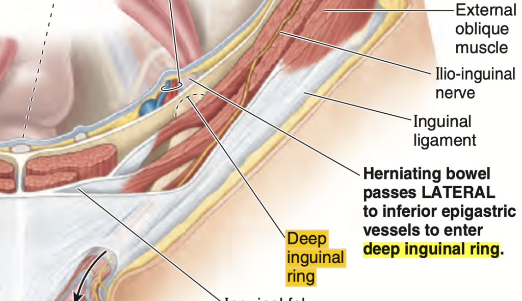
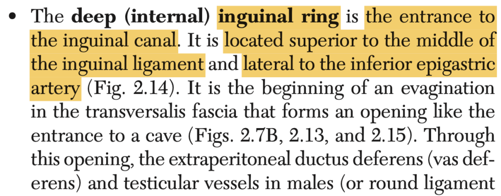
This structure is located superior to the middle part of the inguinal ligament and lateral to the inferior epigastric vessels.
a. Hesselbach’s Triangle
b. Superficial Inguinal Ring
c. Deep Inguinal Ring
d. Myopectineal Orifice
Answer: c. Deep Inguinal Ring
解説: 深鼠径輪(deep inguinal ring)は、鼠径靭帯の中央部の上方に位置し、下腹壁動脈の外側にあります。これは鼠径管の入り口です。
- a. Hesselbach’s Triangle: これは腹直筋の外縁にある三角形領域で、異なる位置にあります。
- b. Superficial Inguinal Ring: これは鼠径管の出口で、深鼠径輪とは異なります。
- d. Myopectineal Orifice: これは鼠径ヘルニアの潜在的な出口となる領域ですが、深鼠径輪とは異なります。
Question 43 ★出た
Which of the following anterior abdominal surface areas is unpaired?
a. Hypochondriac
b. Iliac
c. Lumbar
d. Hypogastric
Answer: d. Hypogastric
解説: 下腹部(hypogastric region)は腹部の中央に位置し、対称ではない単一の領域です。他の選択肢は対称的に左右に分かれています。
- a. Hypochondriac: これは左右対称に存在します。
- b. Iliac: 左右対称に存在します。
- c. Lumbar: 左右対称に存在します。
Question 44 ★出た
Which anterior lateral abdominal wall muscle has its posterior fiber edge span between its costal origin and iliac crest?
a. External Oblique
b. Internal Oblique
c. Transversus Abdominis
d. Rectus Abdominis
Answer: a. External Oblique
解説: 外腹斜筋(external oblique muscle)は、肋骨の起始部と腸骨稜(iliac crest)との間を後部でまたいでいます。この筋肉は最も表層に位置し、体幹の回旋や屈曲に関与します。
- b. Internal Oblique: 内腹斜筋は外腹斜筋の下にありますが、範囲が異なります。
- c. Transversus Abdominis: 横腹筋は腹部の深層に位置しています。
- d. Rectus Abdominis: 腹直筋は中央に位置し、肋骨と腸骨稜には関与しません。
Question 45
What muscle functions to stabilize and control the tilt of the pelvis?
a. Internal Oblique
b. Transversus Abdominis
c. External Oblique
d. Rectus Abdominis
Answer: d. Rectus Abdominis
解説: 腹直筋(rectus abdominis muscle)は骨盤の傾きを安定させ、制御する役割を果たします。これにより、骨盤が前傾または後傾しないようにします。
- a. Internal Oblique: 内腹斜筋も骨盤の安定に寄与しますが、主な筋肉ではありません。
- b. Transversus Abdominis: 横腹筋は主に腹部を圧迫しますが、骨盤の傾きの制御には直接関与しません。
- c. External Oblique: 外腹斜筋も一部関与しますが、腹直筋が主要な役割を果たします。
Question 46 ★出た
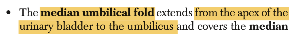
Which umbilical peritoneal fold arises from the apex of the urinary bladder to the umbilicus on the posterior surface of the anterior abdominal wall?
a. Medial Umbilical Fold
b. Inferior Umbilical Fold
c. Lateral Umbilical Fold
d. Median Umbilical Fold
Answer: d. Median Umbilical Fold
解説: 正中臍ヒダ(median umbilical fold)は、膀胱の頂点から臍に向かって走る腹膜のヒダであり、尿膜管(urachus)の遺残を含んでいます。
- a. Medial Umbilical Fold: これは閉鎖された臍動脈の遺残を含むヒダです。
- b. Inferior Umbilical Fold: この名称のヒダは存在しません。
- c. Lateral Umbilical Fold: これは下腹壁動静脈を覆うヒダです。

Question 47 ★出た
The urinary bladder is an example of a visceral organ with what type of peritoneal covering?
a. Extraperitoneal
b. Retroperitoneal
c. Subperitoneal
d. Intraperitoneal
Answer: c. Subperitoneal
解説: 膀胱は腹膜の下に位置するため、腹膜下(subperitoneal)臓器として分類されます。これは腹膜によって完全に覆われていない臓器です。
- a. Extraperitoneal: 腹膜外臓器は腹膜の外に位置しますが、膀胱はこれに該当しません。
- b. Retroperitoneal: 後腹膜臓器は背側に位置しますが、膀胱は該当しません。
- d. Intraperitoneal: 腹膜内臓器は腹膜で完全に覆われますが、膀胱は該当しません。
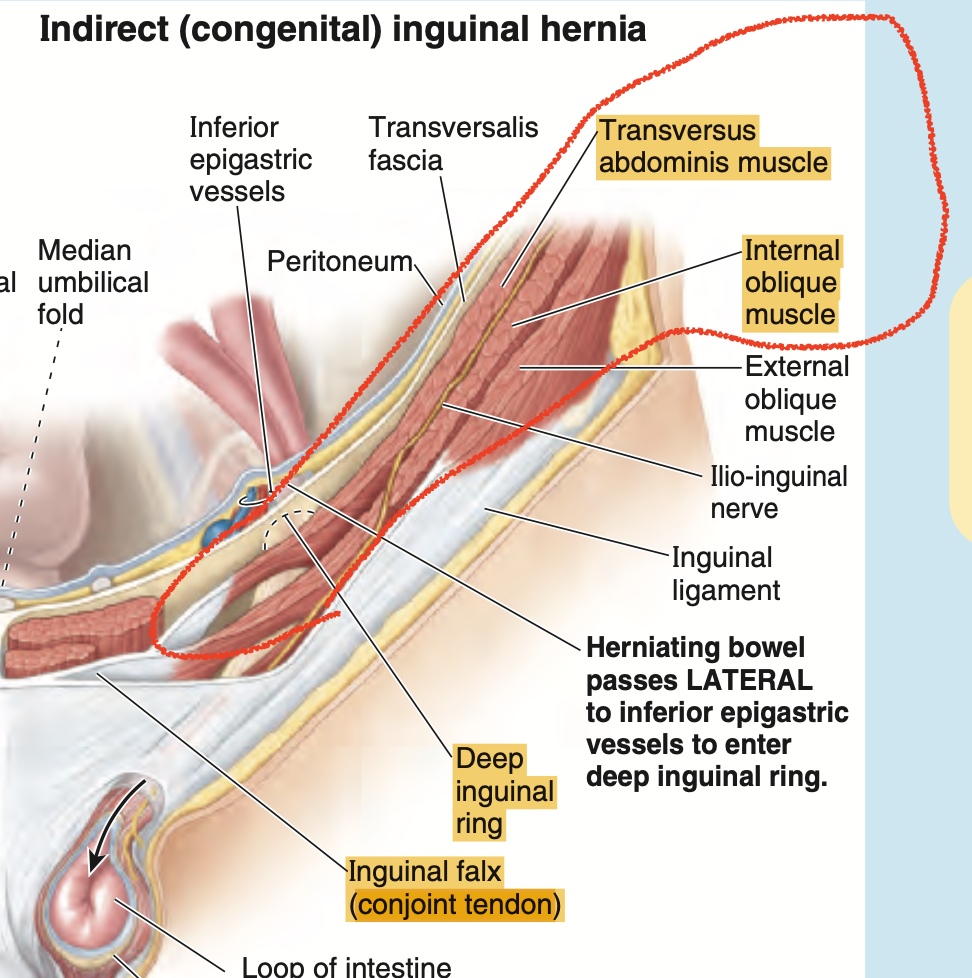
Question 48 ★出た
The conjoined tendon comes from fibers that arise from the internal oblique and aponeurotic fibers of what other muscle?
a. External Oblique
b. Transversus Abdominis
c. Rectus Abdominis
d. Pyramidalis
Answer: b. Transversus Abdominis
解説: 合併腱(conjoined tendon)は、内腹斜筋(internal oblique muscle)と横腹筋(transversus abdominis muscle)の腱膜線維が合わさったものです。この腱は鼠径管の後壁を強化します。
- a. External Oblique: 外腹斜筋は合併腱には関与しません。
- c. Rectus Abdominis: 腹直筋は合併腱の一部ではありません。
- d. Pyramidalis: 錐体筋は腹直筋の前にありますが、合併腱には関与しません。
Question 49 ★出た
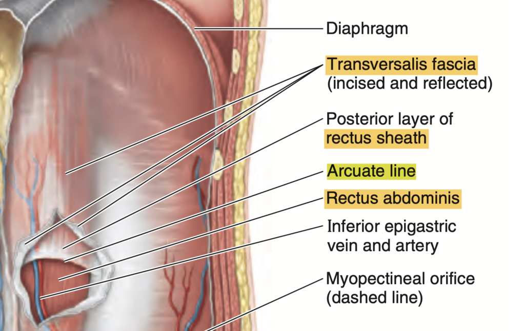
What line demarcates the transition between the aponeurotic posterior wall of the sheath covering the rectus abdominis muscle and transversalis fascia?
a. Spinoumbilical Line
b. Arcuate Line
c. Linea Alba
d. Linea Semilunaris
Answer: b. Arcuate Line
解説: 弓状線(arcuate line)は、腹直筋鞘の後壁が腱膜から横筋筋膜に移行する位置にあります。この位置は腹直筋鞘の下部にあり、臍のすぐ下にあります。
- a. Spinoumbilical Line: これは臍と脊椎を結ぶ仮想の線です。
- c. Linea Alba: 白線は腹直筋を左右に分ける中央の線です。
- d. Linea Semilunaris: 半月線は腹直筋の外側縁に沿った線です。
Question 50
The following are true regarding the inguinal region, EXCEPT:
a. Extends between the ASIS and pubic tubercle
b. All are correct
c. Pathways of entrance and exit into the inguinal canal are potential sites of herniation
d. Relocation of testes from perineum accounts for structural defects
Answer: d. Relocation of testes from perineum accounts for structural defects
解説: 精巣の移動は鼠径管を通じて腹腔から陰嚢へ行われますが、これが会陰から移動するわけではありません。精巣の移動は構造的欠陥を引き起こすことがありますが、腹腔からの移動が原因です。
アセス(29th Oct Dr. Lastimoso)
Question 1(29th Oct)
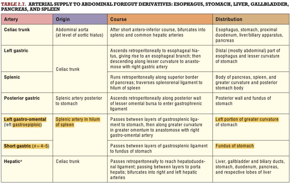
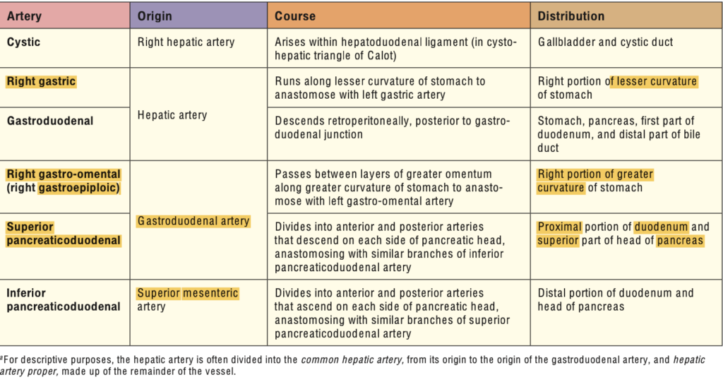
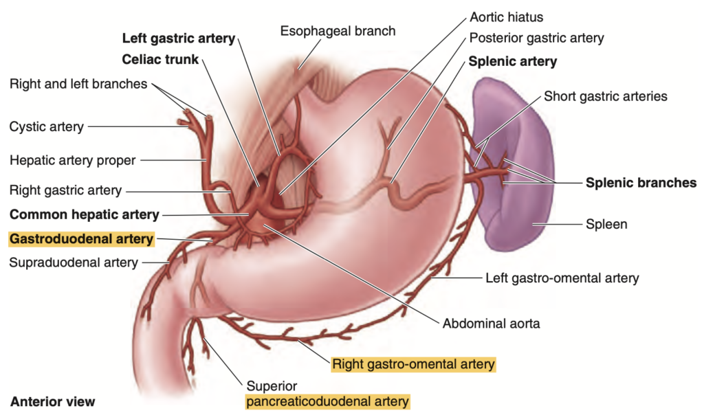
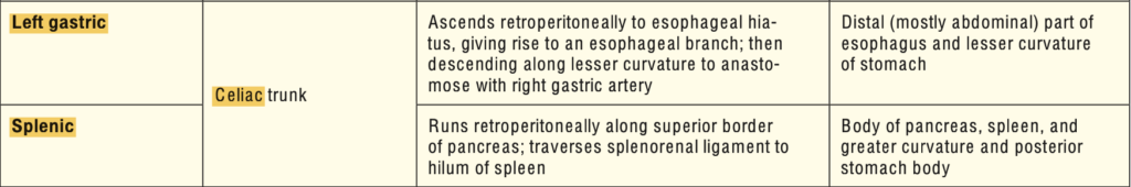
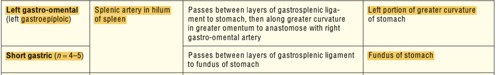
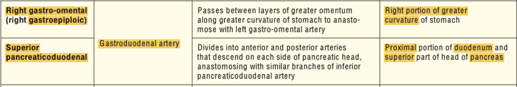
What artery supplies the fundus and upper body of the stomach?
a. Left Gastro-Omental Artery
b. Short Gastric Artery
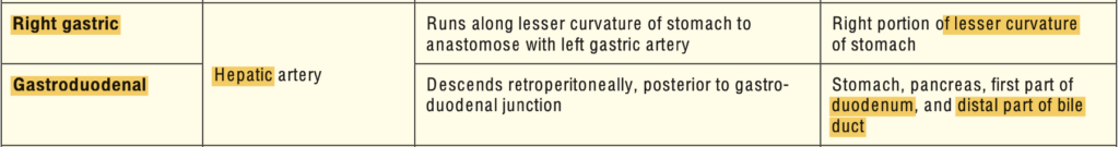
c. Right Gastric Artery
d. Right Gastro-Omental Artery
Answer: b. Short Gastric Artery
解説: 短胃動脈(short gastric artery)は脾動脈から分岐し、胃の上部と底部(fundus)に血液を供給します。他の選択肢は胃の他の部位に血液を供給します。
- a. Left Gastro-Omental Artery: 胃の大弯に沿って血液を供給します。
- c. Right Gastric Artery: 胃の小弯に沿って血液を供給します。
- d. Right Gastro-Omental Artery: 胃の大弯の右側に供給します。
Question 2(5th Nov ブロック)
What do you call an internal part of the cecum described as a ridge formed laterally when the folds meet?
a. Superior Ileocolic Lips
b. Frenula of the Ileocecal Valve
c. Ileal Papilla
d. Inferior Ileocolic Lips
Answer: b. Frenula of the Ileocecal Valve
解説: 回盲弁のヒダ(frenula of the ileocecal valve)は盲腸の内側に形成されるヒダであり、回盲弁の機能に関与します。
- a. Superior Ileocolic Lips: 回盲部の上部ヒダです。
- c. Ileal Papilla: 回腸に関連する小さな隆起です。
- d. Inferior Ileocolic Lips: 回盲部の下部ヒダです。
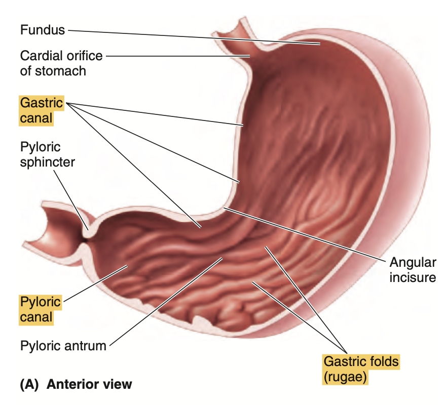
Question 3(29th Oct)
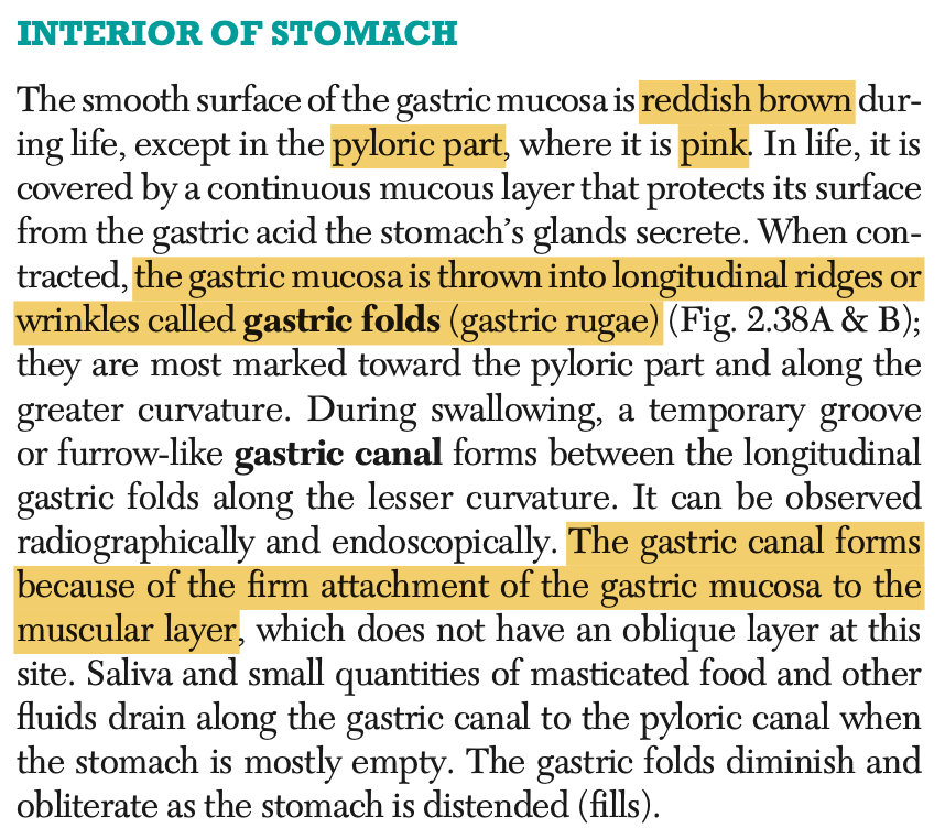
What interior part of the stomach is formed due to the firm attachment of its mucosa to the muscular layer?
a. Gastric Folds
b. Gastric Canal
c. Gastric Rugae
d. Gastric Mucosa
Answer: b
Question 4(29th Oct)
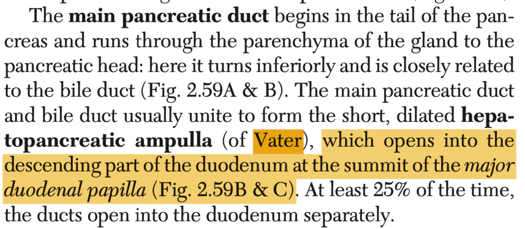
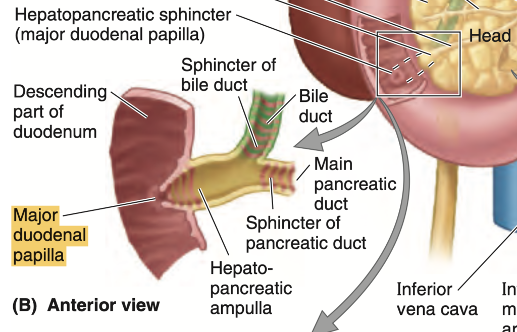
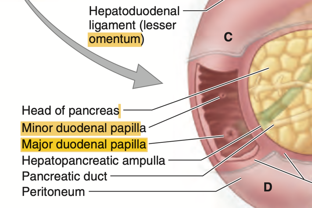
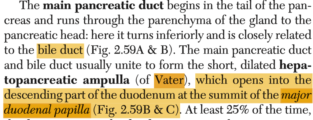
The ampulla of Vater will empty into the duodenum and mark as an eminence known as?
a. Sphincter of Oddi
b. Major Duodenal Valve
c. Minor Duodenal Papilla
d. Major Duodenal Papilla
Answer: d. Major Duodenal Papilla
解説: ファーター膨大部(ampulla of Vater)は、主胆管と膵管が合流し、十二指腸の大十二指腸乳頭(major duodenal papilla)に開口します。
- a. Sphincter of Oddi: この括約筋は胆汁と膵液の流れを調整しますが、膨大部自体ではありません。
- b. Major Duodenal Valve: 十二指腸にそのような「弁」はありません。
- c. Minor Duodenal Papilla: これは副膵管が開口する部位です。
Question 5(5th Nov ブロック)
The fan-shaped fold of peritoneum that attaches the transverse colon to the posterior abdominal wall.
a. Lesser Omentum
b. Mesentery
c. Mesocolon
d. Greater Omentum
Answer: c. Mesocolon
解説: 横行結腸間膜(transverse mesocolon)は、横行結腸を後腹壁に固定する腹膜の折りたたみです。
- a. Lesser Omentum: 小網は肝臓と胃を結びます。
- b. Mesentery: 腸間膜は小腸を支持します。
- d. Greater Omentum: 大網は胃と横行結腸の間にある大きな腹膜のヒダです。
Question 6(29th Oct)
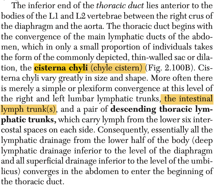
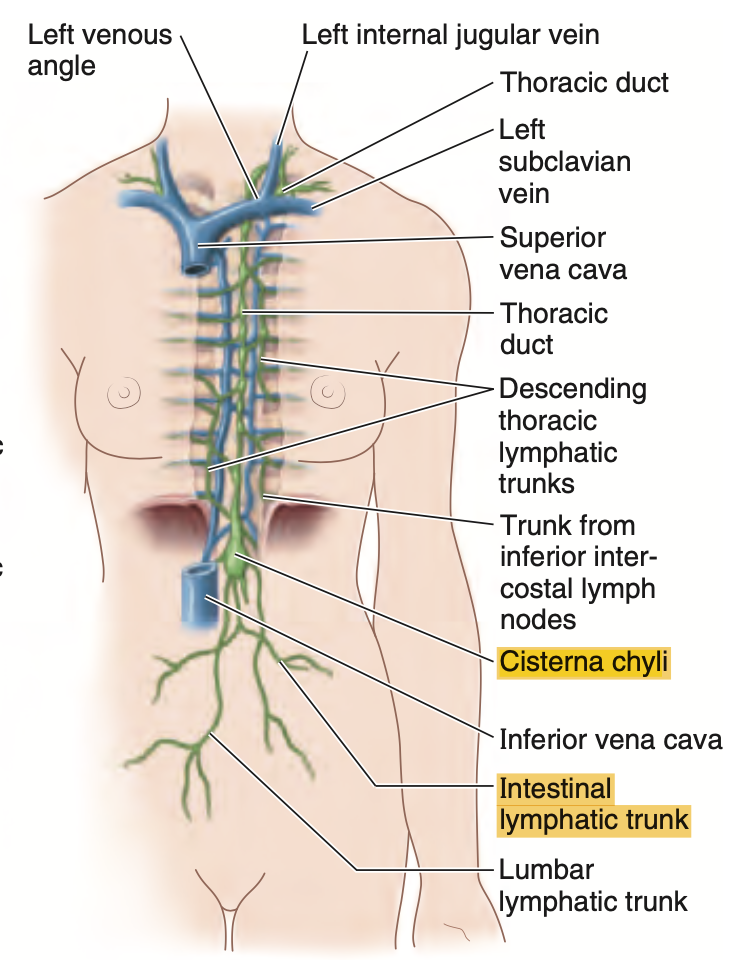
The lymphatic duct dilatation that collects the intestinal lymphatic trunk.
a. Portal Lymphatic Duct
b. Inferior Mesenteric Duct
c. Juxtaintestinal Trunk
d. Cisterna Chyle
Answer: d. Cisterna Chyle
解説: 乳び槽(cisterna chyli)は腸リンパ本幹からリンパを集め、胸管へと続く膨張部です。
- a. Portal Lymphatic Duct: このような構造は存在しません。
- b. Inferior Mesenteric Duct: 下腸間膜静脈はリンパ管ではありません。
- c. Juxtaintestinal Trunk: 腸周囲リンパ管ですが、乳び槽ではありません。
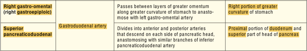
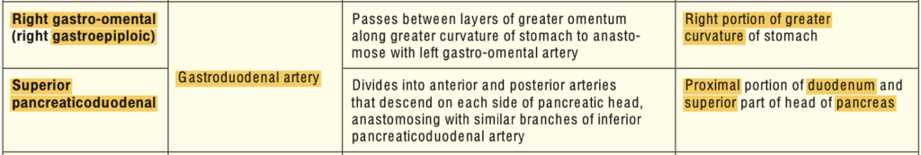
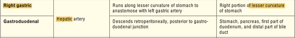
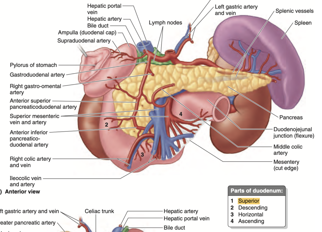
Question 7(29th Oct)
Where does the superior pancreaticoduodenal artery come from?
a. Gastroduodenal Artery
b. Celiac Trunk
c. Superior Mesenteric Artery
d. Direct Abdominal Aorta
Answer: a. Gastroduodenal Artery
解説: 上膵十二指腸動脈(superior pancreaticoduodenal artery)は胃十二指腸動脈(gastroduodenal artery)から分岐し、膵臓と十二指腸の上部を供給します。
- b. Celiac Trunk: 胃十二指腸動脈は腹腔動脈から分岐しますが、直接ではありません。
- c. Superior Mesenteric Artery: 上腸間膜動脈は下膵十二指腸動脈を供給します。
- d. Direct Abdominal Aorta: 腹大動脈は直接関与しません。
Question 8(29th Oct)
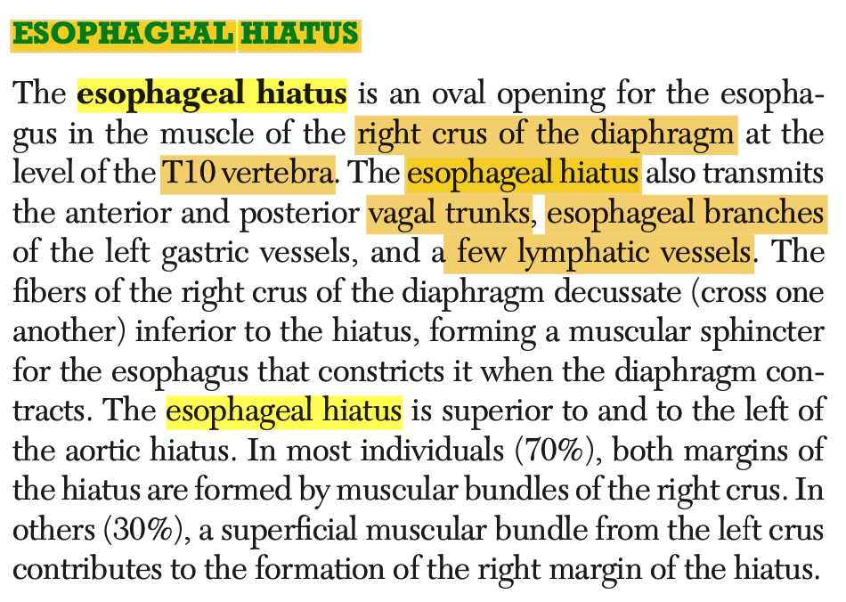
What structure follows & enters the esophageal hiatus along with the esophagus?
a. Azygos Vein
b. Trachea
c. Thoracic Duct
d. Descending Thoracic Aorta
Answer: c. Thoracic Duct
解説: 胸管(thoracic duct)は食道裂孔を通り、食道とともに横隔膜を越えます。
- a. Azygos Vein: 奇静脈は別の経路を通ります。
- b. Trachea: 気管は食道裂孔を通過しません。
- d. Descending Thoracic Aorta: 下行胸大動脈は別の孔を通ります。
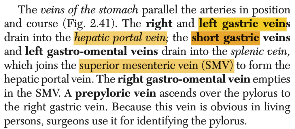
Question 9(29th Oct)
Which of the following veins of the stomach drain directly to the portal vein?
a. Right Gastric Omental Vein
b. Left Gastric Vein
c. Short Gastric Vein
d. Left Gastric Omental Vein
Answer: b. Left Gastric Vein
解説: 左胃静脈(left gastric vein)は直接門脈(portal vein)に排出されます。
- a. Right Gastric Omental Vein: これは門脈に直接排出されません。
- c. Short Gastric Vein: これも脾静脈を介して排出されます。
- d. Left Gastric Omental Vein: 左胃大網静脈は脾静脈に流入します。
Portal veinに直接流れ込む静脈
- 上腸間膜静脈(Superior Mesenteric Vein, SMV)
- 小腸、盲腸、上行結腸、横行結腸の血液を集めて運びます。
- 脾静脈(Splenic Vein)
- 脾臓、膵臓、そして胃の一部からの血液を運びます。脾静脈はまた**下腸間膜静脈(Inferior Mesenteric Vein, IMV)**からの血液を受け取ります。
- 下腸間膜静脈(Inferior Mesenteric Vein, IMV)
- 主に大腸の下部(下行結腸、S状結腸、直腸)からの血液を運びます。IMVはしばしば脾静脈に合流してから門脈に流れ込みます。
- 左胃静脈(Left Gastric Vein)
- 胃の一部(主に小弯部分)と食道の下部からの血液を運びます。
- 右胃静脈(Right Gastric Vein)
- 胃の小弯部分からの血液を運びます。
Question 10(5th Nov ブロック)
What structure is responsible for dividing the abdominal cavity into the supracolic and infracolic compartments?
a. Greater Omentum
b. Lesser Omentum
c. Transverse Mesocolon
d. Sigmoid Mesocolon
Answer: c. Transverse Mesocolon
解説: 横行結腸間膜(transverse mesocolon)は、腹腔を上腹部(supracolic)と下腹部(infracolic)に分けます。
- a. Greater Omentum: 大網は腹部を分けませんが、保護と脂肪貯蔵に関与します。
- b. Lesser Omentum: 小網は肝臓と胃を結びますが、腹腔を分けません。
- d. Sigmoid Mesocolon: S状結腸間膜は下腹部にありますが、腹腔の大きな区分には関与しません。
Question 11(29th Oct)
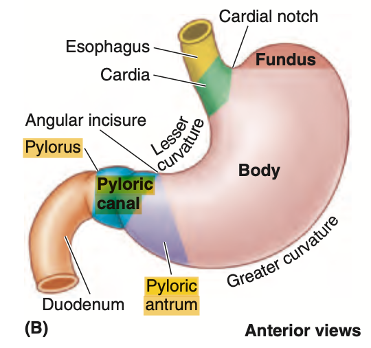
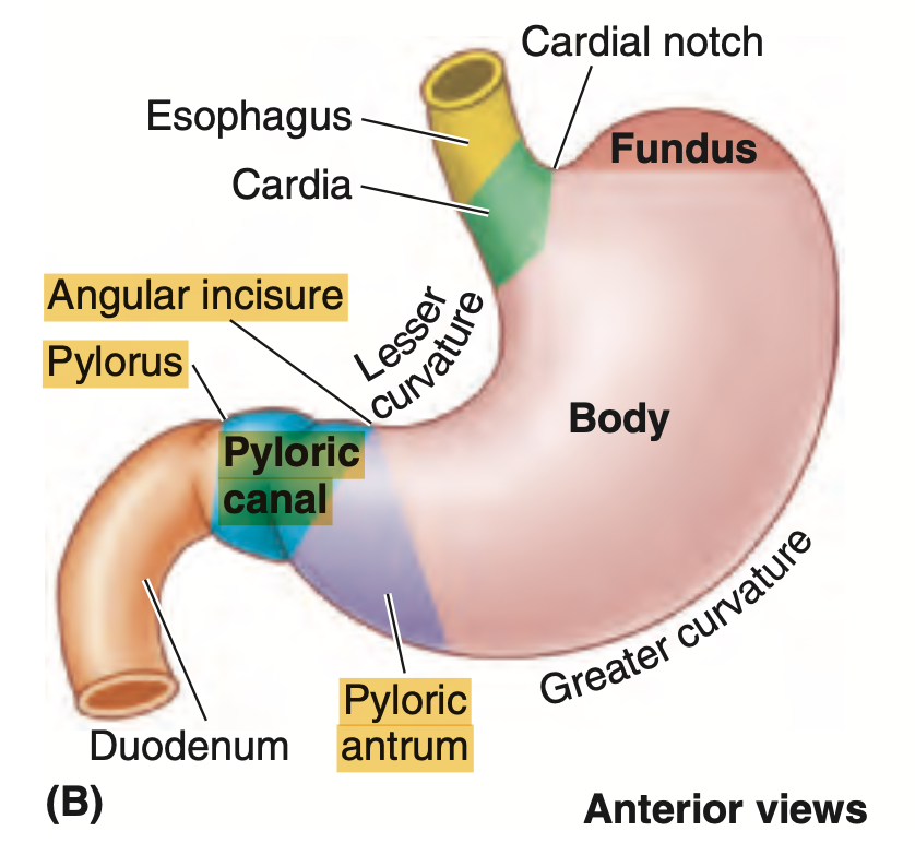
Which of the following statements about the stomach is FALSE?
a. The cardia is the superior opening of the stomach
b. The body of the stomach is between the fundus and pyloric antrum
c. The pyloric antrum is the narrowest part
d. The fundus of the stomach lies posterior to the left 6th rib MCL
Answer: c. The pyloric antrum is the narrowest part
解説: 胃の幽門部(pyloric antrum)は、胃の体部と幽門管の間に位置しますが、胃の最も狭い部分ではありません。幽門括約筋が最も狭い部分です。
- a: カーディアは食道から胃への入り口で正しい。
- b: 胃体部は、胃底と幽門部の間に正しく位置しています。
- d: 胃底は左第6肋骨後方にあります。
Question 12(29th Oct)
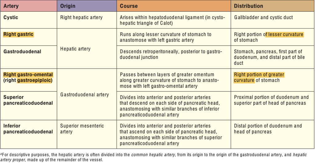
A branch of the gastroduodenal artery that supplies the greater curvature of the stomach:
a. Left Gastro-Omental Artery
b. Right Gastro-Omental Artery
c. Right Gastric Artery
d. Left Gastric Artery
Answer: b. Right Gastro-Omental Artery
解説: 右胃大網動脈(right gastro-omental artery)は胃十二指腸動脈(gastroduodenal artery)の枝であり、胃の大弯を供給します。
- a. Left Gastro-Omental Artery: これは脾動脈から分岐し、胃の大弯を供給します。
- c. Right Gastric Artery: これは胃の小弯を供給します。
- d. Left Gastric Artery: これは胃の小弯を供給します。
Question 13(29th Oct)
What branch of artery mainly supplies the duodenum proximal to the entry of the bile duct?
a. Gastroduodenal Artery
b. Superior Mesenteric Artery
c. Superior Pancreaticoduodenal Artery
d. Inferior Pancreaticoduodenal Artery
Answer: c. Superior Pancreaticoduodenal Artery
解説: 上膵十二指腸動脈(superior pancreaticoduodenal artery)は、胃十二指腸動脈の枝であり、胆管が開口する部位よりも近位の十二指腸を供給します。
- a. Gastroduodenal Artery: これは主要な動脈ですが、上膵十二指腸動脈の枝が近位部に供給します。
- b. Superior Mesenteric Artery: この動脈は主に十二指腸の遠位部を供給します。
- d. Inferior Pancreaticoduodenal Artery: これは十二指腸の遠位部に血液を供給します。
Question 14(29th Oct)
Which of the following is a branch of the celiac artery?
a. Middle Colic Artery
b. Short Gastric Artery
c. Splenic Artery
d. Left Colic Artery
Answer: c. Splenic Artery
解説: 脾動脈(splenic artery)は腹腔動脈(celiac artery)の主要な枝の一つです。
- a. Middle Colic Artery: これは上腸間膜動脈の枝です。
- b. Short Gastric Artery: これは脾動脈の枝です。
- d. Left Colic Artery: これは下腸間膜動脈の枝です。
Question 15(29th Oct)
As the esophagus descends into the abdomen, it is then attached to this part of the stomach:
a. Body
b. Fundus
c. Cardia
d. Pylorus
Answer: c. Cardia
解説: 食道は胃のカーディア部(cardia)に接続します。カーディアは食道から胃への入り口で、逆流を防ぐ役割を果たします。
- a. Body: 胃体部は中央に位置しますが、食道とは接続しません。
- b. Fundus: 胃底は食道の上にあります。
- d. Pylorus: 幽門は胃の出口です。
Question 16(29th Oct)
What is the arterial blood supply of the stomach greater curvature that arises from the splenic artery?
a. Gastroduodenal Artery
b. Short Gastric Artery
c. Left Gastroepiploic Artery
d. Right Gastric Artery
Answer: c. Left Gastroepiploic Artery
解説: 左胃大網動脈(left gastroepiploic artery)は脾動脈から分岐し、胃の大弯に血液を供給します。
- a. Gastroduodenal Artery: これは十二指腸と一部の胃に血液を供給しますが、左胃大網動脈の供給ではありません。
- b. Short Gastric Artery: これは胃の上部に供給しますが、大弯には供給しません。
- d. Right Gastric Artery: これは胃の小弯を供給します。
Question 17(29th Oct)
The right gastro-omental artery originates from?
a. Gastroduodenal Artery
b. Celiac Artery
c. Splenic Artery
d. Common Hepatic Artery
Answer: a. Gastroduodenal Artery
解説: 右胃大網動脈(right gastro-omental artery)は胃十二指腸動脈(gastroduodenal artery)から分岐し、胃の大弯の右側に血液を供給します。
- b. Celiac Artery: これは主要な供給動脈ですが、直接関与しません。
- c. Splenic Artery: これは左胃大網動脈を供給します。
- d. Common Hepatic Artery: これは肝臓と胃の一部に血液を供給しますが、右胃大網動脈の起源ではありません。
Question 18(5th Nov ブロック)
The superior mesenteric artery lies at the level of?
a. T12 vertebra
b. L3 vertebra
c. L2 vertebra
d. L1 vertebra
Answer: d. L1 vertebra
解説: 上腸間膜動脈(superior mesenteric artery)はL1椎骨の高さで腹大動脈から分岐します。
- a. T12 vertebra: これは腹腔動脈の分岐点です。
- b. L3 vertebra: これは下腸間膜動脈の分岐点です。
- c. L2 vertebra: この高さには大きな動脈分岐はありません。
Question 19(5th Nov ブロック)
Which of the following organs is located retroperitoneally(後腹膜)?
a. Sigmoid Colon
b. Transverse Colon
c. Jejunum
d. Ileum
Answer: b. Transverse Colon
解説: 横行結腸(transverse colon)は後腹膜に位置する器官の一つです。
- a. Sigmoid Colon: S状結腸は腹膜内に位置します。
- c. Jejunum: 空腸も腹膜内にあります。
- d. Ileum: 回腸も腹膜内にあります。
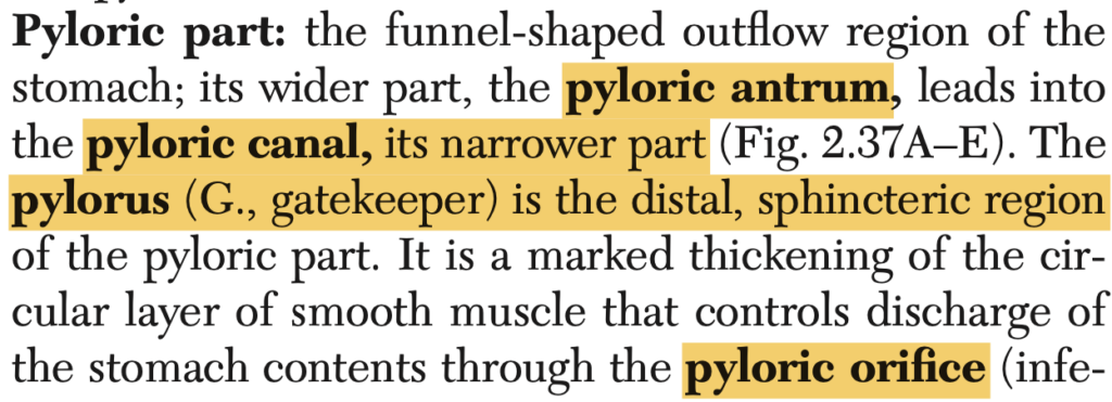
Question 20(29th Oct)
What part of the stomach has a marked thickening of its smooth circular layer muscle that controls discharge of contents?
a. Cardia
b. Fundus
c. Body
d. Pylorus
Answer: d. Pylorus
解説: 幽門部(pylorus)は平滑筋が厚くなっており、胃内容物の十二指腸への排出を制御する役割を果たします。
- a. Cardia: これは胃の入り口であり、主に逆流を防ぎます。
- b. Fundus: 胃底は内容物の貯蔵に関与します。
- c. Body: 胃体部は消化と混合の役割を果たしますが、排出を制御しません。
Question 21(5th Nov ブロック)
The inferior mesenteric artery lies at the level of?
a. L3 vertebra
b. L2 vertebra
c. L1 vertebra
d. L4 vertebra
Answer: a. L3 vertebra
解説: 下腸間膜動脈(inferior mesenteric artery)はL3椎骨の高さで腹大動脈から分岐し、大腸の下部を供給します。
- b. L2 vertebra: このレベルでは下腸間膜動脈は分岐しません。
- c. L1 vertebra: これは上腸間膜動脈の高さです。
- d. L4 vertebra: 腹大動脈の分岐点はL4にあります。
Question 22(29th Oct)
The narrowest part of the stomach:
a. Cardia
b. Gastric Canal
c. Pyloric Canal
d. Fundus
Answer: c. Pyloric Canal
解説: 幽門管(pyloric canal)は胃の最も狭い部分であり、胃の内容物が十二指腸に排出される際に制御されます。
- a. Cardia: これは胃の入り口であり、最も狭い部分ではありません。
- b. Gastric Canal: これは胃の内容物が通過するルートですが、最も狭い部分ではありません。
- d. Fundus: 胃底は胃の上部にあり、最も広い部分です。
Question 23(5th Nov ブロック)
The sympathetic fibers toward the jejunum and ileum come out at what level of the thoracic segments of the spinal cord?
a. T5-T8
b. T6-T9
c. T7-T10
d. T8-T10
Answer: d. T8-T10
解説: 空腸と回腸への交感神経線維はT8〜T10の胸髄セグメントから起こり、腸の運動と血流を調整します。
- a. T5-T8: これは上部消化管に関与する神経です。
- b. T6-T9: これも空腸と回腸には適切ではありません。
- c. T7-T10: これは近いですが、T8-T10が正しい範囲です。
Question 24(29th Oct)
A medical student was allegedly stabbed on his left upper anterior chest by an unknown assailant. During surgery, the esophagus and its related anterior structures were injured. Which structure anterior to the esophagus is most likely injured together with the esophagus?
a. Azygos Vein
b. Thoracic Duct
c. Descending Thoracic Aorta
d. Trachea
Answer: d. Trachea
解説: 気管(trachea)は食道の前に位置し、食道が損傷された場合、同時に損傷される可能性が高いです。
- a. Azygos Vein: 奇静脈は食道の後方にあります。
- b. Thoracic Duct: 胸管も食道の後方にあります。
- c. Descending Thoracic Aorta: 胸大動脈は食道の後ろに位置します。
Question 25(29th Oct)
An elderly patient came with a malignant tumor at the middle third of the esophagus. The cancer cells will likely metastasize to:
a. Deep Cervical Lymph Nodes
b. Tracheobronchial Lymph Nodes
c. Celiac Lymph Nodes
d. Paratracheal Lymph Nodes
Answer: b. Tracheobronchial Lymph Nodes
解説: 食道の中部に位置する腫瘍は、気管支周囲のリンパ節(tracheobronchial lymph nodes)に転移する可能性が高いです。
- a. Deep Cervical Lymph Nodes: 頚部リンパ節は食道の上部の腫瘍に関連します。
- c. Celiac Lymph Nodes: これらは下部食道に関連します。
- d. Paratracheal Lymph Nodes: これらは気管に近いですが、中部食道腫瘍の最も一般的な転移先ではありません。
Question 26(29th Oct)
Supplies the duodenum proximal to the entry of the bile duct in the descending portion of the duodenum:
a. Superior Mesenteric Artery
b. Right Gastro-Omental Artery
c. Gastroduodenal Artery
d. Inferior Pancreaticoduodenal Artery
Answer: c. Gastroduodenal Artery
解説: 胃十二指腸動脈(gastroduodenal artery)は、胆管が十二指腸に入る前の部分を供給します。
- a. Superior Mesenteric Artery: これは主に十二指腸の遠位部を供給します。
- b. Right Gastro-Omental Artery: これは胃の大弯を供給します。
- d. Inferior Pancreaticoduodenal Artery: これは十二指腸の下部を供給します。
Question 27(5th Nov ブロック)
Where does the superior mesenteric vein end before it unites with the splenic vein?
a. Horizontal Part of Duodenum
b. Posterior Neck of Pancreas
c. Pyloric Part of Stomach
d. Proximal Part of Jejunum
Answer: b. Posterior Neck of Pancreas
解説: 上腸間膜静脈(superior mesenteric vein)は膵臓の後部頸部で脾静脈と合流し、門脈を形成します。
- a. Horizontal Part of Duodenum: これは近くにありますが、合流点ではありません。
- c. Pyloric Part of Stomach: 胃の幽門部は関与しません。
- d. Proximal Part of Jejunum: 空腸は関与しません。
Question 28(29th Oct)
What part of the duodenum is closely related to the body of the pancreas superiorly?
a. Descending Duodenum
b. Superior Duodenum
c. Inferior Duodenum
d. Ascending Duodenum
Answer: c
Question 29(5th Nov ブロック)
The blind intestinal diverticulum arising from the posteromedial aspect of the cecum:
a. Hepatic Flexure
b. Appendix
c. Ileocecal Valve
d. Splenic Flexure
Answer: b. Appendix
解説: 虫垂(appendix)は盲腸の後内側から発生する腸の袋状の突出部です。
- a. Hepatic Flexure: これは大腸の右側の屈曲部です。
- c. Ileocecal Valve: これは回盲弁であり、虫垂ではありません。
- d. Splenic Flexure: これは大腸の左側の屈曲部です。
Question 30(5th Nov ブロック)
A space behind the stomach, and between the posterior surface of the stomach and pancreas:
a. Omental Bursa
b. Greater Sac
c. Foramen of Winslow
d. Gastrosplenic Ligament
Answer: a. Omental Bursa
解説: 大網嚢(omental bursa)は胃の後ろにある空間で、胃と膵臓の間に位置します。
- b. Greater Sac: これは腹腔の主要な部分ですが、特定の空間ではありません。
- c. Foramen of Winslow: これは大網嚢への入口ですが、空間そのものではありません。
- d. Gastrosplenic Ligament: これは胃と脾臓を結ぶ靭帯であり、空間ではありません。
Question 31(29th Oct)
The venous drainage of the lower third of the esophagus eventually drains to the:
a. Azygos Vein
b. Inferior Vena Cava
c. Portal Vein
d. Superior Vena Cava
Answer: c. Portal Vein
解説: 食道の下部1/3の静脈は、門脈(portal vein)に排出され、食道静脈瘤の原因となることがあります。
- a. Azygos Vein: 奇静脈は食道の中部や上部の静脈を排出します。
- b. Inferior Vena Cava: 下大静脈は直接的には関与しません。
- d. Superior Vena Cava: 上大静脈も食道の下部には関与しません。
Question 32(29th Oct)
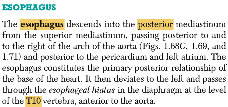
The esophagus pierces the diaphragm and enters the abdominal cavity at the level of:
a. T8
b. T10
c. T12
d. T6
Answer: b. T10
解説: 食道は横隔膜の食道裂孔をT10レベルで通過し、腹腔に入ります。
- a. T8: T8レベルは下大静脈が通過するレベルです。
- c. T12: これは大動脈裂孔のレベルです。
- d. T6: T6レベルでは横隔膜に到達しません。
Question 33(29th Oct)
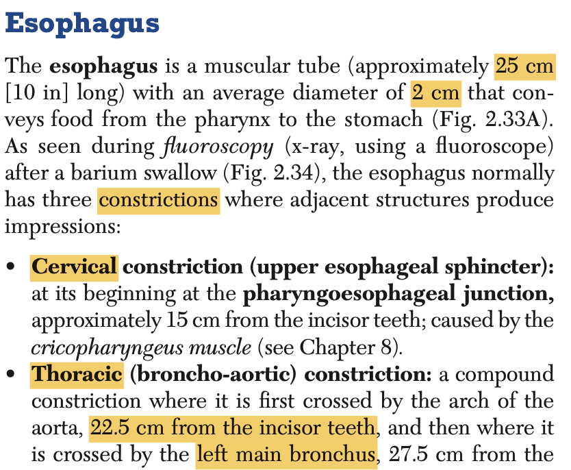
Which part of the esophagus is 22.5 cm from the incisors and is crossed by the left main bronchus?
a. Intra-Abdominal Part
b. Diaphragmatic
c. Thoracic
d. Cervical
Answer: c. Thoracic
解説: 食道の胸部(thoracic esophagus)は切歯から約22.5cmの位置にあり、左主気管支が横切ります。
- a. Intra-Abdominal Part: これは腹腔内の部分です。
- b. Diaphragmatic: これは横隔膜を通過する部分です。
- d. Cervical: 頚部は食道の上部に位置します。
Question 34(5th Nov ブロック)
The 3rd part of the small intestine will terminate at which of the following structures?
a. Ileocecal Junction
b. Ligament of Treitz
c. Hepatic Flexure
d. Middle Part of the Transverse Colon
Answer: a. Ileocecal Junction
解説: 小腸の第3部(回腸)は回盲弁(ileocecal junction)で終わります。
- b. Ligament of Treitz: これは十二指腸の分界を示しますが、小腸の第3部とは関係ありません。
- c. Hepatic Flexure: これは結腸の屈曲部分です。
- d. Middle Part of the Transverse Colon: 結腸の中部であり、回腸とは関係ありません。
Question 35(29th Oct)
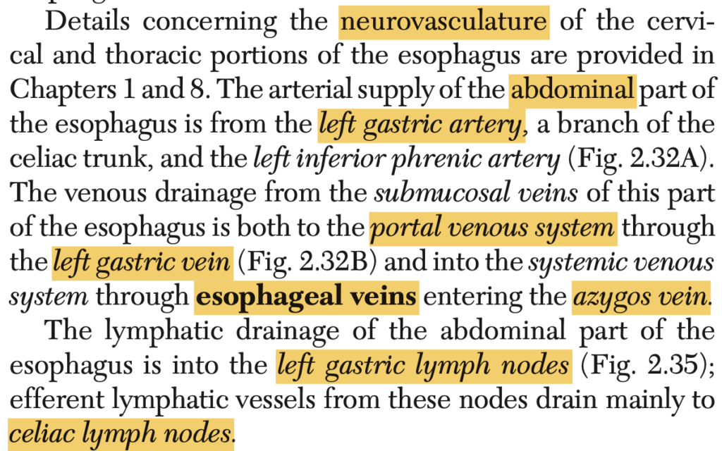
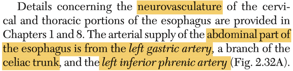
What is the main arterial blood supply of the abdominal part of the esophagus?
a. Posterior Intercostal Arteries
b. Left Gastric Artery
c. Right Inferior Phrenic Artery
d. Musculophrenic Artery
Answer: b. Left Gastric Artery
解説: 腹部の食道の主要な動脈供給は、左胃動脈(left gastric artery)から行われます。
- a. Posterior Intercostal Arteries: これらは主に胸部に供給します。
- c. Right Inferior Phrenic Artery: 横隔膜に供給されます。
- d. Musculophrenic Artery: これも横隔膜と一部の腹壁に供給されます。

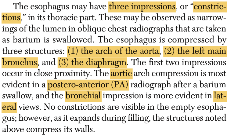
Question 36(29th Oct)
The thoracic esophageal constriction is caused by:
a. Contractions of Cricopharyngeus
b. Esophageal Hiatus
c. External Compression by the Azygos Vein
d. Crossing of Left Main Bronchus
Answer: d. Crossing of Left Main Bronchus
解説: 胸部食道の狭窄部位は、左主気管支が横切る部分で形成されます。
- a. Contractions of Cricopharyngeus: これは咽頭食道の狭窄を引き起こします。
- b. Esophageal Hiatus: これは横隔膜の狭窄部です。
- c. External Compression by the Azygos Vein: 奇静脈は食道の圧迫には関与しません。
Question 37(5th Nov ブロック)
Which of the following characteristics describes the jejunum?
a. Long vasa recta and few large loops of arcades
b. Pale pink in color
c. Many lymphoid nodules
d. Short vasa recta and many short loops of arcades
Answer: a. Long vasa recta and few large loops of arcades
解説: 空腸(jejunum)は長い血管直線(vasa recta)と少ない大きなアーケードを持っています。
- b. Pale pink in color: 空腸は赤みが強いです。
- c. Many lymphoid nodules: これは回腸の特徴です。
- d. Short vasa recta and many short loops of arcades: これも回腸の特徴です。
Question 38(29th Oct)
The celiac artery lies at the level of:
a. T12 vertebra
b. L2 vertebra
c. L3 vertebra
d. T11 vertebra
Answer: a. T12 vertebra
解説: 腹腔動脈(celiac artery)はT12椎骨の高さで腹大動脈から分岐します。
- b. L2 vertebra: これは腎動脈の分岐点です。
- c. L3 vertebra: これは下腸間膜動脈の分岐点です。
- d. T11 vertebra: これは少し高すぎます。
Question 39(29th Oct)
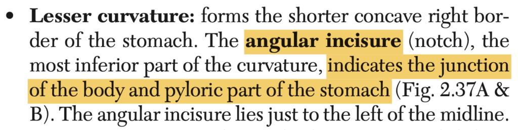
Which anatomical location does the angular incisure or notch represent?
a. Junction between the body & pylorus
b. Junction between the cardia & fundus
c. Distal end of pylorus
d. Superior border of the stomach
Answer: a. Junction between the body & pylorus
解説: 角切痕(angular incisure)は胃体部と幽門部の境界を示します。
- b. Junction between the Cardia & Fundus: これは違う部位です。
- c. Distal End of Pylorus: 幽門の遠位部ではありません。
- d. Superior Border of the Stomach: 角切痕は胃の下部に位置します。
Question 40(29th Oct)
Which of the following branches of the vagus nerve gives rise to the posterior gastric branch?
a. Celiac Branch
b. Splenic Branch
c. Duodenal Branch
d. Hepatic Branch
Answer: a. Celiac Branch
解説: 迷走神経の腹腔枝(celiac branch)は、後胃枝(posterior gastric branch)を派生させ、胃の後面に血液を供給します。
- b. Splenic Branch: これは脾臓に関連しています。
- c. Duodenal Branch: これは十二指腸に関連しています。
- d. Hepatic Branch: これは肝臓に関連しています。
Question 41(5th Nov ブロック)
Which of the following is NOT true about the jejunum?
a. Short vasa recta
b. Large and tall circular folds
c. Thick and heavy wall
d. Less Peyer’s patches
Answer: a. Short vasa recta
解説: 空腸(jejunum)は長い血管直線(vasa recta)を持ち、壁が厚く、大きな輪状ヒダが特徴的です。また、回腸に比べてペイヤー斑(Peyer’s patches)が少ないです。
- b. Large and tall circular folds: これは空腸の特徴です。
- c. Thick and heavy wall: 空腸の壁は厚いです。
- d. Less Peyer’s patches: 空腸には回腸ほどペイヤー斑が多くありません。
Question 42(5th Nov ブロック)
What do you call a structure described as a thickened smooth muscle that represents most of the longitudinal layer of the large bowel?
a. Omental Appendices
b. Haustra
c. Flexure
d. Teniae Coli
Answer: d. Teniae Coli
解説: 結腸帯(teniae coli)は大腸の縦走筋層が厚くなったもので、大腸全体に沿って走行します。
- a. Omental Appendices: これは脂肪がついた腹膜の突起です。
- b. Haustra: これは結腸に見られる袋状の構造です。
- c. Flexure: これは結腸の屈曲部分を指します。
Question 43(5th Nov ブロック)
Which of the following statements is true when one stimulates the parasympathetic nerve fibers of the jejunum and ileum?
a. Decrease Motility and Secretion
b. Reduce or Stop Absorption and Digestion
c. Increase Motility and Secretion
d. Constrict the Blood Supply to the Intestines
Answer: c. Increase Motility and Secretion
解説: 副交感神経の刺激は、空腸と回腸の運動および分泌を増加させ、消化活動を促進します。
- a. Decrease Motility and Secretion: これは交感神経の作用です。
- b. Reduce or Stop Absorption and Digestion: 副交感神経は消化を促進します。
- d. Constrict the Blood Supply to the Intestines: 副交感神経は血流を増加させます。
Question 44(29th Oct)
The major duodenal papilla is located at which part of the duodenum?
a. Superior Part
b. Ascending Part
c. Inferior Part
d. Descending Part
Answer: d. Descending Part
解説: 大十二指腸乳頭(major duodenal papilla)は十二指腸の下降部(descending part)に位置し、胆管と膵管が開口します。
- a. Superior Part: ここには乳頭はありません。
- b. Ascending Part: これは十二指腸の最終部分で、乳頭はありません。
- c. Inferior Part: これは水平部であり、乳頭はありません。
Question 45(29th Oct)
The foregut terminates at which of the following structures and the midgut will start?
a. Descending part of the duodenum
b. Hepatoduodenal ligament
c. Splenic Flexure
d. Hepatic Flexure
Answer: a. Descending part of the duodenum
解説: 前腸(foregut)は十二指腸の下降部で終わり、ここから中腸(midgut)が始まります。
- b. Hepatoduodenal Ligament: これは肝臓と十二指腸を結びますが、前腸と中腸の境界ではありません。
- c. Splenic Flexure: これは結腸の屈曲部です。
- d. Hepatic Flexure: これは右結腸屈曲であり、腸の境界とは関係ありません。
Question 46(5th Nov ブロック)
These are the complex arterial anastomoses in the mesentery before they finally end up in the small intestine.
a. Vasa Recta
b. Arterial Arcade of Drummond
c. Marginal Arteries
d. Superior Mesenteric Artery Anastomosis
Answer: b. Arterial Arcade of Drummond
解説: ドラモンド動脈アーケード(arterial arcade of Drummond)は小腸の供給のために動脈がメセンテリー内で複雑に連結する構造です。
- a. Vasa Recta: これは小腸に直接供給する血管です。
- c. Marginal Arteries: これは結腸の周囲に走る動脈です。
- d. Superior Mesenteric Artery Anastomosis: 上腸間膜動脈の具体的なアナストモーシスの名称ではありません。
Question 47(5th Nov ブロック)
During surgery, the splenic artery was transected at its origin. Which of the following vessels is LESS LIKELY affected?
a. Left Gastro-Omental Artery
b. Posterior Gastric Artery
c. Left Gastric Artery
d. Short Gastric Artery
Answer: b
Question 48(29th Oct)
A paired artery that supplies the esophagus:
a. Superior Thyroid Artery
b. Bronchial Artery
c. Left Gastric Artery
d. Esophageal Artery
Answer: c

Question 49(29th Oct)
Esophageal varices(静脈瘤) develop with portal hypertension because of increased pressure against this vein that drains the distal part of the esophagus:
a. Inferior Thyroid Vein
b. Internal Thoracic Vein
c. Azygos Vein
d. Left Gastric Vein
Answer: d
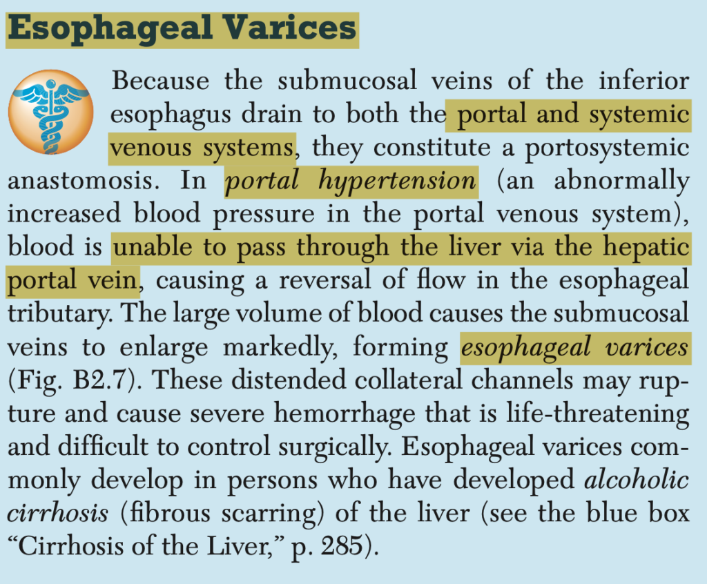
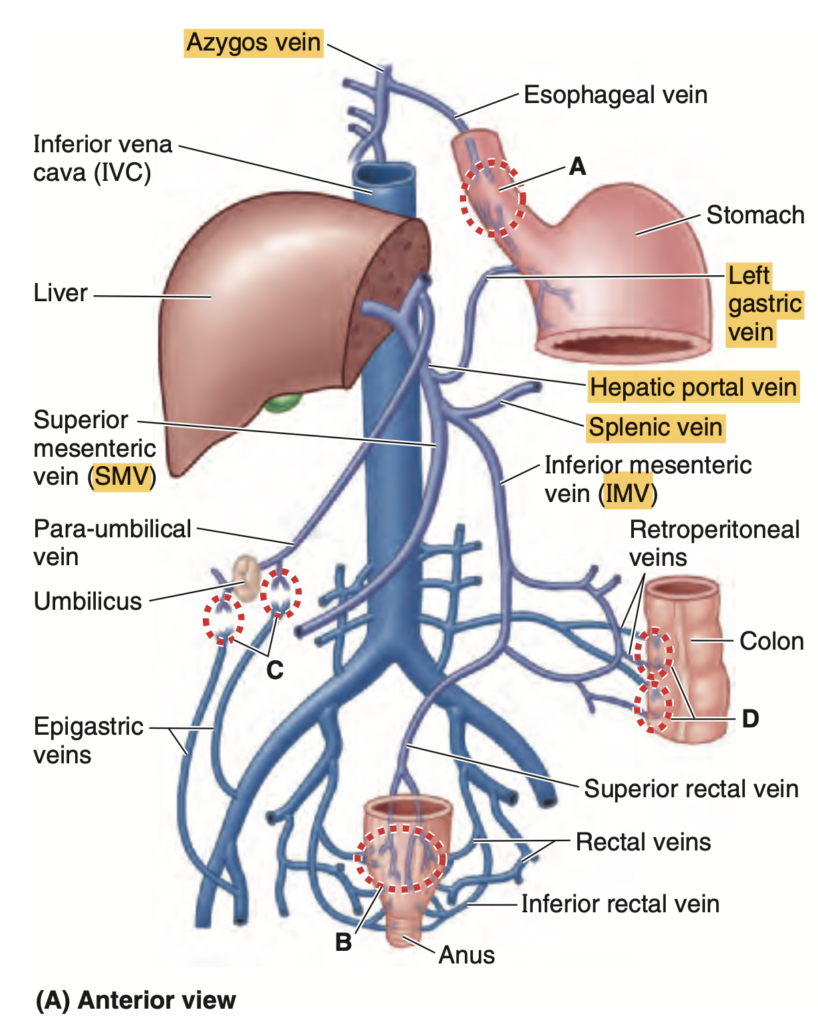
**食道静脈瘤(Esophageal varices)**は、門脈圧亢進症(Portal hypertension)が原因で発生します。これは、食道の下部を排出する静脈に対して圧力が上昇することで起こります。この静脈は、血液が門脈系に流入する経路の一部であり、門脈圧亢進が原因で静脈瘤が形成される可能性があります。
答え: 左胃静脈(Left gastric vein)
左胃静脈(Left gastric vein)(または冠状静脈)は、食道の下部(遠位部)を排出し、門脈系に流れ込む静脈です。門脈圧亢進症では、門脈系にかかる圧力が増加するため、この静脈内の圧力も高まり、静脈が拡張し、食道静脈瘤が形成されることがあります。
解説
門脈圧亢進症は、肝硬変などにより門脈系の血流が妨げられ、門脈にかかる圧力が上昇する状態です。この圧力の上昇が、門脈と全身循環の間にある側副路(collateral circulation)を拡大させ、その結果、**左胃静脈(Left gastric vein)**やその分枝である食道静脈に血液が逆流し、静脈瘤が形成されます。
Question 50(29th Oct)
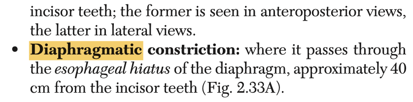
The diaphragmatic esophageal constriction is located at this vertebral level:
a. T8
b. L1
c. T12
d. T10
Answer: d. T10
解説: 食道の横隔膜狭窄はT10レベルで発生します。この部位で食道は横隔膜を貫通します。
- a. T8: これは下大静脈が横隔膜を通過するレベルです。
- b. L1: これは関与しません。
- c. T12: これは大動脈裂孔のレベルです。
ブロック(5th Nov)
Question 1
A male cyclist was brought to the emergency room after he was hit by a speeding car. Based on your examination, you considered a possible splenic injury. Which of the following findings will likely indicate possible injury of the spleen?
a. Fracture on the 8th to 11th ribs on right chest
b. Ultrasound noted fluid at the splenorenal fossa
c. Right shoulder pain
d. Pain on the right hypochondriac area
Answer: b. Ultrasound noted fluid at the splenorenal fossa
解説: 脾臓損傷の兆候として、腹部超音波検査で脾腎窩(splenorenal fossa)に液体が見つかることは重要な所見です。これは、脾臓からの出血を示唆します。
- a. Fracture on the 8th to 11th ribs on right chest: 右側肋骨の骨折は肝臓の損傷を示唆する可能性があり、脾臓の損傷には関連しません。
- c. Right shoulder pain: 右肩の痛みは横隔神経刺激による関連痛で、肝臓や胆嚢の損傷に関連することが多いです。
- d. Pain on the right hypochondriac area: 右季肋部の痛みは通常、肝臓や胆嚢に関連します。
Question 2
In your attempt to remove the spleen, this ligament should be dissected and clamped for it is where the splenic artery & vein traverse towards the splenic hilum. Which of the following structures should you clamp?
a. Splenorenal ligament
b. Gastrosplenic ligament
c. Hepatoduodenal ligament
d. Gastrocolic ligament
Answer: a. Splenorenal ligament
解説: 脾腎靭帯(splenorenal ligament)には脾動脈と脾静脈が通過しており、脾臓の門に達します。脾臓の除去手術では、この靭帯をクランプする必要があります。
- b. Gastrosplenic ligament: 胃脾靭帯は脾臓と胃を結びますが、脾動脈と脾静脈はここを通過しません。
- c. Hepatoduodenal ligament: 肝十二指腸靭帯は胆管、門脈、肝動脈を含み、脾臓とは関係がありません。
- d. Gastrocolic ligament: 胃結腸靭帯は胃と横行結腸を結び、脾臓の血管とは関係がありません。
Question 3
The splenic vein joins this vessel to form the portal vein:
a. Superior mesenteric vein
b. Splenic artery
c. Inferior mesenteric vein
d. Left gastroepiploic vein
Answer: a. Superior mesenteric vein
解説: 脾静脈は上腸間膜静脈(superior mesenteric vein)と合流して門脈(portal vein)を形成します。門脈は肝臓に血液を供給します。
- b. Splenic artery: 脾動脈は動脈であり、静脈との合流には関与しません。
- c. Inferior mesenteric vein: 下腸間膜静脈は脾静脈に流入しますが、門脈の形成には直接関与しません。
- d. Left gastroepiploic vein: 左胃大網静脈は脾静脈に流入しますが、門脈形成には直接関与しません。
Question 4
Partial or subtotal splenectomy may be performed instead of total splenectomy. Which of the following statements supports this?
a. Most patients have an accessory spleen which can replace the resected segment.
b. Splenic artery divides into 5 or more branches inside with avascular plane in between.
c. The spleen can regenerate and become whole again after surgery.
d. Aside from the splenic artery, it is supplied by other branches of celiac trunk.
Answer: b. Splenic artery divides into 5 or more branches inside with avascular plane in between.
解説: 脾動脈は脾臓内で5つ以上の枝に分かれ、それらの間には無血管域が存在します。このため、部分的な脾臓摘出が可能です。
- a. Most patients have an accessory spleen: 副脾がある場合もありますが、脾臓全体を補完することはできません。
- c. The spleen can regenerate: 脾臓が完全に再生することはありません。
- d. Supplied by other branches of celiac trunk: 脾臓は主に脾動脈から供給されており、他の動脈からの供給は少ないです。
Question 5
During the removal of the spleen, the surgeon has opened the gastrosplenic ligament to release the spleen from its gastric attachment. Which of the following vessels will the surgeon likely encounter?
a. Splenic artery
b. Right gastric artery
c. Left gastro-omental artery
d. Left gastric artery
Answer: c. Left gastro-omental artery
解説: 胃脾靭帯には左胃大網動脈(left gastro-omental artery)と短胃動脈が含まれています。これらの血管は脾臓と胃の間を通ります。
- a. Splenic artery: 脾動脈は脾腎靭帯内を通過します。
- b. Right gastric artery: 右胃動脈は胃の小弯に供給され、脾臓には関与しません。
- d. Left gastric artery: 左胃動脈は胃の上部に供給されますが、脾臓には直接関与しません。
Question 6
Ultrasound of the upper abdomen was done which showed a dilated duct with a stone impacted in a pouch along the gallbladder neck. This portion of the gallbladder is called the:
a. Hepatic duct
b. Infundibulum
c. Fundus
d. Cystic duct
Answer: b. Infundibulum
解説: 胆嚢漏斗部(infundibulum)は胆嚢頸部に隣接し、ここに結石が嵌入することがよくあります。
- a. Hepatic duct: 肝管は肝臓から胆汁を運ぶ管であり、胆嚢とは直接関係ありません。
- c. Fundus: 胆嚢底部は胆嚢の最下部に位置し、結石が嵌入する場所ではありません。
- d. Cystic duct: 胆嚢管は胆嚢から胆汁を排出しますが、漏斗部とは異なります。
Question 7
To surgically remove the gallbladder, you need to ligate the:
a. Pancreatic duct
b. Common bile duct
c. Cystic duct
d. Hepatic duct
Answer: c. Cystic duct
解説: 胆嚢を除去するには、胆嚢管(cystic duct)を結紮する必要があります。他の選択肢は胆嚢摘出には関与しません。
- a. Pancreatic duct: 膵管は胆嚢の手術とは無関係です。
- b. Common bile duct: 総胆管は肝臓と十二指腸を結び、結紮すべき管ではありません。
- d. Hepatic duct: 肝管は肝臓から胆汁を運ぶ管であり、胆嚢摘出には関係しません。
Question 8
During surgery, you need to identify and ligate the cystic artery. You are aware that the artery can be located within the hepatocystic triangle bounded by the following structures:
a. Common hepatic duct, visceral surface of the liver, cystic duct.
b. Common bile duct, gallbladder, visceral surface of the liver.
c. Common hepatic duct, gallbladder, right hepatic artery.
d. Common bile duct, visceral surface of the liver, cystic duct.
Answer: d. Common bile duct, visceral surface of the liver, cystic duct.
解説: Calotの三角は総胆管、肝臓の内面、胆嚢管によって形成され、この三角内に胆嚢動脈が存在します。
- a. Common hepatic duct, visceral surface of the liver, cystic duct: 総肝管はCalotの三角の境界には含まれません。
- b. Common bile duct, gallbladder, visceral surface of the liver: 胆嚢自体は三角の境界には含まれません。
- c. Common hepatic duct, gallbladder, right hepatic artery: 右肝動脈はCalotの三角の境界ではありません。
Question 9
Usually, the cystic artery is a branch of:
a. Left gastric artery
b. Right hepatic artery
c. Superior mesenteric artery
d. Common hepatic artery
Answer: b. Right hepatic artery
解説: 胆嚢動脈(cystic artery)は通常、右肝動脈(right hepatic artery)の枝です。
- a. Left gastric artery: 左胃動脈は胃に供給され、胆嚢動脈とは関係ありません。
- c. Superior mesenteric artery: 上腸間膜動脈は腸に供給され、胆嚢動脈とは無関係です。
- d. Common hepatic artery: 総肝動脈は肝臓に供給されますが、胆嚢動脈は右肝動脈の枝です。
Question 10
Patient came in for abdominal pain and mass on his upper abdomen. Ultrasound was done & showed a liver mass medial to the gallbladder & its fossa. The mass is MOST LIKELY located at functional liver segment:
a. Segment IV
b. Segment V
c. Segment III
d. Segment VII
Answer: b. Segment V
解説: 機能的な肝臓の区分において、胆嚢窩の内側に位置する肝区域は第V区域です。
- a. Segment IV: 第IV区域は左葉にあり、胆嚢窩からは外れています。
- c. Segment III: 第III区域はさらに左側にあり、胆嚢窩とは関係ありません。
- d. Segment VII: 第VII区域は右葉の後方にあり、胆嚢窩には位置していません。
Question 11
The venous blood in the liver drains directly to:
a. Portal vein
b. Superior vena cava
c. Inferior mesenteric vein
d. Hepatic veins
Answer: d. Hepatic veins
解説: 肝臓内の静脈血は肝静脈(hepatic veins)を通じて直接下大静脈(inferior vena cava)に排出されます。
- a. Portal vein: 門脈は肝臓に血液を供給する静脈で、排出には関与しません。
- b. Superior vena cava: 上大静脈は肝臓からの血液排出には関与しません。
- c. Inferior mesenteric vein: 下腸間膜静脈は肝臓には関与していません。
Question 12
A liver segment that is supplied by both right & left primary branches of hepatic artery. What is this functional liver segment?
a. Segment III
b. Segment VII
c. Segment V
d. Segment I
Answer: d. Segment I
解説: 肝臓の第I区域(尾状葉)は右肝動脈と左肝動脈の両方から血液を供給されます。他の区域は通常、片方の肝動脈からのみ供給されます。
- a. Segment III: 第III区域は左肝動脈から供給されます。
- b. Segment VII: 第VII区域は右肝動脈から供給されます。
- c. Segment V: 第V区域は右肝動脈から供給されます。
Question 13
Which of the following divides the right lobe of the liver into anterior & posterior divisions?
a. Umbilical fissure
b. Right sagittal fissure
c. Transverse hepatic plane
d. Cantlie’s line
Answer: b. Right sagittal fissure
解説: 右矢状裂(right sagittal fissure)は、肝臓の右葉を前部と後部に分ける構造です。
- a. Umbilical fissure: 臍裂は左葉を分けます。
- c. Transverse hepatic plane: これは肝臓の横断面を示す仮想的な平面です。
- d. Cantlie’s line: カントリー線は右葉と左葉の境界を示します。
Question 14
Divides the liver by marking the diaphragmatic surface of the liver from the fundus of the gallbladder to the inferior vena cava:
a. Right sagittal fissure
b. Transverse hepatic plane
c. Umbilical fissure
d. Cantlie’s line
Answer: d. Cantlie’s line
解説: カントリー線(Cantlie’s line)は、胆嚢底部から下大静脈までを結び、肝臓を右葉と左葉に分けます。
- a. Right sagittal fissure: これは右葉の分割に関連します。
- b. Transverse hepatic plane: 肝臓を横に分ける仮想の平面です。
- c. Umbilical fissure: これは左葉に関連します。
Question 15
Which of the following statements is TRUE about the hepatic artery?
a. Supplies the liver nutrient-rich blood.
b. Provides blood with higher oxygen content than the portal vein.
c. Supplies the same amount of blood to the liver as the portal vein.
d. Provides most of the blood to the liver.
Answer: b
The hepatic artery is a vital blood vessel responsible for delivering oxygen-rich blood to the liver. It has the following characteristics:
- Source of Oxygenated Blood: The hepatic artery primarily supplies the liver with oxygenated blood, which is crucial for the liver’s metabolic and detoxifying functions.
- Branch of the Common Hepatic Artery: The hepatic artery typically branches from the common hepatic artery, which is a branch of the celiac trunk, originating from the abdominal aorta.
- Dual Blood Supply to the Liver: The liver has a unique dual blood supply, receiving blood from both the hepatic artery and the portal vein. The hepatic artery provides about 20-30% of the liver’s blood supply (mainly oxygenated), while the portal vein supplies the remaining 70-80%, which is nutrient-rich but oxygen-poor.
- Branches: The hepatic artery further divides into the left and right hepatic arteries, which supply blood to the left and right lobes of the liver, respectively.
- High-Pressure Vessel: Compared to the portal vein, the hepatic artery operates at a higher pressure, as it carries oxygenated blood directly from the systemic circulation.
Question 16
Which of the following borders of the spleen are notched?
a. Posterior & Inferior
b. Anterior & Superior
c. Posterior & Superior
d. Anterior & Inferior
Answer: b. Anterior & Superior
解説: 脾臓の切痕(notches)は前縁および上縁にあります。これらは脾臓の特徴的な構造です。
- a. Posterior & Inferior: 後部および下部には切痕はありません。
- c. Posterior & Superior: 上縁には切痕がありますが、後部にはありません。
- d. Anterior & Inferior: 下縁には切痕はありません。
Question 17(29th Oct)
Where does the superior pancreatico-duodenal artery come from?
a. Common Hepatic Artery
b. Gastroduodenal Artery
c. Superior Mesenteric Artery
d. Splenic Artery
Answer: b. Gastroduodenal Artery
解説: 上膵十二指腸動脈(superior pancreaticoduodenal artery)は胃十二指腸動脈(gastroduodenal artery)の枝です。
- a. Common Hepatic Artery: 総肝動脈は胃十二指腸動脈の供給源です。
- c. Superior Mesenteric Artery: 下膵十二指腸動脈は上腸間膜動脈から分岐します。
- d. Splenic Artery: 脾動脈は膵臓に供給されますが、十二指腸には供給しません。
Question 18
The spleen lies superficially in the left hypochondriac area between what particular ribs?
a. 7th & 9th
b. 8th & 10th
c. 9th & 11th
d. 10th & 12th
Answer: c. 9th & 11th
解説: 脾臓は左季肋部にあり、9〜11番肋骨の間に位置します。
- a. 7th & 9th: 脾臓の位置には少し高すぎます。
- b. 8th & 10th: これも脾臓の位置には完全には一致しません。
- d. 10th & 12th: これも脾臓の全体的な位置には合いません。
Question 19
What do you call the posterior superior extension of the subhepatic space which is a gravity-dependent part of the peritoneal cavity?
a. Hepatorenal Recess
b. Subphrenic Recess
c. Costodiaphragmatic Recess
d. Superior Recess of Omental Bursa
Answer: a. Hepatorenal Recess
解説: 肝腎陥凹(hepatorenal recess)は、重力によって液体が集まりやすい腹腔の一部です。
- b. Subphrenic Recess: これは肝臓と横隔膜の間の空間です。
- c. Costodiaphragmatic Recess: これは胸腔内の空間です。
- d. Superior Recess of Omental Bursa: これは胃の後ろの空間ですが、液体が集まる部分ではありません。
Question 20
The bare area of the liver is demarcated by the reflections of peritoneum from the diaphragm by what structure?
a. Triangular Ligament
b. Falciform Ligament
c. Groove for the Vena Cava
d. Coronary Ligament
Answer: d. Coronary Ligament
解説: 肝臓の裸区(bare area)は冠状靭帯(coronary ligament)によって横隔膜からの腹膜の反転で区切られています。
- a. Triangular Ligament: これは冠状靭帯の延長部ですが、裸区を定義するものではありません。
- b. Falciform Ligament: これは肝臓を右葉と左葉に分ける靭帯です。
- c. Groove for the Vena Cava: これは下大静脈が通る溝です
Question 21
The right and left sagittal fissures in the visceral surface of the liver are linked centrally by what structure?
a. Round Ligament of Liver
b. Porta Hepatis
c. Fissure for the Ligamentum Venosum
d. Fissure for the Ligamentum Teres
Answer: b. Porta Hepatis
解説: 右矢状裂と左矢状裂は肝門(porta hepatis)によって中央で連結されています。ここには、門脈、肝動脈、および胆管が通ります。
- a. Round Ligament of Liver: 肝円索は左矢状裂に位置しますが、中央での連結には関与しません。
- c. Fissure for the Ligamentum Venosum: 静脈索裂は左側に位置します。
- d. Fissure for the Ligamentum Teres: 肝円索裂は左矢状裂内にあります。
Question 22
What particular lobe of the liver is an elongated papillary process located?
a. Right Lobe of Liver
b. Caudate Lobe
c. Left Lobe of Liver
d. Quadrate Lobe
Answer: b. Caudate Lobe
解説: 尾状葉(caudate lobe)には細長い乳頭状突起があります。これは肝臓の後部に位置し、肝門の背後にあります。
- a. Right Lobe of Liver: 右葉には乳頭状突起はありません。
- c. Left Lobe of Liver: 左葉には乳頭状突起はありません。
- d. Quadrate Lobe: 方形葉には乳頭状突起はありません。
Question 23
In the functional or segmental terms of the liver, segment VIII is also known as what?
a. Posterior Medial Segment
b. Right Anterior Lateral Segment
c. Anterior Medial Segment
d. Posterior Lateral Segment
Answer: c
Question 24
In the functional and surgical terms of the liver, segment V is also known as what?
a. Left Lateral Anterior Segment
b. Anterior Medial Segment
c. Lateral Segment
d. Left Medial Segment
Answer: b. Anterior Medial Segment
解説: 第V区域は前内側区域(anterior medial segment)とも呼ばれ、肝臓の右葉に位置します。
- a. Left Lateral Anterior Segment: これは左葉に位置する別の区域です。
- c. Lateral Segment: これは左葉の側面部分に相当します。
- d. Left Medial Segment: これは左葉の内側部分に相当します。
Question 25
What part of the pancreas does the bile duct lie?
a. Anterior Surface of the Head of Pancreas
b. Posterior Surface of the Head of Pancreas
c. Anterior Surface of the Neck of Pancreas
d. Posterior Surface of the Neck of Pancreas
Answer: b. Posterior Surface of the Head of Pancreas
解説: 胆管(bile duct)は膵頭部の後面に沿って走行し、膵管と合流して十二指腸に開口します。
- a. Anterior Surface of the Head of Pancreas: 胆管は膵頭部の後面にあります。
- c. Anterior Surface of the Neck of Pancreas: 胆管は膵頭部に関連しています。
- d. Posterior Surface of the Neck of Pancreas: 胆管は膵頭部に位置します。
Question 26
What is the main arterial blood supply of the middle part of the bile duct?
a. Gastroduodenal Artery
b. Cystic Artery
c. Right Hepatic Artery
d. Posterior Superior Pancreaticoduodenal Artery
Answer: c
The main arterial blood supply of the middle part of the bile duct primarily comes from branches of the right hepatic artery. Additionally, other nearby arteries, including the gastroduodenal artery and the posterior superior pancreaticoduodenal artery, may provide supplementary blood flow to this part of the bile duct.
The bile duct has a segmental blood supply, with:
- The proximal (upper) part of the bile duct being primarily supplied by the cystic artery and branches of the right hepatic artery.
- The middle part (located near the porta hepatis) mainly receiving blood from branches of the right hepatic artery.
- The distal (lower) part of the bile duct being supplied by branches from the gastroduodenal artery and the posterior superior pancreaticoduodenal artery.
Question 27
Which of the following ducts is the mucosal spiral fold or valve found?
a. Bile Duct
b. Common Hepatic Duct
c. Cystic Duct
d. Pancreatic Duct
Answer: c. Cystic Duct
解説: 胆嚢管(cystic duct)には、螺旋状の粘膜ヒダ(spiral valve)があり、胆汁の流れを調整します。
- a. Bile Duct: 胆管には螺旋状のヒダはありません。
- b. Common Hepatic Duct: 総肝管にも螺旋状のヒダはありません。
- d. Pancreatic Duct: 膵管には螺旋状のヒダはありません。
Question 28(29th Oct)
In the portal-systemic anastomoses, the submucosal esophageal veins drain into either the azygous vein and what other vein?
a. Splenic Vein
b. Short Gastric Vein
c. Distal Esophageal Vein
d. Left Gastric Vein
Answer: d. Left Gastric Vein
解説: 食道の粘膜下静脈は、奇静脈(azygos vein)または左胃静脈(left gastric vein)に排出されます。左胃静脈は門脈系に接続しており、食道静脈瘤の原因となることがあります。
- a. Splenic Vein: 脾静脈は関与しません。
- b. Short Gastric Vein: これは脾静脈に排出されますが、食道静脈とは関係がありません。
- c. Distal Esophageal Vein: これは左胃静脈と関連していますが、正しい選択肢は左胃静脈です。
Moore
FIGURE 2.75. Tributaries of hepatic portal vein and portal–systemic anastomoses. A. Anastomoses provide a collateral circulation in cases of obstruc- tion in the liver or portal vein. Here, the portal tributaries are darker blue and systemic tributaries are lighter blue. A–D indicate sites of anastomoses. A is between the submucosal esophageal veins draining into either the azygos vein (systemic) or the left gastric vein (portal); when dilated these are esophageal varices. B is between the inferior and middle rectal veins draining into the inferior vena cava (systemic) and the superior rectal vein, continuing as the inferior mesenteric vein (portal). The submucosal veins involved are normally dilated (varicose in appearance), even in newborns. When the mucosa containing them prolapses, they form hemorrhoids. (The varicose appearance of the veins and the occurrence of hemorrhoids are not typically related to portal hyperten- sion, as is commonly stated.) C shows para-umbilical veins (portal) anastomosing with small epigastric veins of the anterior abdominal wall (systemic); this may produce the “caput medusae” (Fig. B2.24). D is on the posterior aspects (bare areas) of secondarily retroperitoneal viscera, or the liver, where twigs of visceral veins—for example, the colic vein, splenic veins, or the portal vein itself (portal system)—anastomose with retroperitoneal veins of the posterior abdominal wall or diaphragm (systemic system).
B. Magnetic resonance (MR) angiogram (portal venogram) demonstrating the tributaries and formation of the portal vein in a living person.
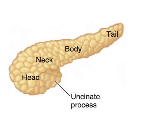
Question 29
What part of the pancreas do the superior mesenteric vessels overlie?
a. Tail
b. Body
c. Neck
d. Head
Answer: c. Neck
解説: 上腸間膜血管(superior mesenteric vessels)は膵臓の頸部(neck)を越えて走行します。
- a. Tail: 膵尾部は脾臓に近いですが、上腸間膜血管はここを通りません。
- b. Body: 膵体部は血管とは関係ありません。
- d. Head: 膵頭部は血管に関与しません。
Question 30
The majority of the pancreatic branches are derived mainly from what artery?
a. Gastroduodenal
b. Splenic
c. Superior Pancreaticoduodenal
d. Superior Mesenteric
Answer: b. Splenic
解説: 膵臓への主な血液供給は脾動脈(splenic artery)からの分枝です。これにより、膵体部と尾部に血液が供給されます。
- a. Gastroduodenal: これは膵頭部に一部供給します。
- c. Superior Pancreaticoduodenal: これは主に膵頭部を供給します。
- d. Superior Mesenteric: これは膵臓の一部に供給されますが、主な供給源ではありません。
Question 31
These are the complex arterial anastomosis in the mesentery before it finally ends up in the small intestine.
a. Superior mesenteric artery anastomosis
b. Marginal arteries
c. Vasa recta
d. Arterial arcade of Drummond
Answer: d. Arterial arcade of Drummond
解説: ドラモンドの動脈アーケード(arterial arcade of Drummond)は、小腸に血液を供給する複雑な動脈吻合です。
- a. Superior mesenteric artery anastomosis: これは具体的な吻合ではなく、上腸間膜動脈からの供給を指します。
- b. Marginal arteries: これらは大腸の周囲に存在します。
- c. Vasa recta: これらは動脈の終末枝であり、小腸に直接供給されますが、複雑な吻合を示すものではありません。
Question 32(29th Oct)
The foregut terminates at which of the following structures and the midgut will start?
a. Hepatoduodenal ligament
b. Splenic flexure
c. Descending part of the duodenum
d. Hepatic flexure
Answer: c. Descending part of the duodenum
解説: 前腸(foregut)は十二指腸の下降部で終わり、そこから中腸(midgut)が始まります。
- a. Hepatoduodenal ligament: これは肝臓と十二指腸を結ぶ靭帯であり、前腸と中腸の境界とは関係ありません。
- b. Splenic flexure: これは大腸の一部で、中腸とは異なります。
- d. Hepatic flexure: これは結腸の右屈曲部で、中腸の構造ではありません。
Question 33
The fan-shaped fold of peritoneum that attaches the transverse colon to the posterior abdominal wall.
a. Lesser omentum
b. Mesocolon
c. Mesentery
d. Greater omentum
Answer: c
Question 34
The 3rd part of the small intestine will terminate at which of the following structures?
a. Middle part of the transverse colon
b. Ileocecal junction
c. Hepatic flexure
d. Ligament of Treitz
Answer: b. Ileocecal junction
解説: 小腸の第3部(回腸)は回盲弁(ileocecal junction)で終わります。
- a. Middle part of the transverse colon: これは結腸の一部です。
- c. Hepatic flexure: これは大腸の屈曲部分です。
- d. Ligament of Treitz: これは十二指腸の分界を示しますが、回腸の終端ではありません。
Question 35(29th Oct)
Which part of the duodenum that exhibits a mesentery?
a. Ascending part
b. Duodenal cap
c. Horizontal part
d. Descending part
Answer: b
The first part of the duodenum, specifically the duodenal bulb (also known as the ampulla, duodenum cap), is the only part of the duodenum that exhibits a mesentery. This segment is intraperitoneal, meaning it is covered by peritoneum and has a mesenteric attachment, allowing it a degree of mobility.
The remaining parts of the duodenum (second, third, and fourth parts) are primarily retroperitoneal (located behind the peritoneum) and thus lack a true mesentery. They are relatively fixed in position due to their retroperitoneal location.
Question 36(29th Oct)
The ampulla of Vater will empty into the duodenum and mark as an eminence known as?
a. Major duodenal valve
b. Sphincter of Oddi
c. Major duodenal papilla
d. Minor duodenal papilla
Answer: c. Major duodenal papilla
解説: ファーター膨大部(ampulla of Vater)は、主胆管と膵管が合流し、十二指腸の大十二指腸乳頭(major duodenal papilla)に開口します。
- a. Major duodenal valve: 十二指腸にそのような「弁」はありません。
- b. Sphincter of Oddi: これは括約筋であり、開口部自体ではありません。
- d. Minor duodenal papilla: これは副膵管が開口する部位です。
Question 37
Which of the following is NOT true to the jejunum?
a. Large and tall circular folds
b. Thick and heavy wall
c. Less Peyer’s patches
d. Short vasa recta
Answer: d. Short vasa recta
解説: 空腸(jejunum)は長い血管直線(vasa recta)を持ち、壁が厚く、大きな輪状ヒダが特徴です。
- a. Large and tall circular folds: これは空腸の特徴です。
- b. Thick and heavy wall: 空腸の壁は厚いです。
- c. Less Peyer’s patches: 空腸には回腸ほどペイヤー斑が多くありません。
Question 38
The lymphatic duct dilatation that collects the intestinal lymphatic trunk.
a. Inferior mesenteric duct
b. Juxtaintestinal trunk
c. Portal lymphatic duct
d. Cisterna chyle
Answer: d. Cisterna chyle
解説: 乳び槽(cisterna chyli)は腸リンパ本幹からリンパを集め、胸管へと続く膨張部です。
- a. Inferior mesenteric duct: このような構造は存在しません。
- b. Juxtaintestinal trunk: 腸周囲リンパ管ですが、乳び槽ではありません。
- c. Portal lymphatic duct: 門脈にはリンパ管はありません。
Question 39
The blind intestinal diverticulum arising from the posteromedial aspect of the cecum.
a. Hepatic flexure
b. Splenic flexure
c. Appendix
d. Ileocecal valve
Answer: c. Appendix
解説: 虫垂(appendix)は盲腸の後内側から発生する腸の袋状の突出部です。
- a. Hepatic flexure: これは大腸の右側の屈曲部です。
- b. Splenic flexure: これは大腸の左側の屈曲部です。
- d. Ileocecal valve: これは回盲弁であり、虫垂ではありません。
Question 40
Which of the following exhibits a mesentery?
a. Transverse part of duodenum
b. Ascending colon
c. Appendix
d. Descending colon
Answer: c. Appendix
解説: 虫垂(appendix)は腸間膜を持ち、自由に動かせます。他の部位は固定されています。
- a. Transverse part of duodenum: 十二指腸の横行部は後腹壁に固定されています。
- b. Ascending colon: 上行結腸も固定されています。
- d. Descending colon: 下行結腸も固定されています。
Question 41
This is called the deep membranous layer of the subcutaneous tissue in the abdomen.
a. Colle’s fascia
b. Camper’s fascia
c. Scamper’s fascia
d. Scarpa’s fascia
Answer: d. Scarpa’s fascia
解説: スカルパ筋膜(Scarpa’s fascia)は腹部の皮下組織の深層膜様層です。これは皮膚の下に位置し、キャンパー筋膜(Camper’s fascia)の下にあります。
- a. Colle’s fascia: これは会陰部にある深層筋膜です。
- b. Camper’s fascia: これは腹部の表層脂肪層です。
- c. Scamper’s fascia: 実際には存在しない構造です。
Question 42
What structure extends from the xiphoid process to the symphysis pubis?
a. External oblique M.
b. Linea alba
c. Tendinous intersection
d. Linea semilunaris
Answer: b. Linea alba
解説: 白線(linea alba)は、剣状突起から恥骨結合まで伸びる線維性の構造で、腹筋の中央に位置します。
- a. External oblique M.: 外腹斜筋は腹部側面の筋肉です。
- c. Tendinous intersection: 腱画は腹直筋に見られる水平の腱線です。
- d. Linea semilunaris: これは腹直筋の外縁に沿った線です。
Question 43
What is the largest and most superficial of the three abdominal muscles?
a. Internal oblique M.
b. External oblique M.
c. Transversus abdominis M.
d. Rectus abdominis M.
Answer: b. External oblique M.
解説: 外腹斜筋(external oblique M.)は、3つの腹筋の中で最も大きく、最も表層に位置する筋肉です。
- a. Internal oblique M.: 内腹斜筋は外腹斜筋の下に位置します。
- c. Transversus abdominis M.: 腹横筋はさらに深層にあります。
- d. Rectus abdominis M.: 腹直筋は中央に位置し、表層ではありません。
Question 44
The internal oblique originates from the ___________?
a. Symphysis pubis
b. 12th rib
c. Anterior iliac crest
d. Xiphoid process
Answer: c. Anterior iliac crest
解説: 内腹斜筋(internal oblique M.)は、腸骨稜の前部(anterior iliac crest)から起始します。
- a. Symphysis pubis: これは恥骨結合であり、筋肉の起始点ではありません。
- b. 12th rib: 12番目の肋骨は他の筋肉の起始点です。
- d. Xiphoid process: これは胸部の構造であり、筋肉の起始点ではありません。
Question 45
What muscle functions to stabilize and control tilt of the pelvis?
a. Internal oblique
b. External oblique
c. Rectus abdominis muscle
d. Transversus abdominis
Answer: c. Rectus abdominis muscle
解説: 腹直筋(rectus abdominis)は骨盤の安定化と傾斜の制御に関与します。特に、骨盤前傾を防ぎます。
- a. Internal oblique: これは回旋と側屈に関与しますが、骨盤の安定化には主要な役割を果たしません。
- b. External oblique: 回旋や側屈に関与します。
- d. Transversus abdominis: 腹圧の増加に寄与しますが、骨盤の傾斜には直接関与しません。
Question 46
The thoraco-abdominal nerves are derived from anterior rami of?
a. T1-6
b. T1-12
c. T12
d. T7-11
Answer: d. T7-11
解説: 胸腹神経(thoraco-abdominal nerves)はT7からT11の前枝から派生し、腹壁に分布しています。
- a. T1-6: これらの神経は主に上部胸部を供給します。
- b. T1-12: T12は胸腹神経の一部ではありません。
- c. T12: これは肋間神経として知られています。
Question 47
This artery that supplies the anterior abdomen originates from the external iliac artery.
a. Superficial circumflex A.
b. Inferior epigastric A.
c. Musculophrenic A.
d. Superior epigastric A.
Answer: d
The artery that supplies the anterior abdominal wall and originates from the external iliac artery is the inferior epigastric artery. This artery branches off from the external iliac artery just above the inguinal ligament and travels upward along the posterior surface of the rectus abdominis muscle.
The inferior epigastric artery supplies blood to the lower part of the anterior abdominal wall and anastomoses (connects) with the superior epigastric artery, which is a branch of the internal thoracic artery. This anastomosis provides collateral circulation to the abdominal wall.
Question 48
It is located between the ASIS and pubic tubercle.
a. Lateral inguinal fossa
b. Inguinal region
c. Medial inguinal fossa
d. Supravesicular fossa
Answer: b. Inguinal region
解説: 鼠径部(inguinal region)は上前腸骨棘(ASIS)と恥骨結節の間に位置します。
- a. Lateral inguinal fossa: これは鼠径部内にありますが、特定の位置ではありません。
- c. Medial inguinal fossa: これも鼠径部内に位置しますが、範囲が異なります。
- d. Supravesicular fossa: これは膀胱の上にあります。
Question 49
It is located between medial and lateral umbilical fold and is also known as the Hesselbach’s triangle.
a. Medial inguinal fossa
b. Inguinal region
c. Lateral inguinal fossa
d. Supravesicular fossa
Answer: a. Medial inguinal fossa
解説: 内側鼠径窩(medial inguinal fossa)は、内外の臍ヒダの間にあり、ヘッセルバッハの三角(Hesselbach’s triangle)としても知られています。
- b. Inguinal region: これは一般的な領域であり、特定の窩ではありません。
- c. Lateral inguinal fossa: これは外側にあります。
- d. Supravesicular fossa: これは膀胱の上にあります。
Question 50
This structure is a double fold of peritoneum that extends from the stomach to the abdominal cavity.
a. Scarpa fascia
b. Omentum
c. Camper fascia
d. Transversalis fascia
Answer: b. Omentum
解説: 大網(omentum)は腹膜の二重層であり、胃から腹腔に伸びています。
- a. Scarpa fascia: これは筋膜であり、腹膜ではありません。
- c. Camper fascia: これは皮下脂肪層です。
- d. Transversalis fascia: これは腹横筋の筋膜です。
Dr Latismoso対策(ソリッド)
脾臓
Question 1
Question: Which of the following statements best describes the primary function of the spleen in prenatal life?
a. Acts as a blood reservoir
b. Serves as a hematopoietic organ
c. Drains lymphatic vessels
d. Produces digestive enzymes
Answer: b. Serves as a hematopoietic organ
Explanation:
脾臓は胎児期には造血器官として重要な役割を果たしています。この時期、脾臓は血液細胞(特に赤血球)を生成し、胎児の発展に必要な血液供給をサポートします。選択肢aの「血液貯蔵庫」としての役割は、主に成人期の脾臓の機能です。選択肢cの「リンパ管の排出」は、脾臓の主な役割ではなく、リンパ節の機能に関連しています。選択肢dの「消化酵素の生成」は膵臓の役割であり、脾臓とは無関係です。
Question 2
Question: The spleen is primarily connected to which two structures through its ligaments?
a. Stomach and pancreas
b. Diaphragm and ribs
c. Stomach and left kidney
d. Pancreas and left kidney
Answer: c. Stomach and left kidney
Explanation:
脾臓は胃と左腎臓にそれぞれ「胃脾靭帯(gastrosplenic ligament)」と「脾腎靭帯(splenorenal ligament)」によって接続されています。これらの靭帯は脾臓の動静脈を含んでおり、脾臓の栄養供給と血液循環に重要な役割を果たします。選択肢aの「胃と膵臓」、選択肢bの「横隔膜と肋骨」、選択肢dの「膵臓と左腎臓」はいずれも脾臓の靭帯と直接的な接続はありません。
Question 3
Question: Which major artery supplies blood to the spleen?
a. Superior mesenteric artery
b. Inferior mesenteric artery
c. Celiac trunk
d. Renal artery
Answer: c. Celiac trunk
Explanation:
脾臓は腹腔動脈(celiac trunk)から分岐する「脾動脈(splenic artery)」によって血液供給を受けます。脾動脈は腹腔動脈の最大の枝であり、膵臓の上縁に沿って走行します。選択肢aの「上腸間膜動脈(superior mesenteric artery)」と選択肢bの「下腸間膜動脈(inferior mesenteric artery)」はそれぞれ腸に血液を供給し、選択肢dの「腎動脈(renal artery)」は腎臓に血液を供給します。脾臓には関与しません。
Question 4
Question: The splenic vein joins which vessel to form the hepatic portal vein?
a. Renal vein
b. Inferior mesenteric vein
c. Superior mesenteric vein
d. Celiac trunk
Answer: c. Superior mesenteric vein
Explanation:
脾静脈(splenic vein)は、上腸間膜静脈(superior mesenteric vein)と合流して門脈(hepatic portal vein)を形成します。この門脈は肝臓に血液を運ぶ重要な経路です。選択肢bの「下腸間膜静脈(inferior mesenteric vein)」は脾静脈に合流しますが、門脈形成には関与しません。選択肢aの「腎静脈(renal vein)」および選択肢dの「腹腔動脈(celiac trunk)」は脾静脈とは無関係であり、門脈形成にも関与しません。
Question 5
Question: Which characteristic best describes the structural nature of the spleen?
a. A dense and robust organ
b. A soft and pulpy mass
c. A fibrous and elastic tissue
d. A solid, muscle-like tissue
Answer: b. A soft and pulpy mass
Explanation:
脾臓は非常に柔らかく、壊れやすい構造を持つ臓器であり、「柔らかい血管(sinusoidal)」組織で構成されています。このため、腹部外傷においては脾臓が損傷を受けやすく、緊急手術が必要になることがあります。選択肢aの「密で頑丈な臓器」や選択肢cの「線維性で弾力性のある組織」、選択肢dの「筋肉のような組織」は脾臓の特徴には合致しません。
Question 6
Question: Which structure is located directly posterior to the spleen?
a. Left kidney
b. Stomach
c. Left colic flexure
d. Pancreas
Answer: a. Left kidney
Explanation:
脾臓の直後(posterior)には左腎が位置しています。これにより、脾臓の位置関係は隣接する臓器の配置を考慮して理解することが重要です。選択肢bの「胃」は脾臓の前方(anterior)に位置し、選択肢cの「左結腸曲」は脾臓の下部(inferior)、選択肢dの「膵臓」は脾動脈と脾静脈が接触する位置であるものの、脾臓の後方ではありません。
Question 7
Question: The spleen receives its vasomotor nerve supply from which plexus?
a. Superior mesenteric plexus
b. Celiac plexus
c. Renal plexus
d. Inferior mesenteric plexus
Answer: b. Celiac plexus
Explanation:
脾臓の神経供給は主に腹腔神経叢(celiac plexus)から供給されます。この神経叢は脾動脈に沿って分布し、血管運動を制御する役割を果たします。選択肢aの「上腸間膜神経叢」や選択肢dの「下腸間膜神経叢」は主に腸に分布し、選択肢cの「腎神経叢」は腎臓の支配に関わりますが、脾臓の神経供給には関与していません。
Question 8
Question: The spleen is located adjacent to which ribs?
a. Ribs 6-8
b. Ribs 7-9
c. Ribs 9-11
d. Ribs 10-12
Answer: c. Ribs 9-11
Explanation:
脾臓は左側の肋骨9-11の範囲に位置しており、これらの肋骨に囲まれるように配置されています。この位置により、外部からの衝撃や圧力に敏感で、外傷を受けやすい臓器です。選択肢a、b、dの肋骨範囲はいずれも脾臓の位置とは一致していません。
膵臓
Question 1
Question: The pancreas is classified as which type of gland?
a. Purely exocrine gland
b. Purely endocrine gland
c. Accessory digestive gland
d. Primary lymphatic gland
Answer: c. Accessory digestive gland
Explanation:
膵臓は「副消化腺(accessory digestive gland)」とされ、消化液を分泌する外分泌機能(膵液)と、血糖値の調節に関わる内分泌機能(インスリンやグルカゴンの分泌)を兼ね備えています。選択肢a「純粋な外分泌腺」や選択肢b「純粋な内分泌腺」は膵臓の複合機能を反映していないため不正解です。また、選択肢d「一次リンパ腺」は膵臓の役割と無関係です。
Question 2
Question: Which of the following statements correctly describes the position of the pancreatic head?
a. Located anterior to the left kidney
b. Embraced by the C-shaped curve of the duodenum
c. Posterior to the pylorus of the stomach
d. Lies anterior to the aorta and L2 vertebra
Answer: b. Embraced by the C-shaped curve of the duodenum
Explanation:
膵頭は十二指腸のC字型の湾曲部分に囲まれており、これは解剖学的な膵臓の位置関係の重要な特徴です。選択肢a「左腎の前に位置する」は膵尾に関する記述であり、選択肢c「胃の幽門の後ろ」は膵頚に関連しています。選択肢d「大動脈およびL2椎体の前」は膵体についての説明です。
Question 3
Question: Which duct combines with the bile duct to form the hepatopancreatic ampulla?
a. Accessory pancreatic duct
b. Main pancreatic duct
c. Cystic duct
d. Common hepatic duct
Answer: b. Main pancreatic duct
Explanation:
主膵管(main pancreatic duct)は胆管と合流して、ファーター膨大部(hepatopancreatic ampulla)を形成し、十二指腸の大乳頭に開口します。選択肢aの「副膵管(accessory pancreatic duct)」は通常、十二指腸の小乳頭に開口します。選択肢c「胆嚢管(cystic duct)」と選択肢d「総肝管(common hepatic duct)」は胆汁系の一部であり、膵管と直接的な接続はありません。
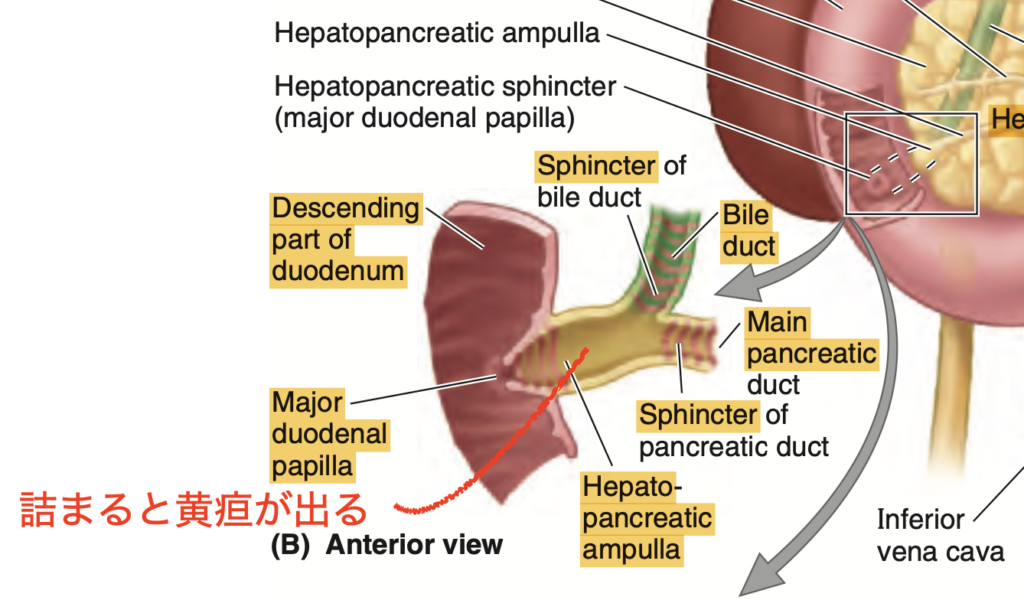
Question 4
Question: Which artery is primarily responsible for the blood supply to the head of the pancreas?
a. Splenic artery
b. Superior mesenteric artery
c. Anterior and posterior superior pancreaticoduodenal arteries
d. Inferior mesenteric artery
Answer: c. Anterior and posterior superior pancreaticoduodenal arteries
Explanation:
膵頭は主に「前上膵十二指腸動脈(anterior superior pancreaticoduodenal artery)」と「後上膵十二指腸動脈(posterior superior pancreaticoduodenal artery)」から血液供給を受けます。これらの動脈は胃十二指腸動脈から分岐します。選択肢a「脾動脈(splenic artery)」は膵体と尾に血液を供給し、選択肢b「上腸間膜動脈(superior mesenteric artery)」は膵頭に接しているものの、直接的な血液供給源ではありません。選択肢dの「下腸間膜動脈(inferior mesenteric artery)」は膵臓に血液を供給しません。
Question 5
Question: Which nerve plexuses provide innervation to the pancreas?
a. Renal plexus and inferior mesenteric plexus
b. Celiac plexus and superior mesenteric plexus
c. Pyloric plexus and hepatic plexus
d. Lumbar plexus and sacral plexus
Answer: b. Celiac plexus and superior mesenteric plexus
Explanation:
膵臓の神経支配は、腹腔神経叢(celiac plexus)および上腸間膜神経叢(superior mesenteric plexus)を介して行われます。これらの神経叢を通じて、膵臓に副交感神経および交感神経線維が供給され、分泌や血管の調節に寄与しています。選択肢a「腎神経叢(renal plexus)と下腸間膜神経叢(inferior mesenteric plexus)」、選択肢c「幽門神経叢(pyloric plexus)と肝神経叢(hepatic plexus)」、選択肢d「腰神経叢(lumbar plexus)と仙骨神経叢(sacral plexus)」はいずれも膵臓に直接関係しません。
Question 6
Question: The tail of the pancreas is closely associated with which of the following structures?
a. Right kidney
b. Left kidney
c. Inferior vena cava
d. Stomach
Answer: b. Left kidney
Explanation:
膵尾は左腎の前に位置しており、脾門や左結腸曲とも密接に関係しています。このため、膵尾の損傷や病変がある場合、これらの周辺臓器にも影響が及ぶ可能性があります。選択肢aの「右腎」は膵頭の後方に位置し、選択肢cの「下大静脈(IVC)」も膵頭の後方にあります。選択肢d「胃」は膵体や膵頭と隣接していますが、膵尾とは直接的な関連がありません。
Question 7
Question: The uncinate process of the pancreas is located in which part of the pancreas?
a. Head
b. Neck
c. Body
d. Tail
Answer: a. Head
Explanation:
膵臓の鈎状突起(uncinate process)は膵頭の一部であり、上腸間膜動脈(SMA)の後方に位置します。この構造は膵頭の下部から左側へ向かって突き出しています。選択肢b「膵頚」、選択肢c「膵体」、選択肢d「膵尾」には鈎状突起は存在しません。
Question 8
Question: Which vertebral levels does the pancreas cross?
a. T12 and L1
b. L1 and L2
c. L3 and L4
d. L2 and L3
Answer: b. L1 and L2
Explanation:
膵臓は主にL1とL2の椎骨レベルに位置しています。この解剖学的位置は、膵臓が胃の後ろで十二指腸から脾臓の間に位置するという特徴を反映しています。選択肢a「T12とL1」および選択肢c「L3とL4」は膵臓の一般的な位置とは異なります。選択肢d「L2とL3」も誤りです。
Question 9
Question: Where does the accessory pancreatic duct typically open into the duodenum?
a. Major duodenal papilla
b. Minor duodenal papilla
c. Pylorus
d. Jejunum
Answer: b. Minor duodenal papilla
Explanation:
副膵管(accessory pancreatic duct)は通常、十二指腸の小乳頭(minor duodenal papilla)に開口します。これは膵液の一部がここを通じて排出されるための経路です。選択肢a「大乳頭(major duodenal papilla)」は主膵管が開口する場所であり、選択肢c「幽門(pylorus)」および選択肢d「空腸(jejunum)」には膵管の開口はありません。
Question 10
Question: The main pancreatic duct unites with which duct to form the hepatopancreatic ampulla?
a. Cystic duct
b. Common bile duct
c. Accessory pancreatic duct
d. Hepatic duct
Answer: b. Common bile duct
Explanation:
主膵管(main pancreatic duct)は総胆管(common bile duct)と合流して、ファーター膨大部(hepatopancreatic ampulla)を形成し、十二指腸に開口します。この膨大部は消化酵素の分泌経路を制御する重要な部位です。選択肢a「胆嚢管(cystic duct)」、選択肢c「副膵管(accessory pancreatic duct)」、選択肢d「肝管(hepatic duct)」は膨大部の形成には直接関与しません。
Question 11
Question: Which structure is located posterior to the head of the pancreas?
a. Superior mesenteric artery
b. Inferior vena cava
c. Left renal artery
d. Splenic vein
Answer: b. Inferior vena cava
Explanation:
膵頭の後方には下大静脈(IVC)が位置しており、これに加えて右腎動脈と静脈、および左腎静脈も膵頭の背後に存在します。選択肢aの「上腸間膜動脈(SMA)」は膵頭の右側に位置し、選択肢cの「左腎動脈」と選択肢dの「脾静脈」は膵頭の背後にはありません。
Question 12
Question: Which sphincter controls the flow of bile into the duodenum?
a. Sphincter of the pancreatic duct
b. Choledochal sphincter
c. Sphincter of Oddi (hepatopancreatic sphincter)
d. Pyloric sphincter
Answer: b. Choledochal sphincter
Explanation:
総胆管括約筋(choledochal sphincter)は胆汁の十二指腸への流入を制御します。選択肢cの「オッディ括約筋(hepatopancreatic sphincter)」はファーター膨大部の周囲に位置しており、膵液と胆汁の流れを調整しますが、胆汁の流入自体は総胆管括約筋が制御します。選択肢aの「膵管括約筋」は膵液の流れを制御し、選択肢dの「幽門括約筋」は胃から十二指腸への食物移動を管理します。
肝臓
Question 1
Question: The liver primarily occupies which regions of the abdomen?
a. Right lumbar and left lumbar regions
b. Epigastrium and right hypochondrium
c. Left hypochondrium and right iliac
d. Umbilical and epigastric
Answer: b. Epigastrium and right hypochondrium
Explanation:
肝臓は主に右季肋部(right hypochondrium)と心窩部(epigastrium)に位置し、左季肋部(left hypochondrium)にわずかに広がっています。選択肢aの「右腰部と左腰部」、選択肢cの「左季肋部と右腸骨部」、選択肢dの「臍部と心窩部」は肝臓の典型的な位置には当てはまりません。
Question 2
Question: Which part of the liver is not covered by visceral peritoneum?
a. Quadrate lobe
b. Bare area
c. Caudate lobe
d. Porta hepatis
Answer: b. Bare area
Explanation:
肝臓の「裸出部(bare area)」は、腹膜が覆っていない唯一の部分で、横隔膜と直接接触しています。選択肢a「方形葉(quadrate lobe)」、選択肢c「尾状葉(caudate lobe)」、選択肢d「門脈域(porta hepatis)」はすべて腹膜で覆われています。
Question 3
Question: Which ligament separates the right and left anatomical lobes of the liver?
a. Coronary ligament
b. Falciform ligament
c. Round ligament
d. Ligamentum venosum
Answer: b. Falciform ligament
Explanation:
鎌状靭帯(falciform ligament)は肝臓の右葉と左葉を解剖学的に分ける構造です。選択肢aの「冠状靭帯(coronary ligament)」は肝臓と横隔膜を連結し、選択肢cの「円索(round ligament)」および選択肢dの「静脈索(ligamentum venosum)」は胎児循環の名残ですが、葉の分割には関与しません。
Question 4
Question: Which structure is a fibrous remnant of the umbilical vein?
a. Falciform ligament
b. Ligamentum venosum
c. Coronary ligament
d. Round ligament
Answer: d. Round ligament
Explanation:
円索(round ligament)は胎児期に酸素化血液を胎盤から肝臓へ運ぶ臍静脈の遺残です。選択肢bの「静脈索(ligamentum venosum)」は、胎児期に静脈管として臍静脈からIVCへの血流を導いた遺残です。選択肢a「鎌状靭帯」や選択肢c「冠状靭帯」は胎児循環には関係ありません。
Question 5
Question: The porta hepatis serves as a transverse fissure for which structures?
a. Hepatic artery, portal vein, bile duct
b. Cystic duct, hepatic veins, portal vein
c. Hepatic artery, hepatic veins, round ligament
d. Bile duct, inferior vena cava, ligamentum venosum
Answer: a. Hepatic artery, portal vein, bile duct
Explanation:
門脈域(porta hepatis)は、肝動脈(hepatic artery)、門脈(portal vein)、胆管(bile duct)が通過する横断裂であり、これらは門脈三つ組(portal triad)と呼ばれます。選択肢b、c、dの構造は、門脈域を通過する主な要素には含まれません。
Question 6
Question: Which lobe of the liver is located next to the inferior vena cava?
a. Right lobe
b. Left lobe
c. Quadrate lobe
d. Caudate lobe
Answer: d. Caudate lobe
Explanation:
尾状葉(caudate lobe)は下大静脈(IVC)の隣に位置し、この位置関係が肝臓の外科的処置や病変診断において重要です。選択肢a「右葉」や選択肢c「方形葉」はIVCの隣にはありません。選択肢b「左葉」もIVCから離れた位置にあります。
Question 7
Question: Which organ’s nutrients, except for fats, are directly transported to the liver?
a. Pancreas
b. Stomach
c. Small intestine
d. Spleen
Answer: c. Small intestine
Explanation:
小腸で吸収された栄養素は、脂肪を除いて肝臓に直接運ばれ、代謝や解毒、栄養素の貯蔵に使用されます。選択肢a「膵臓」や選択肢b「胃」、選択肢d「脾臓」は消化管の一部ではありますが、栄養素の直接的な運搬には関与しません。
Question 8
Question: The hepatorenal recess, also known as Morison’s pouch, is located between which two structures?
a. Liver and stomach
b. Liver and right kidney
c. Liver and spleen
d. Liver and left kidney
Answer: b. Liver and right kidney
Explanation:
肝腎陥凹(hepatorenal recess)は、肝臓の右後部と右腎の間に位置する空間で、腹水が溜まりやすい部位として知られます。選択肢a「肝臓と胃」、選択肢c「肝臓と脾臓」、選択肢d「肝臓と左腎」は正しくありません。
Question 9
Question: The liver’s dual blood supply consists of which two major vessels?
a. Hepatic portal vein and hepatic artery
b. Hepatic portal vein and inferior vena cava
c. Hepatic artery and superior mesenteric artery
d. Hepatic veins and splenic artery
Answer: a. Hepatic portal vein and hepatic artery
Explanation:
肝臓は「門脈(hepatic portal vein)」と「肝動脈(hepatic artery)」という2つの主要な血管から血液供給を受けます。門脈は全体の75〜80%、肝動脈は20〜25%の血液供給を担当します。選択肢b、c、dは肝臓の主要な血液供給源にはなりません。
Question 10
Question: Which lymphatic structures accompany the portal triad within the liver?
a. Superficial lymphatics
b. Deep lymphatics
c. Phrenic lymph nodes
d. Posterior mediastinal lymph nodes
Answer: b. Deep lymphatics
Explanation:
深部リンパ管(deep lymphatics)は、門脈三つ組(portal triad)と一緒に肝臓内で走行し、リンパ液の流れに重要な役割を果たします。選択肢a「浅部リンパ管(superficial lymphatics)」はグリソン鞘に沿って存在し、選択肢cの「横隔膜リンパ節(phrenic lymph nodes)」および選択肢d「後縦隔リンパ節(posterior mediastinal lymph nodes)」は肝臓内には存在しません。
Question 11
Question: The liver spans which ribs on the right side of the body?
a. Ribs 5-8
b. Ribs 7-11
c. Ribs 8-12
d. Ribs 6-10
Answer: b. Ribs 7-11
Explanation:
肝臓は右側の肋骨7〜11にかけて位置しており、これにより外傷からある程度保護されています。選択肢a、c、dはいずれも肝臓の典型的な位置には一致しません。
Question 12
Question: The Morison’s pouch, also known as the hepatorenal recess, is significant in clinical settings because:
a. It is the site where bile is stored.
b. It is a potential space for fluid accumulation.
c. It connects the liver and stomach.
d. It serves as the main lymphatic drainage site for the liver.
Answer: b. It is a potential space for fluid accumulation.
Explanation:
モリソン窩(肝腎陥凹)は腹腔内の液体が溜まりやすい場所として知られ、特に肝臓の右後方と右腎の間に位置しています。選択肢a「胆汁の貯蔵」、選択肢c「肝臓と胃の接続」、選択肢d「肝臓の主なリンパ排出部位」はいずれもモリソン窩の機能には該当しません。
Question 13
Question: Which lobe of the liver is considered functionally independent due to its unique blood supply?
a. Right lobe
b. Left lobe
c. Quadrate lobe
d. Caudate lobe
Answer: d. Caudate lobe
Explanation:
尾状葉(caudate lobe)は他の肝葉とは異なり、独自の血液供給を受けるため、機能的に独立した部位とみなされます。選択肢a「右葉」や選択肢b「左葉」、選択肢c「方形葉」は尾状葉のような独自の血液供給を持っていません。
Question 14
Question: Which structure in the liver is a remnant of the fetal ductus venosus?
a. Round ligament
b. Falciform ligament
c. Ligamentum venosum
d. Coronary ligament
Answer: c. Ligamentum venosum
Explanation:
静脈索(ligamentum venosum)は、胎児期に臍静脈から下大静脈に血液を送る静脈管(ductus venosus)の遺残です。選択肢a「円索(round ligament)」は臍静脈の遺残、選択肢b「鎌状靭帯」および選択肢d「冠状靭帯」は胎児循環とは無関係です。
Question 15
Question: Which space within the liver is primarily responsible for the formation of lymph?
a. Perisinusoidal spaces of Disse
b. Glisson’s capsule
c. Hepatorenal recess
d. Porta hepatis
Answer: a. Perisinusoidal spaces of Disse
Explanation:
ディッセ腔(perisinusoidal spaces of Disse)は、リンパ液の主な生成場所として重要であり、肝臓の微小環境の一部です。選択肢b「グリソン鞘(Glisson’s capsule)」は肝臓の表層で、選択肢c「肝腎陥凹」や選択肢d「門脈域」はリンパ液の生成には直接関係しません。
胆嚢
Question 1
Question: The bile duct is formed by the union of which two ducts?
a. Cystic duct and pancreatic duct
b. Common hepatic duct and cystic duct
c. Hepatic duct and pancreatic duct
d. Gallbladder duct and hepatic duct
Answer: b. Common hepatic duct and cystic duct
Explanation:
胆管(bile duct)は、総肝管(common hepatic duct)と胆嚢管(cystic duct)が合流して形成されます。選択肢a「胆嚢管と膵管」、選択肢c「肝管と膵管」、選択肢d「胆嚢管と肝管」は胆管の形成に関与しません。
Question 2
Question: The bile duct typically lies in a groove on the posterior surface of which organ?
a. Stomach
b. Gallbladder
c. Head of the pancreas
d. Duodenum
Answer: c. Head of the pancreas
Explanation:
胆管は膵頭の後方にある溝に位置しています。選択肢a「胃」、選択肢b「胆嚢」、選択肢d「十二指腸」は胆管が通過する部位ではありません。
Question 3
Question: The hepatopancreatic ampulla is formed by the union of the bile duct and which other duct?
a. Accessory pancreatic duct
b. Cystic duct
c. Main pancreatic duct
d. Common hepatic duct
Answer: c. Main pancreatic duct
Explanation:
ファーター膨大部(hepatopancreatic ampulla)は、胆管(bile duct)と主膵管(main pancreatic duct)が合流して形成され、十二指腸の大乳頭に開口します。選択肢aの「副膵管(accessory pancreatic duct)」や選択肢bの「胆嚢管(cystic duct)」、選択肢dの「総肝管(common hepatic duct)」は膨大部の形成に関与しません。
Question 4
Question: Which artery primarily supplies blood to the proximal part of the bile duct?
a. Cystic artery
b. Right hepatic artery
c. Gastroduodenal artery
d. Left hepatic artery
Answer: a. Cystic artery
Explanation:
胆管の近位部は主に胆嚢動脈(cystic artery)から血液供給を受けます。選択肢bの「右肝動脈(right hepatic artery)」は中間部分に、選択肢cの「胃十二指腸動脈(gastroduodenal artery)」は十二指腸後部に、選択肢dの「左肝動脈(left hepatic artery)」は関与しません。
Question 5
Question: The lymphatic drainage of the bile duct primarily leads to which lymph nodes?
a. Para-aortic lymph nodes
b. Cystic lymph nodes
c. Pancreatic lymph nodes
d. Subphrenic lymph nodes
Answer: b. Cystic lymph nodes
Explanation:
胆管のリンパは、最初に胆嚢リンパ節(cystic lymph nodes)に流れ、次に肝リンパ節や大網孔のリンパ節に移行します。選択肢a「大動脈周囲リンパ節」、選択肢c「膵臓リンパ節」、選択肢d「横隔膜下リンパ節」は胆管のリンパ排出の主経路ではありません。
Question 6
Question: The cystic artery commonly arises from which artery?
a. Left hepatic artery
b. Gastroduodenal artery
c. Right hepatic artery
d. Celiac artery
Answer: c. Right hepatic artery
Explanation:
胆嚢動脈(cystic artery)は一般的に右肝動脈(right hepatic artery)から分岐し、Calot三角(胆嚢管、総肝管、肝臓表面で囲まれる区域)内に位置します。選択肢a「左肝動脈」、選択肢b「胃十二指腸動脈」、選択肢d「腹腔動脈」は通常、胆嚢動脈の起点ではありません。
Question 7
Question: The fundus of the gallbladder typically lies at the tip of which costal cartilage?
a. Right 7th costal cartilage
b. Right 8th costal cartilage
c. Right 9th costal cartilage
d. Right 10th costal cartilage
Answer: c. Right 9th costal cartilage
Explanation:
胆嚢の底(fundus)は右第9肋軟骨の先端付近に位置しています。選択肢a「右第7肋軟骨」、選択肢b「右第8肋軟骨」、選択肢d「右第10肋軟骨」は胆嚢底の典型的な位置ではありません。
Question 8
Question: The spiral valves in the cystic duct have which function?
a. Prevent bile from entering the gallbladder
b. Keep the cystic duct open for bile flow
c. Control the release of bile into the duodenum
d. Close the cystic duct to prevent infection
Answer: b. Keep the cystic duct open for bile flow
Explanation:
胆嚢管(cystic duct)の螺旋状弁(spiral valves)は、胆嚢管が閉塞しないようにし、胆汁が胆嚢に流れるようにする役割を果たします。選択肢a、c、dの機能は螺旋状弁には関係ありません。
Question 9
Question: The cystic vein primarily drains into which structure?
a. Inferior vena cava
b. Portal vein
c. Right hepatic vein
d. Left hepatic vein
Answer: b. Portal vein
Explanation:
胆嚢静脈(cystic vein)は主に門脈(portal vein)に流れ込み、そこから肝臓に血液が送られます。選択肢a「下大静脈」、選択肢c「右肝静脈」、選択肢d「左肝静脈」は通常、胆嚢静脈の排出先ではありません。
Question 10
Question: Which nerve supplies somatic afferent fibers that may carry pain from gallbladder inflammation?
a. Vagus nerve
b. Right phrenic nerve
c. Celiac plexus
d. Left phrenic nerve
Answer: b. Right phrenic nerve
Explanation:
右横隔神経(right phrenic nerve)は、胆嚢の炎症によって引き起こされる痛みを伝える体性感覚神経線維を供給しています。選択肢a「迷走神経(vagus nerve)」、選択肢c「腹腔神経叢(celiac plexus)」、選択肢d「左横隔神経(left phrenic nerve)」はこの痛みの伝達には直接関与していません。
Question 11
Question: What is the typical length range of the bile duct?
a. 3-8 cm
b. 5-15 cm
c. 10-20 cm
d. 2-5 cm
Answer: b. 5-15 cm
Explanation:
胆管(bile duct)の長さは通常5〜15 cmと変動があり、個人差がみられます。選択肢a「3-8 cm」や選択肢c「10-20 cm」、選択肢d「2-5 cm」は胆管の典型的な長さには該当しません。
Question 12
Question: The sphincter of the bile duct is located around which part of the bile duct?
a. Proximal end
b. Middle portion
c. Distal end
d. Opening of the cystic duct
Answer: c. Distal end
Explanation:
胆管括約筋(sphincter of the bile duct)は胆管の遠位端に位置し、胆汁の流れを調整しています。選択肢a「近位端」、選択肢b「中間部」、選択肢d「胆嚢管の開口部」には括約筋は存在しません。
Question 13
Question: Which lymph nodes do the lymphatics of the bile duct primarily drain into after passing through the cystic lymph nodes?
a. Mesenteric lymph nodes
b. Hepatic lymph nodes
c. Inguinal lymph nodes
d. Para-aortic lymph nodes
Answer: b. Hepatic lymph nodes
Explanation:
胆管のリンパは、胆嚢リンパ節(cystic lymph nodes)を通過した後、主に肝リンパ節(hepatic lymph nodes)へと流れます。選択肢a「腸間膜リンパ節」、選択肢c「鼠径リンパ節」、選択肢d「大動脈周囲リンパ節」は胆管のリンパ排出経路の主な経路には関与しません。
Question 14
Question: The neck of the gallbladder typically makes an S-shaped bend before joining which duct?
a. Common hepatic duct
b. Right hepatic duct
c. Left hepatic duct
d. Cystic duct
Answer: d. Cystic duct
Explanation:
胆嚢頸部(neck of the gallbladder)は通常S字形の屈曲を形成し、胆嚢管(cystic duct)と接続します。選択肢a「総肝管」、選択肢b「右肝管」、選択肢c「左肝管」は胆嚢頸部の直接的な接続先ではありません。
自作100問(22nd Oct 2024)
Question 1
Which structure separates the abdominal cavity from the thoracic cavity?
a. Pelvic girdle
b. Diaphragm
c. Endoabdominal fascia
d. Transpyloric plane
Answer: b. Diaphragm
解説:
横隔膜(diaphragm)は腹腔と胸腔を分離する主要な筋肉構造です。aのpelvic girdleは骨盤を形成し、下腹部のサポートを行います。cのendoabdominal fasciaは腹壁の内部を覆う膜であり、dのtranspyloric planeは解剖学的な平面であって、体腔の分離には関与しません。
Question 2
Which of the following best describes the abdominal cavity?
a. It is separated from the pelvic cavity by the inguinal ligament
b. It contains the majority of the urogenital system
c. It is continuous with the pelvic cavity inferiorly
d. It is located entirely below the diaphragm
Answer: c. It is continuous with the pelvic cavity inferiorly
解説:
腹腔は下方で骨盤腔と連続しており、これにより消化器官や泌尿生殖器の一部が腹腔から骨盤腔へ移行します。aの鼠径靭帯は腹腔を分けるものではなく、bの泌尿生殖器系の一部は骨盤腔にあります。dは誤りで、腹腔は胸腔に近い部分にまで伸びています。
Question 3
Which region is associated with the transpyloric plane?
a. T12 vertebral level
b. L1 vertebral level
c. L4 vertebral level
d. S1 vertebral level
Answer: b. L1 vertebral level
解説:
幽門平面(transpyloric plane)はL1椎骨の高さにあり、胸骨の上部と恥骨結合の間に位置します。aのT12は胃と関連する高さですが、pyloric planeとは異なります。cのL4は臍平面であり、dのS1は骨盤に近い位置にあります。
Question 4
Which plane passes through the iliac tubercle and the body of the L5 vertebra?
a. Subcostal plane
b. Transtubercular plane
c. Interspinous plane
d. Transpyloric plane
Answer: b. Transtubercular plane
解説:
腸骨結節(iliac tubercle)とL5椎骨の間を通過する平面は、腸骨結節平面(transtubercular plane)です。aのsubcostal planeは肋骨の下を通過し、cのinterspinous planeはASISの間を通過します。dのtranspyloric planeはL1椎骨の高さに位置します。
Question 5
What is the name of the fascia layer located deep to the Camper’s fascia?
a. Scarpa’s fascia
b. Endoabdominal fascia
c. Colle’s fascia
d. Rectus sheath
Answer: a. Scarpa’s fascia
解説:
Scarpa’s fasciaは、Camper’s fasciaの下に位置する膜性の層です。これは腹壁の一部であり、コラゲンや弾性繊維で強化されています。bのendoabdominal fasciaは内腹壁を裏打ちする膜で、cのColle’s fasciaは会陰部の浅い部分の膜です。dのrectus sheathは腹直筋を包む膜です。
Question 6
Which layer of fascia is continuous inferiorly with the perineal region as Colle’s fascia?
a. Camper’s fascia
b. Scarpa’s fascia
c. Endoabdominal fascia
d. Investing fascia
Answer: b. Scarpa’s fascia
解説:
Scarpa’s fasciaは下方で会陰部に移行し、Colle’s fasciaとして知られる膜に続いています。aのCamper’s fasciaは浅い脂肪層で、cのendoabdominal fasciaは腹壁の内面を覆う膜です。dのinvesting fasciaは筋肉の外側にある膜です。
Question 7
Which plane passes through the umbilicus and divides the abdomen into upper and lower quadrants?
a. Transtubercular plane
b. Transumbilical plane
c. Subcostal plane
d. Transpyloric plane
Answer: b. Transumbilical plane
解説:
臍平面(transumbilical plane)は、L3とL4の椎間板を通り、腹部を上部と下部に分けます。aのtranstubercular planeは腸骨結節の高さを通り、cのsubcostal planeは第10肋軟骨を通ります。dのtranspyloric planeはL1の高さに位置します。
Question 8
What structure supports and partially protects the lower abdominal viscera inferiorly?
a. Greater pelvis
b. Pelvic girdle
c. Thoracic cage
d. Diaphragm
Answer: a. Greater pelvis
解説:
大骨盤(greater pelvis)は、下腹部の臓器を支持し、一部を保護する役割を果たします。bのpelvic girdleは骨盤を形成し、cのthoracic cageは上腹部を保護します。dのdiaphragmは胸腔と腹腔を隔てる筋膜です。
Question 9
Which part of the abdominal wall is musculoaponeurotic, except for the posterior wall?
a. Anterior wall
b. Lateral wall
c. Inferior wall
d. Posterior wall
Answer: d. Posterior wall
解説:
後腹壁(posterior wall)は筋膜層が少なく、主に骨格で形成されています。前壁や側壁は筋膜性構造を持ちますが、後腹壁は異なります。
Question 10
Which of the following planes is easily palpated at the anterior superior iliac spines (ASIS)?
a. Subcostal plane
b. Transtubercular plane
c. Interspinous plane
d. Transpyloric plane
Answer: c. Interspinous plane
解説:
前上腸骨棘(ASIS)は簡単に触知でき、棘間平面(interspinous plane)はこれを通過します。aのsubcostal planeは第10肋軟骨、bのtranstubercular planeは腸骨結節を通過し、dのtranspyloric planeはL1椎骨を通ります。
Question 11
Which plane joins the midclavicular points to the mid-inguinal points?
a. Transumbilical plane
b. Midclavicular planes
c. Subcostal plane
d. Transtubercular plane
Answer: b. Midclavicular planes
解説:
鎖骨中線(midclavicular planes)は、鎖骨中点から鼠径靭帯の中点までを結び、解剖学的領域を分ける平面です。aのtransumbilical planeは臍を通過し、cのsubcostal planeは肋骨下部を通過します。dのtranstubercular planeは腸骨結節を通過します。
Question 12
Which of the following structures is part of the musculoaponeurotic abdominal wall?
a. Diaphragm
b. Rectus abdominis
c. Endoabdominal fascia
d. Greater pelvis
Answer: b. Rectus abdominis
解説:
腹直筋(rectus abdominis)は筋膜性腹壁の一部であり、体幹をサポートし、腹部内圧を調整します。aのdiaphragmは胸腔と腹腔を隔てる筋膜です。cのendoabdominal fasciaは内側の膜で、dのgreater pelvisは下腹部を支持しますが、筋膜性構造ではありません。
Question 13
Which type of tissue is primarily stored in the Camper’s fascia?
a. Collagen fibers
b. Elastic fibers
c. Fat
d. Muscle
Answer: c. Fat
解説:
Camper’s fasciaは浅い脂肪層で、特に下腹部では脂肪の貯蔵が顕著です。aのコラーゲン繊維やbの弾性繊維はScarpa’s fasciaに多く含まれます。dの筋肉はCamper’s fasciaには含まれません。
Question 14
What is the primary function of the musculoaponeurotic abdominal wall?
a. To protect the thoracic cavity
b. To aid in voluntary and reflexive contraction
c. To store abdominal fat
d. To regulate blood pressure
Answer: b. To aid in voluntary and reflexive contraction
解説:
筋膜性腹壁は、自発的および反射的な収縮を助け、腹部内圧を上昇させることで、排泄や呼吸補助に寄与します。aの胸腔保護は主に肋骨や横隔膜が行い、cは脂肪貯蔵に関与しますが、腹壁の主な機能ではありません。dは血圧調整とは無関係です。
Question 15
Which of the following describes the fascia that covers the internal aspects of the abdominal wall?
a. Endoabdominal fascia
b. Scarpa’s fascia
c. Camper’s fascia
d. Investing fascia
Answer: a. Endoabdominal fascia
解説:
Endoabdominal fasciaは腹壁の内部を覆う膜で、筋膜や骨と内臓を分けています。bのScarpa’s fasciaやcのCamper’s fasciaは皮下層にあり、dのinvesting fasciaは筋肉を覆う膜です。
Question 16
Which region is defined by the subcostal plane?
a. Superior to the 10th costal cartilage
b. Through the iliac tubercle
c. Between the ASIS on each side
d. Below the diaphragm
Answer: a. Superior to the 10th costal cartilage
解説:
肋骨下平面(subcostal plane)は第10肋軟骨の下端を通過し、上腹部を定義します。bの腸骨結節平面は腸骨結節を通り、cの棘間平面はASIS間を通過します。dの横隔膜下ではありません。
Question 17
Which of the following regions is part of the nine-region abdominal division?
a. Right upper quadrant
b. Left lower quadrant
c. Epigastric region
d. Hypogastric region
Answer: c. Epigastric region
解説:
上腹部(epigastric region)は9つの腹部領域の1つであり、胸骨の下に位置します。aとbの四分割法は異なる分類法であり、dのhypogastric regionは正中下部に位置しますが、上腹部には含まれません。
Question 18
Which quadrant is defined by the transumbilical plane and the median plane?
a. Right lower quadrant
b. Left upper quadrant
c. Epigastric region
d. Umbilical region
Answer: a. Right lower quadrant
解説:
右下腹部(right lower quadrant)は臍平面と正中平面によって定義されます。bのleft upper quadrantは左上部で、cとdの領域は異なる分類法で定義されます。
Question 19
Which structure in the abdominal cavity aids in expulsion of air, fluids, and feces?
a. Diaphragm
b. Abdominal wall
c. Greater pelvis
d. Thoracic cage
Answer: b. Abdominal wall
解説:
腹壁(abdominal wall)は、腹腔内圧を上昇させることで、空気や体液、糞便の排出を助けます。aのdiaphragmは呼吸における主役であり、cとdはサポート構造ですが、直接排出には関与しません。
Question 20
Which fascia is continuous with the superficial perineal fascia in the perineal region?
a. Endoabdominal fascia
b. Scarpa’s fascia
c. Camper’s fascia
d. Investing fascia
Answer: b. Scarpa’s fascia
解説:
Scarpa’s fasciaは下方で会陰部の浅層筋膜(Colle’s fascia)に連続しています。aのendoabdominal fasciaは腹壁の内層にあり、cのCamper’s fasciaは浅い脂肪層、dのinvesting fasciaは筋肉を覆います。
Question 1
Which muscle is the largest and most superficial of the three flat abdominal muscles?
a. Internal oblique muscle
b. External oblique muscle
c. Transversus abdominis muscle
d. Rectus abdominis muscle
Answer: b. External oblique muscle
解説:
外腹斜筋(external oblique muscle)は、三つの平らな腹筋の中で最も大きく、最も表層に位置します。aの内腹斜筋(internal oblique muscle)は中間層にあり、cの腹横筋(transversus abdominis muscle)は最も深層にあります。dの腹直筋(rectus abdominis muscle)は縦に走る筋肉で、他の3つの筋肉とは異なります。
Question 2
Which structure is formed by the aponeuroses of the three flat muscles and runs from the xiphoid process to the pubic symphysis?
a. Linea alba
b. Arcuate line
c. Inguinal ligament
d. Rectus sheath
Answer: a. Linea alba
解説:
白線(linea alba)は、三つの平坦な筋肉の腱膜が中央で交差して形成され、剣状突起から恥骨結合まで走ります。bの弓状線(arcuate line)は下部の構造であり、cの鼠径靭帯(inguinal ligament)は下腹部にあります。dの腹直筋鞘(rectus sheath)は腹直筋を包む腱膜です。
Question 3
Which abdominal muscle has fibers that run transversally and is primarily responsible for compressing the abdominal contents?
a. Rectus abdominis
b. Internal oblique
c. External oblique
d. Transversus abdominis
Answer: d. Transversus abdominis
解説:
腹横筋(transversus abdominis)は、腹部の内容物を圧縮するために、横方向に走る筋線維を持ち、腹圧を増加させる役割を果たします。aの腹直筋は縦に走り、bの内腹斜筋やcの外腹斜筋は斜めに走っています。
Question 4
Which ligament is formed by the thickened inferior margin of the external oblique aponeurosis?
a. Inguinal ligament
b. Arcuate line
c. Linea alba
d. Rectus sheath
Answer: a. Inguinal ligament
解説:
鼠径靭帯(inguinal ligament)は、外腹斜筋の腱膜が下方で厚くなり、前上腸骨棘(ASIS)と恥骨結節(pubic tubercle)を結んで形成されています。bの弓状線は腹直筋の下端に関連し、cの白線は中央に位置します。dの腹直筋鞘は腹直筋を包む構造です。
Question 5
What is the function of the rectus abdominis muscle?
a. Compress the abdominal contents
b. Flex the trunk
c. Rotate the trunk
d. Maintain posture
Answer: b. Flex the trunk
解説:
腹直筋(rectus abdominis)は、体幹の屈曲を行います。aの腹部内容物の圧縮は腹横筋が主に担当し、cの体幹の回旋は内腹斜筋と外腹斜筋が担当します。dの姿勢維持には複数の筋肉が関与しますが、主に脊柱起立筋群が担います。
Question 6
Which structure contains the superior and inferior epigastric vessels?
a. Linea alba
b. Rectus sheath
c. Arcuate line
d. Inguinal ligament
Answer: b. Rectus sheath
解説:
腹直筋鞘(rectus sheath)は、腹直筋を包み、上腹壁と下腹壁の血管(epigastric vessels)を含みます。aの白線は腱膜が交差する線であり、cの弓状線は腹直筋鞘の構造に関係し、dの鼠径靭帯は鼠径部に存在します。
Question 7
Which muscle lies within the rectus sheath and is absent in about 20% of people?
a. Pyramidalis muscle
b. Internal oblique muscle
c. Transversus abdominis muscle
d. External oblique muscle
Answer: a. Pyramidalis muscle
解説:
錐体筋(pyramidalis muscle)は、腹直筋の下部に位置する小さな三角形の筋肉で、約20%の人に欠如しています。bの内腹斜筋やcの腹横筋、dの外腹斜筋は、他の主要な腹筋です。
Question 8
What is the name of the crescent-shaped line that demarcates the transition between the aponeurotic posterior wall of the rectus sheath and the transversalis fascia?
a. Linea alba
b. Arcuate line
c. Inguinal ligament
d. Semilunar line
Answer: b. Arcuate line
解説:
弓状線(arcuate line)は、腹直筋鞘の後壁と横筋膜(transversalis fascia)の間の移行を示す半月状の線です。aの白線は中央に位置し、cの鼠径靭帯は下腹部に存在し、dの半月線(semilunar line)は腹直筋の外側境界を示します。
Question 9
Which layer of the abdominal wall is continuous inferiorly with the superficial perineal fascia (Colle’s fascia)?
a. Camper’s fascia
b. Scarpa’s fascia
c. Endoabdominal fascia
d. Transversalis fascia
Answer: b. Scarpa’s fascia
解説:
Scarpa’s fasciaは、浅い膜性の層で、下方では会陰部の浅筋膜(Colle’s fascia)と連続しています。aのCamper’s fasciaは脂肪層で、cのEndoabdominal fasciaやdの横筋膜(transversalis fascia)は内層の膜です。
Question 10
What is the primary function of the transversus abdominis muscle?
a. Flex the trunk
b. Rotate the trunk
c. Compress the abdominal contents
d. Extend the trunk
Answer: c. Compress the abdominal contents
解説:
腹横筋(transversus abdominis)は、腹部の内容物を圧縮し、腹圧を増加させることで、排泄や呼吸補助に寄与します。aの屈曲は腹直筋が主に担当し、bの回旋は内腹斜筋と外腹斜筋が行い、dの伸展は脊柱起立筋が行います。
Question 11
Which of the following muscles contributes to the formation of the inguinal ligament?
a. Internal oblique muscle
b. Transversus abdominis muscle
c. External oblique muscle
d. Rectus abdominis muscle
Answer: c. External oblique muscle
解説:
外腹斜筋(external oblique muscle)は、腱膜が下方で厚くなり、鼠径靭帯(inguinal ligament)を形成します。aの内腹斜筋やbの腹横筋は、他の筋肉として機能し、dの腹直筋は縦に走る筋肉です。
Question 12
Which structure tenses the linea alba?
a. Pyramidalis muscle
b. Rectus abdominis muscle
c. External oblique muscle
d. Transversus abdominis muscle
Answer: a. Pyramidalis muscle
解説:
錐体筋(pyramidalis muscle)は、白線(linea alba)を緊張させる役割を持っています。bの腹直筋は体幹の屈曲を行い、cの外腹斜筋やdの腹横筋は腹部を支える役割を果たしますが、白線の緊張には関与しません。
Question 13
Which layer of the rectus sheath covers only the anterior part of the rectus abdominis below the arcuate line?
a. External oblique aponeurosis
b. Internal oblique aponeurosis
c. Transversus abdominis aponeurosis
d. Transversalis fascia
Answer: d. Transversalis fascia
解説:
弓状線(arcuate line)以下では、腹直筋の後部は横筋膜(transversalis fascia)のみが覆っており、前部は他の筋腱膜が覆います。aからcの筋腱膜は腹直筋鞘の他の部分を形成しています。
Question 14
Which of the following is a function of the abdominal muscles?
a. Extension of the spine
b. Flexion of the trunk
c. Stabilization of the shoulder
d. Protection of the heart
Answer: b. Flexion of the trunk
解説:
腹筋は体幹の屈曲(flexion of the trunk)を行い、姿勢維持にも関与します。aの脊柱の伸展は背部筋が行い、cの肩の安定は肩筋群が行います。dの心臓の保護は胸郭によるものです。
Question 15
Which muscle has a transverse orientation that is ideal for increasing intra-abdominal pressure (IAP)?
a. Internal oblique
b. External oblique
c. Transversus abdominis
d. Rectus abdominis
Answer: c. Transversus abdominis
解説:
腹横筋(transversus abdominis)は、横方向に走る筋繊維を持ち、腹圧を増加させるのに理想的な筋肉です。aとbの斜筋は斜めに走り、dの腹直筋は縦に走っています。
Question 16
Which muscle is located in the rectus sheath and anchored by tendinous intersections?
a. Rectus abdominis
b. External oblique
c. Internal oblique
d. Pyramidalis
Answer: a. Rectus abdominis
解説:
腹直筋(rectus abdominis)は、腹直筋鞘の中にあり、腱の交差点によって固定されています。bの外腹斜筋やcの内腹斜筋は腹壁の外側に位置し、dの錐体筋は腹直筋の下にあります。
Question 17
Which fascia layer is located deep to the Scarpa’s fascia?
a. Camper’s fascia
b. Transversalis fascia
c. Endoabdominal fascia
d. Investing fascia
Answer: c. Endoabdominal fascia
解説:
内腹壁筋膜(Endoabdominal fascia)は、Scarpa’s fasciaの深部に位置する膜で、内臓を包みます。aのCamper’s fasciaは浅い脂肪層で、bの横筋膜はさらに内側に位置します。dのinvesting fasciaは筋肉を包む膜です。
Question 18
Which of the following is a fibrous compartment that contains the rectus abdominis and pyramidalis muscles?
a. Rectus sheath
b. Linea alba
c. Arcuate line
d. Semilunar line
Answer: a. Rectus sheath
解説:
腹直筋鞘(rectus sheath)は、腹直筋と錐体筋を含む腱膜性の鞘です。bの白線は中央の線であり、cの弓状線は腹直筋鞘の後壁に関連し、dの半月線は腹直筋の外側境界です。
Question 19
Which muscle is anchored transversely by tendinous intersections?
a. Rectus abdominis
b. Internal oblique
c. Transversus abdominis
d. Pyramidalis
Answer: a. Rectus abdominis
解説:
腹直筋(rectus abdominis)は、横方向の腱の交差点によって固定され、これにより筋の分割された外観(シックスパック)が形成されます。b、c、dの筋肉は異なる役割を持ちます。
Question 20
Which layer of the abdominal wall contributes to increasing intra-abdominal pressure and aids in respiration, defecation, and childbirth?
a. Transversus abdominis
b. Internal oblique
c. External oblique
d. Rectus abdominis
Answer: a. Transversus abdominis
解説:
腹横筋(transversus abdominis)は、腹圧を増加させることによって、呼吸、排便、分娩などの機能に重要な役割を果たします。bとcの斜筋やdの腹直筋も貢献しますが、主に腹横筋が担当します。
Question 1
Which nerve supplies the skin superior to the umbilicus?
a. Thoracoabdominal nerve T7-T9
b. Subcostal nerve
c. Iliohypogastric nerve
d. Ilioinguinal nerve
Answer: a. Thoracoabdominal nerve T7-T9
解説:
T7からT9までの胸腹神経(thoracoabdominal nerves)は、臍より上の皮膚に分布しています。bのsubcostal nerve(T12)は臍の下の皮膚に、cとdの腸骨下腹神経および腸骨鼠径神経は臍より下の部分に分布しています。
Question 2
Which nerve is a large anterior ramus of spinal nerve T12?
a. Subcostal nerve
b. Iliohypogastric nerve
c. Ilioinguinal nerve
d. Lateral cutaneous nerve
Answer: a. Subcostal nerve
解説:
T12の大きな前枝であるのが肋間下神経(subcostal nerve)です。bとcの腸骨下腹神経と腸骨鼠径神経はL1の枝であり、dの外側皮枝は胸神経の分枝です。
Question 3
Which nerves supply the skin inferior to the umbilicus?
a. Thoracoabdominal nerve T7-T9
b. Subcostal nerve and ilioinguinal nerve
c. Lateral cutaneous nerve
d. Iliohypogastric and thoracoabdominal nerves T10
Answer: b. Subcostal nerve and ilioinguinal nerve
解説:
臍より下の皮膚には、肋間下神経(T12)および腸骨鼠径神経(L1)が主に分布しています。aのT7-T9は臍より上を支配し、cの外側皮枝は胸郭の皮膚に分布します。dは一部正しいが、全ての分布を網羅していません。
Question 4
Which nerve innervates the muscles of the anterolateral abdominal wall?
a. Thoracoabdominal nerve
b. Phrenic nerve
c. Femoral nerve
d. Sciatic nerve
Answer: a. Thoracoabdominal nerve
解説:
胸腹神経(thoracoabdominal nerve)は、前外側腹壁の筋肉を支配しています。bの横隔神経は横隔膜を支配し、cの大腿神経やdの坐骨神経は腹壁の筋肉には関与しません。
Question 5
Which vessels supply the superior part of the rectus abdominis muscle?
a. Inferior epigastric vessels
b. Superior epigastric vessels
c. Musculophrenic vessels
d. Femoral vessels
Answer: b. Superior epigastric vessels
解説:
上腹壁動静脈(superior epigastric vessels)は、腹直筋の上部を供給します。aの下腹壁動静脈は下部を供給し、cの筋横隔動静脈は横隔膜付近を供給します。dの大腿動静脈は下肢に血液を供給します。
Question 6
What is the role of the thoracoepigastric vein?
a. Drains blood from the thorax
b. Provides collateral circulation between the superficial epigastric and lateral thoracic veins
c. Supplies oxygenated blood to the rectus abdominis
d. Drains blood into the inferior vena cava
Answer: b. Provides collateral circulation between the superficial epigastric and lateral thoracic veins
解説:
胸腹壁静脈(thoracoepigastric vein)は、浅腹壁静脈と外側胸壁静脈の間で側副循環を提供します。aやcはこの静脈の役割ではなく、dも誤りです。
Question 7
Which artery enters the rectus sheath below the arcuate line?
a. Superior epigastric artery
b. Inferior epigastric artery
c. Femoral artery
d. Musculophrenic artery
Answer: b. Inferior epigastric artery
解説:
下腹壁動脈(inferior epigastric artery)は、弓状線の下で腹直筋鞘に入り、上腹壁動脈と吻合します。aの上腹壁動脈は上部から供給し、cとdは異なる供給領域を持ちます。
Question 8
Which lymph nodes do the superficial lymphatic vessels superior to the transumbilical plane drain into?
a. Axillary nodes
b. Superficial inguinal nodes
c. Parasternal nodes
d. Lumbar nodes
Answer: a. Axillary nodes
解説:
臍平面の上方にある表在リンパ管は、主に腋窩リンパ節(axillary nodes)へ排出されます。bの浅鼠径リンパ節は臍平面の下方の排出を受け、cの胸骨傍リンパ節は一部関与します。dの腰リンパ節は深部リンパ管が排出します。
Question 9
Which structure does the median umbilical fold cover?
a. Inferior epigastric vessels
b. Median umbilical ligament
c. Umbilical arteries
d. Urachus
Answer: d. Urachus
解説:
正中臍ヒダ(median umbilical fold)は、胎児期の尿膜管(urachus)の遺残である正中臍靭帯を覆います。aの下腹壁動脈は外側臍ヒダに、bの正中臍靭帯はこのヒダの構成要素です。cの臍動脈は内側臍ヒダに覆われています。
Question 10
What is the potential site for direct inguinal hernias?
a. Lateral inguinal fossa
b. Medial inguinal fossa
c. Supravesical fossa
d. Peritoneal fossa
Answer: b. Medial inguinal fossa
解説:
内側鼠径窩(medial inguinal fossa)は、直接鼠径ヘルニアの発生しやすい部位で、Hesselbach三角とも呼ばれます。aの外側鼠径窩は間接ヘルニアが発生する部位であり、cの膀胱上窩は膀胱の前方に位置します。dの腹膜窩は広い概念です。
Question 11
Which fold covers the inferior epigastric vessels?
a. Median umbilical fold
b. Medial umbilical fold
c. Lateral umbilical fold
d. Inguinal fold
Answer: c. Lateral umbilical fold
解説:
外側臍ヒダ(lateral umbilical fold)は、下腹壁血管(inferior epigastric vessels)を覆います。aの正中臍ヒダは尿膜管の遺残を覆い、bの内側臍ヒダは閉塞した臍動脈を覆います。dの鼠径ヒダは存在しません。
Question 12
Which ligament is enclosed in the falciform ligament of the liver?
a. Median umbilical ligament
b. Round ligament of the liver
c. Lateral umbilical ligament
d. Ligamentum venosum
Answer: b. Round ligament of the liver
解説:
肝円索(round ligament of the liver)は、肝鎌状間膜(falciform ligament)に包まれており、胎児期の臍静脈の遺残です。aとcは臍ヒダに関連し、dの静脈管索は別の構造です。
Question 13
Which vessels are the tributaries of the portal vein that parallel the obliterated umbilical vein?
a. Inferior epigastric vessels
b. Paraumbilical veins
c. Subcostal vessels
d. Femoral vessels
Answer: b. Paraumbilical veins
解説:
臍傍静脈(paraumbilical veins)は、閉鎖した臍静脈に沿って走り、門脈系の一部を形成します。a、c、dは異なる部位に関連する血管です。
Question 14
Which structure is a potential site for indirect inguinal hernias?
a. Supravesical fossa
b. Medial inguinal fossa
c. Lateral inguinal fossa
d. Peritoneal fossa
Answer: c. Lateral inguinal fossa
解説:
外側鼠径窩(lateral inguinal fossa)は、間接鼠径ヘルニアが発生する可能性がある部位です。bの内側鼠径窩は直接ヘルニアの部位であり、aの膀胱上窩は膀胱の上に位置します。
Question 15
Which lymph nodes do the superficial lymphatic vessels inferior to the transumbilical plane drain into?
a. Axillary nodes
b. Superficial inguinal nodes
c. Parasternal nodes
d. Lumbar nodes
Answer: b. Superficial inguinal nodes
解説:
臍平面の下方の表在リンパ管は、主に浅鼠径リンパ節(superficial inguinal nodes)に排出されます。aは上方に関連し、cとdは深部の排出に関連します。
Question 16
What is the source of the inferior epigastric artery?
a. Internal thoracic artery
b. External iliac artery
c. Femoral artery
d. Subclavian artery
Answer: b. External iliac artery
解説:
下腹壁動脈(inferior epigastric artery)は外腸骨動脈(external iliac artery)から分岐します。aの内胸動脈は上腹壁動脈に分岐し、cの大腿動脈は下肢を供給し、dは上肢に関連します。
Question 17
Which structure is covered by the medial umbilical folds?
a. Inferior epigastric vessels
b. Umbilical arteries
c. Urachus
d. Femoral vessels
Answer: b. Umbilical arteries
解説:
内側臍ヒダ(medial umbilical folds)は、閉鎖した臍動脈(umbilical arteries)を覆います。aの下腹壁動脈は外側臍ヒダに覆われ、cの尿膜管は正中臍ヒダに覆われます。
Question 18
Which ligament runs between the anterior abdominal wall and the liver?
a. Falciform ligament
b. Round ligament of the liver
c. Median umbilical ligament
d. Ligamentum venosum
Answer: a. Falciform ligament
解説:
肝鎌状間膜(falciform ligament)は、前腹壁と肝臓を結ぶ縦の膜です。bの肝円索はその中にあり、cとdは他の構造です。
Question 19
Which veins drain blood superiorly to the internal thoracic vein?
a. Thoracoepigastric veins
b. Superficial epigastric veins
c. Femoral veins
d. Paraumbilical veins
Answer: a. Thoracoepigastric veins
解説:
胸腹壁静脈(thoracoepigastric veins)は、上方で内胸静脈に血液を排出します。bの浅腹壁静脈は下方で排出され、cは下肢に関連し、dは門脈系に関連します。
Question 20
Which nerve is the terminal branch of the anterior ramus of spinal nerve L1?
a. Subcostal nerve
b. Iliohypogastric nerve
c. Ilioinguinal nerve
d. Femoral nerve
Answer: c. Ilioinguinal nerve
解説:
腸骨鼠径神経(ilioinguinal nerve)は、L1の前枝の終末枝です。bの腸骨下腹神経もL1の枝ですが、aの肋間下神経はT12に由来し、dの大腿神経はL2-L4から分岐します。
Question 1
Which structure forms the floor of the inguinal canal?
a. Internal oblique aponeurosis
b. Transversalis fascia
c. Inguinal ligament
d. Pectineal ligament
Answer: c. Inguinal ligament
解説:
鼠径管(inguinal canal)の床は、外腹斜筋腱膜の下縁である**鼠径靭帯(inguinal ligament)**によって形成されます。aの内腹斜筋腱膜は上部の一部を形成し、bの横筋膜(transversalis fascia)は背側に位置します。dの恥骨靭帯(pectineal ligament)は側方の支持構造です。
Question 2
Which ligament extends from the anterior superior iliac spine (ASIS) to the pubic tubercle?
a. Lacunar ligament
b. Inguinal ligament
c. Pectineal ligament
d. Iliopubic tract
Answer: b. Inguinal ligament
解説:
鼠径靭帯(inguinal ligament)は、前上腸骨棘(ASIS)から恥骨結節(pubic tubercle)にかけて延び、鼠径管の床を形成します。aの鎌状靭帯(lacunar ligament)は内側に位置し、cの恥骨靭帯(pectineal ligament)は更に側方に位置します。dの腸骨恥骨路(iliopubic tract)は、横筋膜の下縁にあります。
Question 3
What is the anatomical function of the inguinal region?
a. To serve as a passage for blood vessels to the lower limb
b. To separate the pelvis from the thoracic cavity
c. To allow structures to exit and enter the abdominal cavity
d. To provide protection for the hip joint
Answer: c. To allow structures to exit and enter the abdominal cavity
解説:
鼠径部(inguinal region)は、腹腔から構造が出入りする通路として機能します。aの選択肢は下肢への血管通路に関するものですが、鼠径部の主な役割ではありません。bは胸腔との分離に関係なく、dは直接的には鼠径部の機能とは関係ありません。
Question 4
Which structure is a potential site for herniation in the inguinal region?
a. Superficial inguinal ring
b. Iliopubic tract
c. Lacunar ligament
d. Conjoined tendon
Answer: a. Superficial inguinal ring
解説:
浅鼠径輪(superficial inguinal ring)は、鼠径部のヘルニアが発生しやすい部位であり、構造が腹腔から突き出ることがあります。bの腸骨恥骨路やcの鎌状靭帯は補強構造であり、dの結合腱は腹壁を強化しますが、直接的なヘルニアの部位ではありません。
Question 5
Which muscle forms the cremasteric fascia and contributes to the cremaster reflex?
a. Transversus abdominis
b. Internal oblique
c. External oblique
d. Rectus abdominis
Answer: b. Internal oblique
解説:
内腹斜筋(internal oblique muscle)は、**精巣挙筋反射(cremaster reflex)**を担う精巣挙筋(cremaster muscle)を形成し、その筋膜(cremasteric fascia)に貢献しています。aの腹横筋やcの外腹斜筋、dの腹直筋は、この反射には関与していません。
Question 6
Which structure forms the entrance to the inguinal canal?
a. Lacunar ligament
b. Superficial inguinal ring
c. Deep inguinal ring
d. Iliopubic tract
Answer: c. Deep inguinal ring
解説:
深鼠径輪(deep inguinal ring)は、鼠径管の入口を形成します。bの浅鼠径輪(superficial inguinal ring)は出口であり、aの鎌状靭帯とdの腸骨恥骨路は補強構造です。
Question 7
Which ligament is located posteriorly and attaches to the superior ramus of the pubis?
a. Lacunar ligament
b. Inguinal ligament
c. Pectineal ligament
d. Iliopubic tract
Answer: a. Lacunar ligament
解説:
鎌状靭帯(lacunar ligament)は後方に位置し、恥骨の上枝に付着します。bの鼠径靭帯は前方にあり、cの恥骨靭帯(pectineal ligament)は側方に位置します。dの腸骨恥骨路は横筋膜の一部です。
Question 8
What is the clinical significance of the inguinal canal in males?
a. Site of undescended testes
b. Site of femoral hernias
c. Passage for the ovarian vessels
d. Formation of the pubic symphysis
Answer: a. Site of undescended testes
解説:
鼠径管は男性において、**停留精巣(undescended testes)**が発生することがあり、これは精巣が腹腔から陰嚢に降りてこない状態です。bは大腿ヘルニアに関するものですが、鼠径管とは関係ありません。cは卵巣の血管に関するものであり、dは恥骨結合の形成に関係しません。
Question 9
Which muscle’s aponeurosis forms the majority of the anterior wall of the inguinal canal?
a. External oblique
b. Internal oblique
c. Transversus abdominis
d. Rectus abdominis
Answer: a. External oblique
解説:
外腹斜筋(external oblique muscle)の腱膜は、鼠径管の前壁を大部分形成します。bの内腹斜筋とcの腹横筋は後壁や補強構造を形成し、dの腹直筋は前壁には関与しません。
Question 10
Which structure supplies blood to the scrotum?
a. Superior epigastric artery
b. Inferior epigastric artery
c. Posterior scrotal artery
d. Femoral artery
Answer: c. Posterior scrotal artery
解説:
後陰嚢動脈(posterior scrotal artery)は、陰嚢に血液を供給する主要な血管です。aの上腹壁動脈やbの下腹壁動脈は腹壁に血液を供給し、dの大腿動脈は下肢に血液を送ります。
Question 11
What is the role of the dartos muscle in the scrotum?
a. Elevates the testes
b. Contracts in response to temperature changes
c. Separates the spermatic cord from the testes
d. Provides blood supply to the testes
Answer: b. Contracts in response to temperature changes
解説:
ダートス筋(dartos muscle)は、温度変化に反応して収縮し、陰嚢の表面を縮小させることで体温調節を行います。aの精巣の挙上は精巣挙筋が担い、cとdは筋の役割ではありません。
Question 12
Which structure is commonly involved in hydrocele, a condition where excess fluid accumulates?
a. Pampiniform plexus
b. Tunica vaginalis
c. Cremaster muscle
d. Spermatic cord
Answer: b. Tunica vaginalis
解説:
水腫(hydrocele)は、**精巣鞘膜(tunica vaginalis)**に余分な液体が蓄積する状態です。aの静脈叢(pampiniform plexus)は血液循環に関係し、cの精巣挙筋は筋肉であり、dの精索(spermatic cord)は内容物を運びますが、水腫には関係ありません。
Question 13
Which structure is the primary blood supply to the testes?
a. Cremasteric artery
b. Testicular artery
c. Inferior epigastric artery
d. Femoral artery
Answer: b. Testicular artery
解説:
精巣動脈(testicular artery)は、精巣と精巣上体への主な血液供給を行います。aの精巣挙筋動脈やcの下腹壁動脈、dの大腿動脈は異なる部位に血液を供給します。
Question 14
Which muscle in the scrotum is controlled by the autonomic nervous system?
a. Cremaster muscle
b. Dartos muscle
c. External oblique
d. Rectus abdominis
Answer: b. Dartos muscle
解説:
ダートス筋(dartos muscle)は自律神経系によって制御され、陰嚢の収縮を調整します。aの精巣挙筋は体性神経系によって制御され、cとdの筋肉は腹壁に関連します。
Question 15
What is the function of the cremaster muscle in males?
a. Contracts in response to heat
b. Elevates the testes
c. Separates the scrotum from the perineum
d. Compresses the spermatic cord
Answer: b. Elevates the testes
解説:
精巣挙筋(cremaster muscle)は、温度変化や体性感覚刺激に応じて精巣を挙上し、精巣の温度を調節します。aの選択肢は誤りであり、cやdも正しくありません。
Question 16
Which condition involves blood accumulating in the tunica vaginalis?
a. Hydrocele
b. Hematocele
c. Spermatic cord torsion
d. Hernia
Answer: b. Hematocele
解説:
**血腫(hematocele)**は、**精巣鞘膜(tunica vaginalis)**に血液がたまる状態です。aの水腫(hydrocele)は液体の蓄積、cの精索捻転(spermatic cord torsion)は精索が捻転する状態で、dのヘルニアは異常な臓器突出です。
Question 17
Which nerve supplies the anterior scrotum?
a. Pudendal nerve
b. Ilioinguinal nerve
c. Genitofemoral nerve
d. Femoral nerve
Answer: b. Ilioinguinal nerve
解説:
腸骨鼠径神経(ilioinguinal nerve)は、陰嚢前部に感覚を供給します。aの陰部神経(pudendal nerve)は後陰嚢に関与し、cの生殖大腿神経は一部陰嚢を支配します。dの大腿神経は下肢に関係します。
Question 18
Which structure allows the testes to descend into the scrotum during development?
a. Inguinal ligament
b. Inguinal canal
c. Pampiniform plexus
d. Cremaster muscle
Answer: b. Inguinal canal
解説:
精巣は、発達中に鼠径管(inguinal canal)を通って陰嚢に降下します。aの鼠径靭帯やcの静脈叢はこれに関与せず、dの精巣挙筋は精巣の挙上に関与しますが、降下には関係しません。
Question 19
Which structure is at risk of torsion, resulting in compromised blood flow to the testes?
a. Spermatic cord
b. Dartos muscle
c. Cremaster muscle
d. External oblique muscle
Answer: a. Spermatic cord
解説:
精索捻転(spermatic cord torsion)は、精索がねじれて血流が遮断される状態です。bやcの筋肉、dの外腹斜筋は捻転には関与しません。
Question 20
Which artery supplies blood to the cremaster muscle?
a. Testicular artery
b. Inferior epigastric artery
c. Cremasteric artery
d. Femoral artery
Answer: c. Cremasteric artery
解説:
精巣挙筋動脈(cremasteric artery)は、精巣挙筋に血液を供給します。aの精巣動脈は精巣に、bの下腹壁動脈やdの大腿動脈は他の領域に血液を供給します。
Question 1
Which membrane covers the internal surface of the abdominal-pelvic cavity?
a. Visceral peritoneum
b. Parietal peritoneum
c. Mesothelium
d. Omentum
Answer: b. Parietal peritoneum
解説:
**壁側腹膜(parietal peritoneum)**は、腹腔と骨盤腔の内面を覆う膜です。aの臓側腹膜(visceral peritoneum)は臓器を覆い、cの中皮(mesothelium)は腹膜の構成要素であり、dのオメンタム(omentums)は胃や十二指腸から延びる腹膜のひだです。
Question 2
What is the function of the fluid within the peritoneal cavity?
a. Transport nutrients
b. Lubricate the surfaces of organs
c. Aid in digestion
d. Supply blood to the abdominal organs
Answer: b. Lubricate the surfaces of organs
解説:
腹膜腔内の液体は、臓器の表面を滑らかにし、摩擦を減らして臓器が動く際にスムーズに動作できるようにします。aの栄養運搬やcの消化支援は液体の主な機能ではなく、dは血液供給に関するものです。
Question 3
Which of the following organs is almost completely covered by visceral peritoneum?
a. Kidney
b. Urinary bladder
c. Stomach
d. Pancreas
Answer: c. Stomach
解説:
**胃(stomach)は、ほぼ完全に臓側腹膜で覆われた腹膜内臓器(intraperitoneal organ)**です。aの腎臓やbの膀胱は部分的に覆われた後腹膜臓器(retroperitoneal organs)、dの膵臓も後腹膜臓器です。
Question 4
What is the term for the excess fluid accumulation in the peritoneal cavity?
a. Peritonitis
b. Ascites
c. Adhesions
d. Paracentesis
Answer: b. Ascites
解説:
**腹水(ascites)**は、腹膜腔内に過剰な液体が蓄積する状態を指します。aの腹膜炎(peritonitis)は感染による炎症であり、cの癒着(adhesions)は腹膜の損傷により繊維組織が形成される状態です。dの腹腔穿刺(paracentesis)は、液体を排出するための外科的手法です。
Question 5
Which structure forms a double layer of peritoneum connecting organs to the abdominal wall?
a. Omentum
b. Mesentery
c. Peritoneal ligament
d. Mesocolon
Answer: b. Mesentery
解説:
**腸間膜(mesentery)**は、臓側腹膜と壁側腹膜を連続させ、臓器を腹壁に接続する二重の腹膜層です。aのオメンタムは胃から延びる腹膜のひだであり、cの腹膜靭帯(peritoneal ligament)は臓器間の接続に関連し、dの結腸間膜(mesocolon)は結腸に関連します。
Question 6
Which organ is connected to the liver by the hepatogastric ligament?
a. Spleen
b. Stomach
c. Kidney
d. Small intestine
Answer: b. Stomach
解説:
**肝胃靭帯(hepatogastric ligament)**は、肝臓と胃を結ぶ腹膜のひだです。aの脾臓やcの腎臓、dの小腸とは関係ありません。
Question 7
Which structure connects the lesser curvature of the stomach to the liver?
a. Greater omentum
b. Lesser omentum
c. Transverse mesocolon
d. Gastrosplenic ligament
Answer: b. Lesser omentum
解説:
**小網(lesser omentum)**は、胃の小彎(lesser curvature)と肝臓を結ぶ腹膜のひだです。aの大網(greater omentum)は胃の大彎から延び、cの横結腸間膜は結腸に関連し、dの胃脾靭帯(gastrosplenic ligament)は胃と脾臓を接続します。
Question 8
Which of the following forms the posterior boundary of the omental foramen?
a. Hepatoduodenal ligament
b. Inferior vena cava
c. Portal vein
d. Stomach
Answer: b. Inferior vena cava
解説:
網嚢孔(omental foramen)の後部境界は、**下大静脈(inferior vena cava)**によって形成されます。aの肝十二指腸靭帯(hepatoduodenal ligament)は前部境界、cの門脈(portal vein)も関与しますが、dの胃は直接的には関与しません。
Question 9
Which of the following is a potential space behind the stomach and lesser omentum?
a. Greater peritoneal sac
b. Lesser peritoneal sac
c. Supracolic compartment
d. Paracolic gutter
Answer: b. Lesser peritoneal sac
解説:
**小嚢(lesser peritoneal sac)**は、胃と小網の後方に位置する空間です。aの大嚢(greater peritoneal sac)は腹膜腔の主要部分であり、cの上結腸区画やdの傍結腸溝は異なる部位に関連します。
Question 10
Which condition is caused by bacterial contamination and inflammation of the peritoneum?
a. Ascites
b. Adhesions
c. Peritonitis
d. Paracentesis
Answer: c. Peritonitis
解説:
**腹膜炎(peritonitis)**は、腹膜が細菌による感染で炎症を起こす状態です。aの腹水(ascites)は液体の過剰蓄積、bの癒着(adhesions)は炎症による癒着、dの腹腔穿刺(paracentesis)は診断または治療のための手法です。
Question 11
Which peritoneal formation is a double-layered fold that connects the stomach to the proximal duodenum?
a. Greater omentum
b. Mesentery
c. Lesser omentum
d. Falciform ligament
Answer: c. Lesser omentum
解説:
**小網(lesser omentum)**は、胃の小彎と肝臓および十二指腸を結ぶ二重の腹膜層です。aの大網(greater omentum)は胃の大彎から延び、bの腸間膜は小腸に関連し、dの肝鎌状間膜(falciform ligament)は肝臓を前腹壁に結びます。
Question 12
What structure connects the greater curvature of the stomach to the transverse colon?
a. Gastrosplenic ligament
b. Transverse mesocolon
c. Gastrophrenic ligament
d. Gastocolic ligament
Answer: d. Gastocolic ligament
解説:
**胃結腸靭帯(gastocolic ligament)**は、胃の大彎と横行結腸を結びます。aの胃脾靭帯(gastrosplenic ligament)は胃と脾臓を結び、bの横結腸間膜は結腸を支える役割を果たし、cの胃横隔靭帯(gastrophrenic ligament)は胃と横隔膜を結びます。
Question 13
Which compartment contains the stomach, liver, and spleen?
a. Supracolic compartment
b. Infracolic compartment
c. Paracolic gutter
d. Omental bursa
Answer: a. Supracolic compartment
解説:
**上結腸区画(supracolic compartment)**には、胃、肝臓、脾臓が含まれます。bの下結腸区画(infracolic compartment)は小腸や結腸を含み、cの傍結腸溝(paracolic gutter)は液体や感染の移動経路となり、dの網嚢(omental bursa)は胃の後方に位置します。
Question 14
Which ligament connects the liver to the anterior abdominal wall?
a. Hepatogastric ligament
b. Falciform ligament
c. Gastrosplenic ligament
d. Hepatoduodenal ligament
Answer: b. Falciform ligament
解説:
**肝鎌状間膜(falciform ligament)**は、肝臓と前腹壁を結ぶ構造です。aの肝胃靭帯(hepatogastric ligament)は肝臓と胃を、cの胃脾靭帯(gastrosplenic ligament)は胃と脾臓を結び、dの肝十二指腸靭帯(hepatoduodenal ligament)は肝臓と十二指腸を結びます。
Question 15
Which part of the peritoneal cavity is posterior to the stomach?
a. Greater peritoneal sac
b. Lesser peritoneal sac
c. Supracolic compartment
d. Paracolic gutter
Answer: b. Lesser peritoneal sac
解説:
**小嚢(lesser peritoneal sac)**は、胃の後方にある腹膜腔の一部です。aの大嚢(greater peritoneal sac)は腹膜腔の大部分を占め、cの上結腸区画やdの傍結腸溝は異なる部分に位置します。
Question 16
Which of the following is a function of the greater omentum?
a. To protect abdominal organs from infection
b. To facilitate nutrient absorption
c. To supply blood to the intestines
d. To regulate digestion
Answer: a. To protect abdominal organs from infection
解説:
**大網(greater omentum)**は、腹部臓器を感染や炎症から保護する役割を果たします。また、炎症がある部分を包み込むことで、感染が広がるのを防ぐ働きもあります。b、c、dは大網の機能ではありません。
Question 17
What is the clinical procedure called where fluid is drained from the peritoneal cavity?
a. Paracentesis
b. Laparotomy
c. Endoscopy
d. Peritonitis
Answer: a. Paracentesis
解説:
**腹腔穿刺(paracentesis)**は、腹膜腔から液体を排出するための外科的処置です。bの開腹術(laparotomy)は腹部を切開する手術、cの内視鏡検査(endoscopy)は内視鏡を使った診断方法で、dの腹膜炎(peritonitis)は感染による炎症です。
Question 18
Which structure forms the inferior boundary of the omental foramen?
a. Hepatoduodenal ligament
b. Inferior vena cava
c. Portal vein
d. First part of the duodenum
Answer: d. First part of the duodenum
解説:
網嚢孔(omental foramen)の下部境界は、十二指腸の最初の部分が形成します。aの肝十二指腸靭帯は前部、bの下大静脈は後部、cの門脈は前部の一部です。
Question 19
Which condition involves the formation of fibrous tissue in response to peritoneal inflammation?
a. Ascites
b. Adhesions
c. Peritonitis
d. Hydrocele
Answer: b. Adhesions
解説:
**癒着(adhesions)**は、腹膜の炎症や損傷により、繊維組織が形成される状態です。aの腹水(ascites)は液体の過剰蓄積、cの腹膜炎(peritonitis)は感染による炎症、dの水腫(hydrocele)は液体が蓄積する状態です。
Question 20
Which ligament connects the stomach to the spleen?
a. Gastrosplenic ligament
b. Hepatoduodenal ligament
c. Falciform ligament
d. Gastrophrenic ligament
Answer: a. Gastrosplenic ligament
解説:
**胃脾靭帯(gastrosplenic ligament)**は、胃と脾臓を結ぶ腹膜のひだです。bの肝十二指腸靭帯は肝臓と十二指腸を、cの肝鎌状間膜は肝臓と前腹壁を、dの胃横隔靭帯は胃と横隔膜を結びます。

コメント