
Contents
- 1 アセスメント
- 1.1 Question 1
- 1.2 Question 2
- 1.3 Question 3
- 1.4 Question 4
- 1.5 Question 5
- 1.6 Question 6(10/1出た)
- 1.7 Question 7(10/1出た)
- 1.8 Question 8(10/1出た)
- 1.9 Question 9(10/1出た)
- 1.10 Question 10
- 1.11 Question 11
- 1.12 Question 12
- 1.13 Question 13
- 1.14 Question 14(10/1出た)
- 1.15 Question 15
- 1.16 Question 16(10/1出た)
- 1.17 Question 17
- 1.18 Question 18
- 1.19 Question 19
- 1.20 Question 20
- 1.21 Question 21
- 1.22 Question 22
- 1.23 Question 23
- 1.24 Question 24(10/1出た)
- 1.25 Question 25(10/1出た)
- 1.26 Question 26
- 1.27 Question 27
- 1.28 Question 28(10/1出た)
- 1.29 Question 29
- 1.30 Question 30
- 1.31 Question 31
- 1.32 Question 32
- 1.33 Question 33(10/1出た)
- 1.34 Question 34(10/1出た)
- 1.35 Question 35
- 1.36 Question 36
- 1.37 Question 37
- 1.38 Question 38
- 1.39 Question 39
- 1.40 Question 40
- 1.41 Question 41(10/1出た)
- 1.42 Question 42
- 1.43 Question 43
- 1.44 Question 44
- 1.45 Question 45(10/1出た)
- 1.46 Question 46
- 1.47 Question 47
- 1.48 Question 48(10/1出た)
- 1.49 Question 49(10/1出た)
- 1.50 Question 50
- 2 ブロック
- 2.1 Question 1(10/1出た)
- 2.2 Question 2(10/1出た)
- 2.3 Question 3(10/1出た)
- 2.4 Question 4(10/1出た)
- 2.5 Question 5(10/1出た)
- 2.6 Question 6(10/1出た)
- 2.7 Question 7(10/1出た)
- 2.8 Question 8(10/1出た)
- 2.9 Question 9(10/1出た)
- 2.10 Question 10(10/1出た)
- 2.11 Question 11(10/1出た)
- 2.12 Question 12(10/1出た)
- 2.13 Question 13(10/1出た)
- 2.14 Question 14
- 2.15 Question 15
- 2.16 Question 16
- 2.17 Question 17
- 2.18 Question 18
- 2.19 Question 19
- 2.20 Question 20
- 2.21 Question 21
- 2.22 Question 22
- 2.23 Question 23
- 2.24 Question 24
- 2.25 Question 25
- 2.26 Question 26
- 2.27 Question 27
- 2.28 Question 28
- 2.29 Question 29(10/1出た)
- 2.30 Question 30
- 2.31 Question 31
- 2.32 Question 32
- 2.33 Question 33(10/1出た)
- 2.34 Question 34(10/1出た)
- 2.35 Question 35(10/1出た)
- 2.36 Question 36(10/1出た)
- 2.37 Question 37(10/1出た)
- 2.38 Question 38(10/1出た)
- 2.39 Question 39(10/1出た)
- 2.40 Question 40(10/1出た)
- 2.41 Question 41
- 2.42 Question 42
- 2.43 Question 43
- 2.44 Question 44(10/1出た)
- 2.45 Question 45
- 2.46 Question 46
- 2.47 Question 47
- 2.48 Question 48
- 2.49 Question 49
- 2.50 Question 50
- 3 追加問題(10/8対策)
- 3.1 Question 1
- 3.2 Question 2
- 3.3 Question 3
- 3.4 Question 4
- 3.5 Question 5
- 3.6 Question 6
- 3.7 Question 7
- 3.8 Question 8
- 3.9 Question 9
- 3.10 Question 10
- 3.11 Question 11
- 3.12 Question 12
- 3.13 Question 13
- 3.14 Question 14
- 3.15 Question 15
- 3.16 Question 16
- 3.17 Question 17
- 3.18 Question 18
- 3.19 Question 19
- 3.20 Question 20
- 3.21 Question 21
- 3.22 Question 22
- 3.23 Question 23
- 3.24 Question 24
- 3.25 Question 25
- 3.26 Question 26
- 3.27 Question 27
- 3.28 Question 28
- 3.29 Question 29
- 3.30 Question 30
- 3.31 Question 31
- 3.32 Question 32
- 3.33 Question 33
- 3.34 Question 34
- 3.35 Question 35
- 3.36 Question 36
- 3.37 Question 37
- 3.38 Question 38
- 3.39 Question 39
- 3.40 Question 40
- 3.41 Question 45
- 3.42 Question 46
- 3.43 Question 48
- 3.44 Question 49
- 3.45 Question 50
アセスメント
Question 1
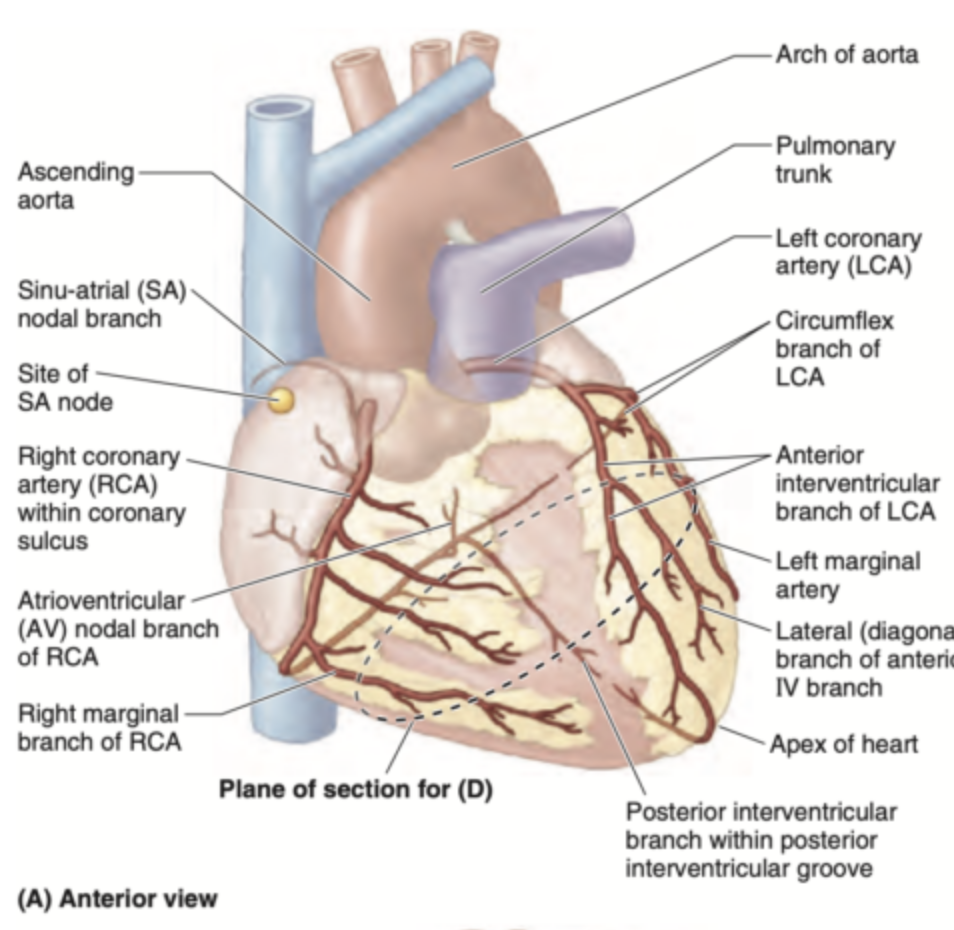
What branch of the coronary artery accompanies the great cardiac vein?
a. Left circumflex artery
b. Posterior descending artery
c. Anterior interventricular branch of RCA
d. Anterior interventricular branch of LCA
Answer: d. Anterior interventricular branch of LCA
解説:
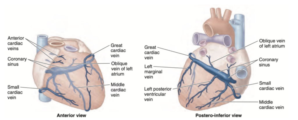
大心静脈(Great cardiac vein)は、左冠動脈前下行枝(Anterior interventricular branch of LCA)に沿って走行します。これは、左前下行枝が心臓の前部で酸素供給を行う主要な動脈であり、大心静脈は酸素を使用した血液を回収して冠状静脈洞に流れ込みます。
他の選択肢が間違っている理由:
- a. Left circumflex artery: 左回旋枝は心臓の後面や側面を供給する動脈であり、大心静脈とは関係がありません。
- b. Posterior descending artery: 後下行枝は主に心臓の下部を供給し、中心静脈と関係していますが、大心静脈と関わりません。
- c. Anterior interventricular branch of RCA: 右冠動脈の前下行枝は、通常存在せず、
- この部位は主に左冠動脈によって供給されています。
Question 2
This cardiac vein accompanies the posterior interventricular branches that usually arise from the RCA.
a. Great cardiac vein
b. Small cardiac vein
c. Marshall vein
d. Middle cardiac vein
Answer: d. Middle cardiac vein
解説:
中心静脈(Middle cardiac vein)は、通常、右冠動脈の後下行枝(Posterior interventricular branch of RCA)に沿って走行します。この静脈は心臓の後面にある酸素を使用した血液を回収し、冠状静脈洞に流れ込みます。
他の選択肢が間違っている理由:
- a. Great cardiac vein: 大心静脈は左前下行枝に沿って走行し、心臓の前部に位置します。
- b. Small cardiac vein: 小心静脈は右縁動脈に沿って走行し、主に心臓の右側を流れるため、後下行枝とは関係がありません。
- c. Marshall vein: マーシャル静脈(左心房斜静脈)は左心房の領域にあり、後下行枝とは関係がありません。
Question 3
The majority of the arterial blood supply towards the sinoatrial node comes from what particular source?
a. Right marginal artery
b. Right coronary artery
c. Left anterior descending artery
d. Left coronary artery
Answer: b. Right coronary artery
解説:
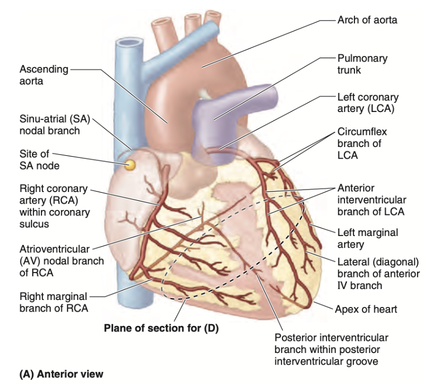
洞房結節(Sinoatrial node)への主要な血液供給は、右冠動脈(Right coronary artery)から供給されます。洞房結節は心臓のペースメーカーとしての役割を果たしており、その動脈供給は正常な心拍の維持にとって重要です。
他の選択肢が間違っている理由:
- a. Right marginal artery: 右縁動脈は右冠動脈の枝の一つですが、主に心臓の側面を供給し、洞房結節への直接的な供給源ではありません。
- c. Left anterior descending artery: 左前下行枝は心臓の前部を供給しますが、洞房結節への血液供給には関与しません。
- d. Left coronary artery: 左冠動脈は主に左側の心臓の血液供給を担っていますが、洞房結節の血液供給には直接関与しません。
Question 4
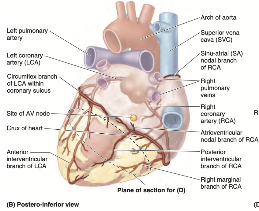
What branch of the coronary artery supplies the diaphragmatic surface of the heart?
a. Right marginal artery
b. Left ventricular artery
c. Posterior interventricular artery
d. Atrioventricular artery
Answer: c. Posterior interventricular artery
解説:
後下行枝(Posterior interventricular artery)は、心臓の横隔面(diaphragmatic surface)を供給します。右冠動脈から分岐し、心臓の後面と下部を供給する重要な動脈です。
他の選択肢が間違っている理由:
- a. Right marginal artery: 右縁動脈は心臓の右側面を供給しますが、横隔面の血液供給には関与しません。
- b. Left ventricular artery: 左室動脈は心臓の左側を供給しますが、横隔面の供給には直接関与していません。
- d. Atrioventricular artery: 房室動脈は房室結節や心房に関連する構造を供給しますが、横隔面の血液供給には関与しません。
Question 5
What cardiac vein empties directly into the right atrium?
a. Great cardiac vein
b. Middle cardiac vein
c. Small cardiac vein
d. Anterior cardiac vein
Answer: d. Anterior cardiac vein
解説:
前心静脈(Anterior cardiac vein)は、右心房(Right atrium)に直接流れ込みます。他の多くの心静脈は冠状静脈洞を経由して右心房に流れ込みますが、前心静脈は直接的に右心房に入ります。
他の選択肢が間違っている理由:
- a. Great cardiac vein: 大心静脈は冠状静脈洞に流れ込み、右心房には直接流れ込みません。
- b. Middle cardiac vein: 中心静脈もまた冠状静脈洞に流れ込み、右心房に直接流れ込むことはありません。
- c. Small cardiac vein: 小心静脈も冠状静脈洞に流れ込み、右心房に直接流れ込むことはありません。
Question 6(10/1出た)
The anterior sternocostal surface of the heart is formed by what chamber?
a. Left atrium
b. Right ventricle
c. Left ventricle
d. Right atrium
Answer: b. Right ventricle
解説:
心臓の前胸骨肋骨面(anterior sternocostal surface)は主に右心室(Right ventricle)によって形成されます。右心室は心臓の前部に位置し、胸骨に近接しています。この位置により、胸郭の前方に面しています。
他の選択肢が間違っている理由:
- a. Left atrium: 左心房は心臓の背側に位置し、前胸骨肋骨面の形成には関与していません。主に心臓の基底部を形成しています。
- c. Left ventricle: 左心室は心臓の左側および後方に位置し、主に横隔膜面を形成しますが、前面には影響しません。
- d. Right atrium: 右心房は心臓の右側に位置しており、前面の形成には関与しません。
Question 7(10/1出た)
What valve is located at the left of the sternum at the level of the 3rd intercostal space?
a. Mitral valve
b. Tricuspid valve
c. Pulmonic valve
d. Aortic valve
Answer: d. Aortic valve
解説:
大動脈弁(Aortic valve)は胸骨の左側、第3肋間に位置しています。これは、心臓の左心室と大動脈の間に位置し、血液が左心室から大動脈へ流れる際に重要な役割を果たします。
他の選択肢が間違っている理由:
- a. Mitral valve: 僧帽弁(Mitral valve)は左心房と左心室の間に位置し、第3肋間ではなく、主に左側の第5肋間で聞こえます。
- b. Tricuspid valve: 三尖弁(Tricuspid valve)は右心房と右心室の間に位置し、胸骨の右側第4肋間に位置します。
- c. Pulmonic valve: 肺動脈弁(Pulmonic valve)は胸骨の左第2肋間に位置しますが、第3肋間には位置しません。
Question 8(10/1出た)
The base of the heart is formed mainly by what chamber?
a. Right ventricle
b. Left atrium
c. Left ventricle
d. Right atrium
Answer: b. Left atrium
解説:
心臓の基底部(Base of the heart)は主に左心房(Left atrium)によって形成されます。左心房は肺静脈から酸素化された血液を受け取り、心臓の背側に位置しています。
他の選択肢が間違っている理由:
- a. Right ventricle: 右心室は心臓の前面に位置し、基底部の形成には関与していません。
- c. Left ventricle: 左心室は心臓の横隔膜面に面しており、基底部の形成には関与していません。
- d. Right atrium: 右心房は心臓の右側に位置していますが、主に基底部を形成するのは左心房です。
Question 9(10/1出た)
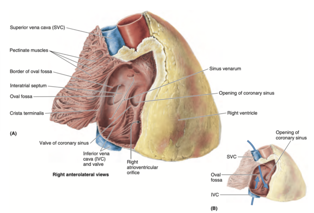
The internal landmark in the right atrium that divides the smooth and rough portions of the wall internally.
a. Crista terminalis
b. Central sulcus
c. Auricle
d. Sulcus terminalis
Answer: a. Crista terminalis
解説:
クリスタ・ターミナリス(Crista terminalis)は右心房の内壁にある構造で、滑らかな部分(静脈洞の部分)と粗い部分(心房筋の部分)を内部で区分します。この構造は、右心房の解剖学的ランドマークとして非常に重要です。
他の選択肢が間違っている理由:
- b. Central sulcus: 中央溝(Central sulcus)は主に脳に関連する構造で、心臓の解剖とは関係がありません。
- c. Auricle: 耳介(Auricle)は心房の外部に存在する小さな構造であり、内部の滑らかさや粗さを区分するものではありません。
- d. Sulcus terminalis: 終末溝(Sulcus terminalis)は外部の構造であり、右心房の外側を区切りますが、内部の区分には関与していません。
Question 10
Where does the anterior cardiac vein drain directly?
a. Right atrium
b. Coronary sinus
c. Left atrium
d. Middle cardiac vein
Answer: a. Right atrium
解説:
前心静脈(Anterior cardiac vein)は直接右心房(Right atrium)に流れ込みます。他のほとんどの心静脈は冠状静脈洞を通じて右心房に入りますが、前心静脈はこれを経由せずに直接右心房に入ります。
他の選択肢が間違っている理由:
- b. Coronary sinus: 冠状静脈洞は多くの心静脈が流れ込む場所ですが、前心静脈はこれを通りません。
- c. Left atrium: 左心房は静脈から血液を受け取りますが、前心静脈は右心房に直接流れ込みます。
- d. Middle cardiac vein: 中心静脈は冠状静脈洞を経由して右心房に流れ込みますが、前心静脈とは異なります。
Question 11
The fetal remnant of the foramen ovale is:
a. Ligamentum arteriosum
b. Fossa ovalis
c. Ligamentum teres hepatis
d. Ligamentum venosum
Answer: b. Fossa ovalis
解説:
卵円窩(Fossa ovalis)は、胎児期において心房間の血流を可能にしていた卵円孔(Foramen ovale)の遺残です。出生後、肺循環が始まると卵円孔が閉じ、卵円窩という凹みが右心房の壁に残ります。
他の選択肢が間違っている理由:
- a. Ligamentum arteriosum: 動脈管索(Ligamentum arteriosum)は動脈管(ductus arteriosus)の遺残であり、卵円孔とは関係がありません。
- c. Ligamentum teres hepatis: 肝円索(Ligamentum teres hepatis)は臍静脈(umbilical vein)の遺残であり、心臓とは関係がありません。
- d. Ligamentum venosum: 静脈管索(Ligamentum venosum)は胎児期における静脈管(ductus venosus)の遺残であり、卵円孔とは無関係です。
Question 12
Where does the other remaining blood supply towards the atrioventricular node come from?
a. Left anterior descending
b. Right marginal
c. Posterior interventricular branch
d. Circumflex
Answer: d. Circumflex
解説:
房室結節(Atrioventricular node)への血液供給は主に右冠動脈からですが、残りの血液供給は左冠動脈の回旋枝(Circumflex)から供給されることがあります。これは解剖学的な変異の一部としてみられる場合があります。
他の選択肢が間違っている理由:
- a. Left anterior descending: 左前下行枝(Left anterior descending)は主に心臓の前面を供給しますが、房室結節への血液供給には直接関与していません。
- b. Right marginal: 右縁動脈(Right marginal)は心臓の右側を供給しますが、房室結節への主要な供給源ではありません。
- c. Posterior interventricular branch: 後下行枝(Posterior interventricular branch)は房室結節への供給には直接関与していません。
Question 13
Lymphatics follow the coronary arteries. What particular group of lymph nodes do they drain?
a. Superior tracheobronchial, left
b. Inferior tracheobronchial, right
c. Superior tracheobronchial, right
d. Inferior tracheobronchial, left
Answer: b
解説:
心臓からのリンパ液は冠状動脈に沿って流れ、最終的に胸部のリンパ節に到達します。特に、心臓からのリンパ液は右の下気管支リンパ節(inferior tracheobronchial lymph nodes, right)に排出されます。これらのリンパ節は、気管の分岐部付近(気管支分岐部、carina)に位置しており、心臓を含む胸部の様々な組織からのリンパ液を集める役割を担っています。
Question 14(10/1出た)
What valve is located at the left of the sternum at the level of the 3rd intercostal space?
a. Mitral valve
b. Pulmonic valve
c. Aortic valve
d. Tricuspid valve
Answer: c. Aortic valve
解説:
大動脈弁(Aortic valve)は胸骨の左側、第3肋間に位置しています。この位置は、心音を聴取する際に重要な解剖学的ランドマークとなります。
他の選択肢が間違っている理由:
- a. Mitral valve: 僧帽弁は左心房と左心室の間に位置し、第3肋間ではなく第5肋間で聞こえます。
- b. Pulmonic valve: 肺動脈弁は胸骨の左第2肋間に位置します。
- d. Tricuspid valve: 三尖弁は胸骨の右側、第4肋間に位置します。
Question 15
Which blood vessel runs along the coronary sulcus?
a. Great cardiac vein
b. Left coronary artery
c. Middle cardiac vein
d. Right coronary artery
Answer: d. Right coronary artery
解説:
冠状動脈溝(Coronary sulcus)は心臓を取り巻く溝で、右冠動脈(Right coronary artery)がその溝に沿って走行します。右冠動脈は心臓の右側および後面に血液を供給します。
他の選択肢が間違っている理由:
- a. Great cardiac vein: 大心静脈は左前下行枝に沿って走行し、冠状動脈溝には位置しません。
- b. Left coronary artery: 左冠動脈は主に心臓の前面および左側に沿って走行し、冠状動脈溝には関与しません。
- c. Middle cardiac vein: 中心静脈は後下行枝に沿って走行しますが、冠状動脈溝には位置しません。

Question 16(10/1出た)
Which of the following papillary muscles of the right ventricle is considered the largest and also attached to the cusps?
a. Septal
b. Anterior
c. Lateral
d. Posterior
Answer: b. Anterior
解説:
右心室の前乳頭筋(Anterior papillary muscle)は最も大きく、三尖弁の小尖(cusps)に付着しています。この筋肉は心室収縮時に弁が逆流しないように機能します。
他の選択肢が間違っている理由:
- a. Septal: 中隔乳頭筋(Septal papillary muscle)は右心室内の中隔に近い位置にありますが、前乳頭筋ほど大きくはありません。
- c. Lateral: 側方乳頭筋(Lateral papillary muscle)は右心室には存在しません。
- d. Posterior: 後乳頭筋(Posterior papillary muscle)は前乳頭筋ほど大きくはなく、三尖弁の後方部分に付着しています。
Question 17
What chamber of the heart do you find the remnant of the oval foramen that is present during fetal life?
a. Left ventricle
b. Right ventricle
c. Left atrium
d. Right atrium
Answer: d. Right atrium
解説:
胎児期に心房間で血流を通していた卵円孔(Foramen ovale)の遺残である卵円窩(Fossa ovalis)は、出生後に右心房(Right atrium)の壁に見られます。卵円孔は胎児期に肺を通過せずに血液を左心房へ送る役割を果たしていましたが、出生後は閉じます。
他の選択肢が間違っている理由:
- a. Left ventricle: 左心室には卵円孔の遺残は存在せず、卵円窩は右心房にのみ見られます。
- b. Right ventricle: 右心室は血液を肺動脈に送り出す部屋であり、卵円窩の遺残とは無関係です。
- c. Left atrium: 左心房には卵円孔の出口は存在しますが、遺残としては右心房にのみ卵円窩が見られます。
Question 18
All of the following supply the left ventricle EXCEPT:
a. Posterior branch of the RCA
b. Diagonal branch of the LCA
c. Posterior branch of the LCA
d. Marginal branch of RCA
Answer: d. Marginal branch of RCA
解説:
右冠動脈の縁枝(Marginal branch of RCA)は右心室や心臓の右側の供給を担当し、左心室(Left ventricle)には供給しません。左心室は主に左冠動脈の前下行枝(LAD)や回旋枝(Circumflex artery)から供給を受けます。
他の選択肢が間違っている理由:
- a. Posterior branch of the RCA: 右冠動脈の後枝は、一部左心室の後方も供給する場合があります。
- b. Diagonal branch of the LCA: 左冠動脈の対角枝は左心室の前側を供給します。
- c. Posterior branch of the LCA: 左冠動脈の後枝も左心室に血液を供給します。
Question 19
What is considered as the 1st and main tributary of the coronary sinus?
a. Anterior cardiac
b. Middle cardiac
c. Oblique vein of the left atrium
d. Great cardiac vein
Answer: d. Great cardiac vein
解説:
大心静脈(Great cardiac vein)は冠状静脈洞(Coronary sinus)に流れ込む最初かつ主要な静脈です。この静脈は心臓の前部と左側の酸素を使用した血液を集め、冠状静脈洞に排出します。
他の選択肢が間違っている理由:
- a. Anterior cardiac: 前心静脈は冠状静脈洞を経由せず、直接右心房に流れ込みます。
- b. Middle cardiac: 中心静脈も冠状静脈洞に流れ込みますが、最初の主要な静脈ではありません。
- c. Oblique vein of the left atrium: 左心房斜静脈も冠状静脈洞に流れ込みますが、主要な流入源ではありません。
Question 20
Which of the following aortic sinuses is considered as non-coronary, where no arteries arise?
a. Posterior
b. Right
c. Anterior
d. Left
Answer: a. Posterior
解説:
大動脈弁の後洞(Posterior aortic sinus)は「非冠動脈洞(Non-coronary sinus)」と呼ばれ、ここからは冠動脈が発生しません。右冠動脈は右洞から、左冠動脈は左洞から発生します。
他の選択肢が間違っている理由:
- b. Right: 右洞(Right aortic sinus)からは右冠動脈が発生します。
- c. Anterior: 「前洞」という表現は一般的ではなく、実際には「右洞」や「左洞」が使用されます。
- d. Left: 左洞(Left aortic sinus)からは左冠動脈が発生します。
Question 21
What vein is considered as the first and main tributary of the coronary sinus?
a. Middle cardiac
b. Anterior cardiac
c. Great cardiac
d. Oblique vein
Answer: c. Great cardiac
解説:
大心静脈(Great cardiac vein)は冠状静脈洞に流れ込む最初の主要な静脈です。心臓の前面から酸素を使用した血液を収集し、冠状静脈洞に運びます。
他の選択肢が間違っている理由:
- a. Middle cardiac: 中心静脈は冠状静脈洞に流れ込みますが、最初の主要な静脈ではありません。
- b. Anterior cardiac: 前心静脈は冠状静脈洞を経由せず、右心房に直接流れ込みます。
- d. Oblique vein: 左心房斜静脈は冠状静脈洞に流れ込みますが、主要な流入源ではありません。
Question 22
All of the following cardiac veins empty into the coronary sinus EXCEPT:
a. Small cardiac vein
b. Oblique cardiac vein
c. Middle cardiac vein
d. Smallest cardiac vein
Answer: d. Smallest cardiac vein
解説:
最小心静脈(Smallest cardiac vein)は冠状静脈洞に流れ込まず、直接右心房に流れ込みます。他の心静脈は冠状静脈洞を通じて右心房に血液を供給します。
他の選択肢が間違っている理由:
- a. Small cardiac vein: 小心静脈は冠状静脈洞に流れ込みます。
- b. Oblique cardiac vein: 左心房斜静脈も冠状静脈洞に流れ込みます。
- c. Middle cardiac vein: 中心静脈も冠状静脈洞に流れ込みます。
Question 23
What cardiac vein runs in the posterior interventricular groove?
a. Oblique cardiac vein
b. Small cardiac vein
c. Posterior cardiac vein
d. Middle cardiac vein
Answer: d. Middle cardiac vein
解説:
中心静脈(Middle cardiac vein)は、後室間溝(posterior interventricular groove)を走行します。この静脈は、右冠動脈の後下行枝(posterior interventricular branch of the RCA)に沿っており、心臓の後面から血液を集めます。
他の選択肢が間違っている理由:
- a. Oblique cardiac vein: 斜静脈(Oblique cardiac vein)は左心房に沿って走行し、後室間溝には位置しません。
- b. Small cardiac vein: 小心静脈(Small cardiac vein)は右冠動脈に沿って走行しますが、後室間溝には位置しません。
- c. Posterior cardiac vein: 後心静脈(Posterior cardiac vein)は左心室の後側を走行しますが、後室間溝には位置しません。
Question 24(10/1出た)
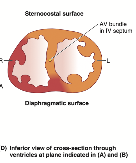
What structure in the right ventricle is described as a muscular bundle that traverses the chamber from the inferior part of the interventricular septum?
a. Tendinous cords
b. Trabeculae carneae
c. Septomarginal trabecula (moderator band)
d. Supraventricular crest
Answer: c. Septomarginal trabecula (moderator band)
解説:
中隔縁柱(Septomarginal trabecula)、別名モデレーター帯(Moderator band)は、右心室の内壁に存在する筋束で、心室中隔(interventricular septum)の下部から右心室壁へと伸びています。この構造は右房室束(right branch of the AV bundle)の一部を伝導し、前乳頭筋に信号を伝えます。
他の選択肢が間違っている理由:
- a. Tendinous cords: 腱索(Tendinous cords)は弁を支持する線維ですが、筋束ではありません。
- b. Trabeculae carneae: 肉柱(Trabeculae carneae)は心室の壁に存在する筋の隆起ですが、中隔縁柱のように部屋全体を横断することはありません。
- d. Supraventricular crest: 超室稜(Supraventricular crest)は右心室内の他の構造であり、筋束ではありません。
Question 25(10/1出た)
What valve is located to the right of the sternum at the level of the 3rd intercostal space?
a. Pulmonic valve
b. Mitral valve
c. Aortic valve
d. Tricuspid valve
Answer: d. Tricuspid valve
解説:
三尖弁(Tricuspid valve)は胸骨の右側、第3肋間に位置します。この弁は右心房と右心室の間にあり、心臓に戻ってきた静脈血が右心房から右心室へ流れる際に開きます。
他の選択肢が間違っている理由:
- a. Pulmonic valve: 肺動脈弁(Pulmonic valve)は胸骨の左側、第2肋間に位置します。
- b. Mitral valve: 僧帽弁(Mitral valve)は左心房と左心室の間に位置し、胸骨の第5肋間に近いです。
- c. Aortic valve: 大動脈弁(Aortic valve)は胸骨の左側、第3肋間に位置しますが、右側ではありません。
Question 26
The circumflex artery comes from what other branch of the coronaries?
a. Left coronary artery
b. Lateral branch from the anterior interventricular branch
c. Right coronary artery
d. Left anterior descending
Answer: a. Left coronary artery
解説:
回旋枝(Circumflex artery)は左冠動脈(Left coronary artery)から分岐します。この動脈は心臓の左側面および後面を供給します。
他の選択肢が間違っている理由:
- b. Lateral branch from the anterior interventricular branch: 左前下行枝(Left anterior descending)から分岐する枝は回旋枝ではなく、通常対角枝(Diagonal branch)です。
- c. Right coronary artery: 右冠動脈(Right coronary artery)は心臓の右側と後方を供給しますが、回旋枝は左冠動脈から分岐します。
- d. Left anterior descending: 左前下行枝(Left anterior descending)は左冠動脈から分岐しますが、回旋枝はこの動脈からは分岐しません。
Question 27
Which of the branches come from the right coronary artery?
a. Diagonal
b. Circumflex
c. Posterior interventricular
d. Terminal
Answer: c. Posterior interventricular
解説:
後下行枝(Posterior interventricular branch)は右冠動脈(Right coronary artery)から分岐します。この動脈は心臓の後面と下部を供給し、右冠動脈の重要な枝の一つです。
他の選択肢が間違っている理由:
- a. Diagonal: 対角枝(Diagonal branch)は左前下行枝から分岐し、右冠動脈からは分岐しません。
- b. Circumflex: 回旋枝(Circumflex artery)は左冠動脈から分岐します。
- d. Terminal: 終末枝(Terminal branch)という特定の用語は、冠動脈において使用されることは一般的ではありません。
Question 28(10/1出た)
The valve located along the oblique line of the heart, immediately below the aortic chamber and at the level of the 4th costal cartilage, is:
a. Aortic valve
b. Pulmonic valve
c. Mitral valve
d. Tricuspid valve
Answer: d. Tricuspid valve
解説:
三尖弁(Tricuspid valve)は心臓の右側にあり、大動脈弁の直下、心臓の斜めのラインに沿って第4肋軟骨のレベルに位置します。右心房と右心室の間に位置する重要な弁です。
他の選択肢が間違っている理由:
- a. Aortic valve: 大動脈弁は第3肋間のレベルにあり、心臓の上部に位置します。
- b. Pulmonic valve: 肺動脈弁は大動脈弁のすぐ左側にあり、通常第2肋間のレベルに位置します。
- c. Mitral valve: 僧帽弁は左心房と左心室の間に位置し、右心室には関与していません。
Question 29
Which of the following branches of the coronaries follows the course of the great cardiac vein?
a. Posterior interventricular
b. Circumflex
c. Right marginal
d. Left anterior descending
Answer: d. Left anterior descending
解説:
左前下行枝(Left anterior descending, LAD)は、大心静脈(Great cardiac vein)と並行して走行します。LADは心臓の前面を供給する主要な動脈で、大心静脈は酸素を使用した血液を回収し、冠状静脈洞に流れ込みます。
他の選択肢が間違っている理由:
- a. Posterior interventricular: 後下行枝(Posterior interventricular artery)は心臓の後面を供給し、大心静脈とは無関係です。
- b. Circumflex: 回旋枝(Circumflex artery)は左冠動脈から分岐し、心臓の側面を供給しますが、大心静脈とは並行していません。
- c. Right marginal: 右縁動脈(Right marginal artery)は右冠動脈の枝であり、右側面を供給しますが、大心静脈と関係はありません。
Question 30
The coronary sinus drains into which particular heart chamber?
a. Right atrium
b. Left ventricle
c. Right ventricle
d. Left atrium
Answer: a. Right atrium
解説:
冠状静脈洞(Coronary sinus)は心臓の酸素を使用した血液を集めて右心房(Right atrium)に流れ込みます。右心房は静脈血を受け取り、右心室に送る役割を果たします。
他の選択肢が間違っている理由:
- b. Left ventricle: 左心室は酸素化された血液を全身に送り出す部屋であり、冠状静脈洞の排出先ではありません。
- c. Right ventricle: 右心室は肺に血液を送り出す部屋であり、冠状静脈洞に関与していません。
- d. Left atrium: 左心房は肺静脈から酸素化された血液を受け取りますが、冠状静脈洞の排出先ではありません。
Question 31
The coronaries arise from what specific structure in the ascending aorta?
a. Sinus
b. Hiatus
c. Lunule
d. Nodule
Answer: a. Sinus
解説:
冠動脈(Coronary arteries)は、大動脈弁の上に位置する大動脈洞(Aortic sinus)から発生します。右冠動脈は右大動脈洞から、左冠動脈は左大動脈洞から発生し、心臓に酸素化された血液を供給します。
他の選択肢が間違っている理由:
- b. Hiatus: 「Hiatus」は一般的に開口部や孔を指し、冠動脈の起始部には関係ありません。
- c. Lunule: 「Lunule」は大動脈弁の小さな構造ですが、冠動脈の起始部には関係しません。
- d. Nodule: 「Nodule」は大動脈弁の中心部にある小さな結節ですが、冠動脈の発生源ではありません。
Question 32
The atrioventricular node receives most of its blood supply from which of the following coronary arteries?
a. Anterior interventricular branch
b. Posterior interventricular branch
c. Right coronary artery
d. Left coronary artery
Answer: c. Right coronary artery
解説:
房室結節(Atrioventricular node)は、主に右冠動脈(Right coronary artery)から血液供給を受けます。右冠動脈は心臓の右側に沿って走行し、房室結節や洞房結節などの重要な構造に酸素化された血液を供給します。
他の選択肢が間違っている理由:
- a. Anterior interventricular branch: 左前下行枝(Anterior interventricular branch)は心臓の前面を供給しますが、房室結節には直接関与しません。
- b. Posterior interventricular branch: 後下行枝(Posterior interventricular branch)は心臓の後面を供給しますが、房室結節の主要な供給源ではありません。
- d. Left coronary artery: 左冠動脈は主に左心房と左心室を供給しますが、房室結節の供給源ではありません。
Question 33(10/1出た)
What do you call a dilated portion of the heart that is superior to each cusp and that prevents it from sticking to the wall of the vessel?
a. Nodule
b. Lunule
c. Valve
d. Sinus
Answer: d. Sinus
解説:
大動脈洞(Aortic sinus)は大動脈弁の各小尖(cusp)の上に存在し、弁が血管壁に貼り付くのを防ぎます。この洞は冠動脈の起始部としても重要です。
他の選択肢が間違っている理由:
- a. Nodule: 大動脈弁の結節(Nodule)は、小尖の中心にある小さな構造で、洞ではありません。
- b. Lunule: 小尖の辺縁にある小さな半月状の部分を指しますが、洞とは異なります。
- c. Valve: 弁(Valve)は心臓内の構造全体を指す一般的な用語で、洞とは異なります。
Question 34(10/1出た)
What structure in the right atrium separates the smooth muscle from its rough parts?
a. Auricle
b. Oval fossa
c. Sulcus terminalis
d. Crista terminalis
Answer: d. Crista terminalis
解説:
クリスタ・ターミナリス(Crista terminalis)は右心房の内壁に存在し、滑らかな部分(静脈洞由来の部分)と粗い部分(心房筋由来の部分)を分ける重要なランドマークです。
他の選択肢が間違っている理由:
- a. Auricle: 心耳(Auricle)は右心房の外側に位置する小さな構造であり、内部の区分には関与していません。
- b. Oval fossa: 卵円窩(Oval fossa)は胎児期の遺残であり、右心房の区分には関係しません。
- c. Sulcus terminalis: 終末溝(Sulcus terminalis)は右心房の外部にある溝で、内部の構造を区分するものではありません。
Question 35
An embolism has lodged inside the pulmonary circuitry. The most likely formation of this embolism occurred in:
a. RCA
b. Coronary sinus
c. Circumflex arteries
d. LCA
Answer: b. Coronary sinus
解説:
肺循環に栓塞が生じた場合、その栓塞は通常、静脈系から来るため、冠状静脈洞(Coronary sinus)などの静脈系で形成された可能性があります。血栓が静脈を経由して心臓に入り、右心室を通過して肺へ向かいます。
他の選択肢が間違っている理由:
- a. RCA: 右冠動脈(Right coronary artery)は動脈系であり、栓塞が肺循環に入ることは通常ありません。
- c. Circumflex arteries: 回旋枝は動脈であり、静脈系を経由する肺栓塞の原因とはなりません。
- d. LCA: 左冠動脈(Left coronary artery)は動脈系であり、肺への栓塞の原因にはなりません。
Question 36
The sinoatrial nodal branch comes from what other specific branch of the left coronary artery?
a. Left marginal
b. Circumflex
c. Left anterior descending
d. Diagonal
Answer: b. Circumflex
解説:
洞房結節枝(Sinoatrial nodal branch)は、左冠動脈の回旋枝(Circumflex artery)から分岐することがあります。洞房結節枝は、心臓の自然なペースメーカーである洞房結節に酸素化された血液を供給します。
他の選択肢が間違っている理由:
- a. Left marginal: 左縁枝(Left marginal artery)は左冠動脈の枝ですが、洞房結節に直接血液を供給することはありません。
- c. Left anterior descending: 左前下行枝(Left anterior descending artery)は心臓の前面を供給しますが、洞房結節には直接関与しません。
- d. Diagonal: 対角枝(Diagonal branch)は左前下行枝の枝であり、洞房結節への供給には関与しません。
Question 37
What valve is located between the right atrium and right ventricle?
a. Mitral valve
b. Aortic valve
c. Pulmonic valve
d. Tricuspid valve
Answer: d. Tricuspid valve
解説:
三尖弁(Tricuspid valve)は、右心房(Right atrium)と右心室(Right ventricle)の間に位置する弁です。この弁は、右心房から右心室へ血液が流れる際に開閉を制御し、逆流を防ぎます。
他の選択肢が間違っている理由:
- a. Mitral valve: 僧帽弁(Mitral valve)は左心房と左心室の間に位置する弁で、右心房や右心室には関与しません。
- b. Aortic valve: 大動脈弁(Aortic valve)は左心室と大動脈の間に位置します。
- c. Pulmonic valve: 肺動脈弁(Pulmonic valve)は右心室と肺動脈の間に位置しますが、右心房には関与しません。
Question 38
What other vein merges with the great cardiac vein to form the coronary sinus?
a. Anterior cardiac
b. Oblique vein
c. Small cardiac
d. Middle cardiac
Answer: b. Oblique vein
解説:
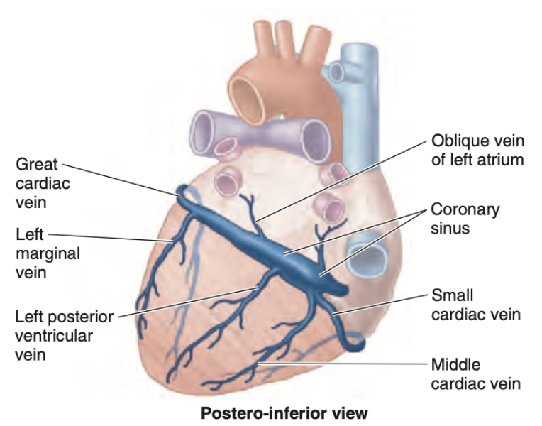
左心房斜静脈(Oblique vein of the left atrium)は大心静脈(Great cardiac vein)と合流して冠状静脈洞(Coronary sinus)を形成します。冠状静脈洞は、心臓の酸素を使用した血液を右心房に排出します。
他の選択肢が間違っている理由:
- a. Anterior cardiac: 前心静脈(Anterior cardiac vein)は右心房に直接流れ込み、冠状静脈洞とは関係がありません。
- c. Small cardiac: 小心静脈(Small cardiac vein)は冠状静脈洞に流れ込みますが、大心静脈とは合流しません。
- d. Middle cardiac: 中心静脈(Middle cardiac vein)も冠状静脈洞に流れ込みますが、大心静脈とは合流しません。
Question 39
What branch of the coronary artery supplies mainly the diaphragmatic surface of the heart?
a. Left ventricular
b. Right marginal
c. Posterior interventricular
d. Diagonal
Answer: c. Posterior interventricular
解説:
後下行枝(Posterior interventricular artery)は、心臓の横隔面(diaphragmatic surface)を供給します。この枝は通常、右冠動脈から分岐し、心臓の下部に血液を供給します。
他の選択肢が間違っている理由:
- a. Left ventricular: 左室枝(Left ventricular branch)は心臓の左側を供給しますが、横隔面を主に供給するわけではありません。
- b. Right marginal: 右縁枝(Right marginal artery)は心臓の右側面を供給しますが、横隔面の主要な供給源ではありません。
- d. Diagonal: 対角枝(Diagonal branch)は左前下行枝の枝で、主に左心室の前面を供給します。
Question 40
What cardiac vein runs in the anterior interventricular groove of the heart?
a. Small cardiac vein
b. Smallest cardiac vein
c. Great cardiac vein
d. Middle cardiac vein
Answer: c. Great cardiac vein
解説:
大心静脈(Great cardiac vein)は前室間溝(anterior interventricular groove)を走行します。この静脈は、心臓の前面で酸素を使用した血液を集め、冠状静脈洞に流れ込みます。
他の選択肢が間違っている理由:
- a. Small cardiac vein: 小心静脈(Small cardiac vein)は右冠動脈に沿って走行し、前室間溝には位置しません。
- b. Smallest cardiac vein: 最小心静脈(Smallest cardiac vein)は心臓の壁に直接排出される微小な静脈であり、前室間溝には位置しません。
- d. Middle cardiac vein: 中心静脈(Middle cardiac vein)は後室間溝に位置します。
Question 41(10/1出た)
The membrane layer that covers the heart and is continuous with the central tendon of the diaphragm is:
a. Fibrous pericardium
b. Visceral layer of pericardium
c. Parietal layer of serous pericardium
d. Serous pericardium
Answer: a. Fibrous pericardium
解説:
心膜の線維性層(Fibrous pericardium)は、心臓を覆う外側の強靭な膜で、横隔膜の中央腱(central tendon of the diaphragm)と連続しています。この膜は、心臓の位置を安定させるとともに、外部の衝撃から心臓を保護します。
他の選択肢が間違っている理由:
- b. Visceral layer of pericardium: 心膜の臓側層(Visceral pericardium)は心臓の表面に直接接している層で、横隔膜とは直接連続していません。
- c. Parietal layer of serous pericardium: 心膜の壁側層(Parietal pericardium)は心臓の外側を覆っていますが、線維性心膜とは異なり、横隔膜と連続していません。
- d. Serous pericardium: 漿膜性心膜(Serous pericardium)は臓側層と壁側層から構成されますが、線維性心膜のように横隔膜とは直接連続していません。
Question 42
Which of the following cardiac veins drains the areas supplied by the right coronary artery?
a. Anterior and small cardiac veins
b. Middle and small cardiac veins
c. Great cardiac and oblique veins
d. Posterior interventricular and oblique veins
Answer: b. Middle and small cardiac veins
解説:
右冠動脈(Right coronary artery)から供給される領域を排出する主な静脈は、中心静脈(Middle cardiac vein)と小心静脈(Small cardiac vein)です。これらの静脈は右側と後面の酸素を使用した血液を集め、冠状静脈洞に流れ込みます。
他の選択肢が間違っている理由:
- a. Anterior and small cardiac veins: 前心静脈(Anterior cardiac vein)は右心房に直接流れ込み、冠状静脈洞には流れ込みません。
- c. Great cardiac and oblique veins: 大心静脈(Great cardiac vein)と斜静脈(Oblique vein)は左冠動脈領域を排出します。
- d. Posterior interventricular and oblique veins: 後室間静脈は右冠動脈領域を排出しますが、斜静脈は左心房の領域を排出します。
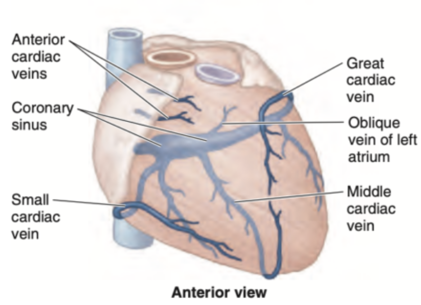
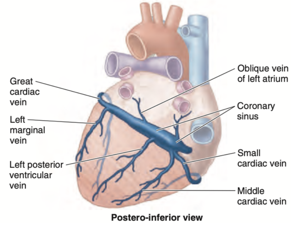
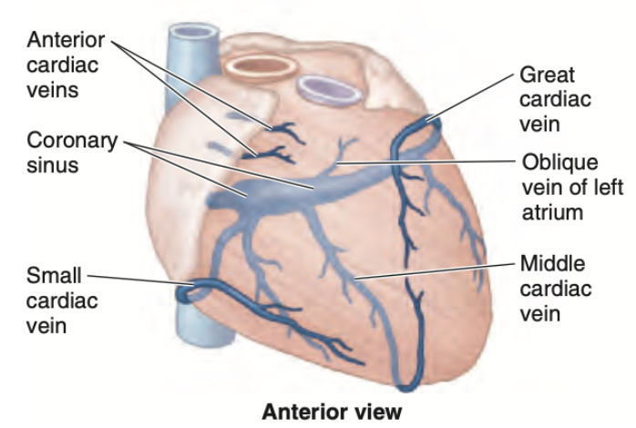
| 静脈の名前 | 説明 |
|---|---|
| 冠状静脈洞 (Coronary Sinus) | 心臓の主な静脈であり、冠状溝に沿って走り、心臓からの血液を右心房に排出します。 |
| 大心臓静脈 (Great Cardiac Vein) | 前室間静脈として始まり、冠状静脈洞に流入する。左冠状動脈が供給する領域を排出します。 |
| 中心臓静脈 (Middle Cardiac Vein) | 右冠状動脈の後室間枝に沿って走り、冠状静脈洞に流入します。 |
| 小心臓静脈 (Small Cardiac Vein) | 冠状静脈洞に流入し、右冠状動脈の右辺縁枝に沿って走ります。 |
| 前心臓静脈 (Anterior Cardiac Vein) | 右心室からの血液を直接右心房に流入します。 |
| 左後心室静脈 (Left Posterior Ventricular Vein) | 左後室領域からの血液を冠状静脈洞に流入します。 |
| 左辺縁静脈 (Left Marginal Vein) | 左心臓の辺縁から冠状静脈洞に流入します。 |
| 左心房斜静脈 (Oblique Vein of Left Atrium) | 左心房から冠状静脈洞に流入します。 |
Question 43
What branch of the coronary artery accompanies the middle cardiac vein?
a. Posterior interventricular branch of LCA
b. Left anterior descending artery
c. Posterior interventricular branch of RCA
d. Right marginal artery
Answer: c. Posterior interventricular branch of RCA
解説:
中心静脈(Middle cardiac vein)は通常、右冠動脈(RCA)の後下行枝(Posterior interventricular branch of RCA)に沿って走行します。この動脈と静脈は、心臓の後面と下部を供給および排出する役割を担っています。
他の選択肢が間違っている理由:
- a. Posterior interventricular branch of LCA: 左冠動脈(LCA)の後下行枝は通常存在せず、この領域は右冠動脈が供給します。
- b. Left anterior descending artery: 左前下行枝(LAD)は心臓の前面を供給し、中心静脈とは関係がありません。
- d. Right marginal artery: 右縁枝(Right marginal artery)は右冠動脈の枝ですが、後下行枝とは異なる領域を供給します。
Question 44
The arterial blood supply of both sinoatrial and atrioventricular nodes usually comes from what source?
a. Posterior interventricular branch
b. Left coronary
c. Right coronary
d. Left anterior descending
Answer: c. Right coronary
解説:
洞房結節(Sinoatrial node)と房室結節(Atrioventricular node)の血液供給は、通常、右冠動脈(Right coronary artery)から供給されます。右冠動脈は心臓の右側を走行し、これらの重要な伝導系を支える酸素化された血液を提供します。
他の選択肢が間違っている理由:
- a. Posterior interventricular branch: 後下行枝は右冠動脈の枝ですが、通常、心臓の後部を供給し、結節への供給は行いません。
- b. Left coronary: 左冠動脈は心臓の左側を供給しますが、通常は結節への血液供給には関与しません。
- d. Left anterior descending: 左前下行枝(LAD)は心臓の前面を供給しますが、結節の主要な供給源ではありません。
Question 45(10/1出た)
What structure carries part of the right branch of the AV bundle, relaying conduction impulses to the anterior papillary muscle?
a. Trabeculae carneae
b. Pectinate muscle
c. Moderator band
d. Interventricular septum
Answer: c. Moderator band
解説:
モデレーター帯(Moderator band)、別名中隔縁柱(Septomarginal trabecula)は、右房室束(Right branch of AV bundle)の一部を運び、右心室の前乳頭筋(Anterior papillary muscle)へ伝導信号を送ります。この構造は右心室内での伝導系の一部です。
他の選択肢が間違っている理由:
- a. Trabeculae carneae: 肉柱(Trabeculae carneae)は心室壁の筋肉の隆起であり、伝導系には直接関与しません。
- b. Pectinate muscle: 櫛状筋(Pectinate muscle)は心房に見られる筋肉であり、心室の伝導には関与しません。
- d. Interventricular septum: 心室中隔(Interventricular septum)は心室を分ける壁であり、伝導路としての役割を果たすことはありますが、モデレーター帯のように直接乳頭筋に信号を伝えることはありません。
Question 46
What embryonic dilatation of the heart would give rise to the aorta?
a. Truncus arteriosus
b. Bulbous cordis
c. Primitive ventricle
d. Sinus venosus
Answer: a. Truncus arteriosus
解説:
動脈幹(Truncus arteriosus)は、胚発生中に大動脈(Aorta)および肺動脈(Pulmonary artery)を形成する部分です。この構造は、胎児の心臓の血液を全身と肺に分配する大動脈と肺動脈に分化します。
他の選択肢が間違っている理由:
- b. Bulbous cordis: 心臓円錐部(Bulbous cordis)は、主に心室の一部を形成しますが、大動脈そのものにはなりません。
- c. Primitive ventricle: 原始心室(Primitive ventricle)は、心臓の左右の心室を形成しますが、大動脈には関与しません。
- d. Sinus venosus: 静脈洞(Sinus venosus)は心房の一部を形成し、血液の流入部門を担当しますが、大動脈の形成には関与しません。
Question 47
The coronary sinus drains into what particular chamber of the heart?
a. Right atrium
b. Left atrium
c. Left ventricle
d. Right ventricle
Answer: a. Right atrium
解説:
冠状静脈洞(Coronary sinus)は右心房(Right atrium)に直接流れ込みます。冠状静脈洞は心臓の酸素を使用した血液を集め、右心房に送る役割を果たします。
他の選択肢が間違っている理由:
- b. Left atrium: 左心房は肺静脈から酸素化された血液を受け取りますが、冠状静脈洞の排出先ではありません。
- c. Left ventricle: 左心室は全身に血液を送り出す部屋であり、冠状静脈洞とは関係がありません。
- d. Right ventricle: 右心室は肺に血液を送り出す部屋であり、冠状静脈洞とは関係がありません。
Question 48(10/1出た)
Which chamber of the heart occupies the entire inferior border?
a. Left ventricle
b. Right atrium
c. Right ventricle
d. Left atrium
Answer: c. Right ventricle
解説:
右心室(Right ventricle)は心臓の全ての下縁(Inferior border)を占めています。右心室は、肺動脈に血液を送り出す役割を果たし、心臓の前面および下部に位置します。
他の選択肢が間違っている理由:
- a. Left ventricle: 左心室は主に心臓の左側面と後部に位置しており、下縁の全てを占めているわけではありません。
- b. Right atrium: 右心房は右側の上部に位置し、下縁を占めることはありません。
- d. Left atrium: 左心房は主に心臓の後面に位置し、下縁の形成には関与していません。
Question 49(10/1出た)
What structure in the left ventricle mostly covers it and is described as finer and more numerous than the rest?
a. Papillary muscle
b. Interventricular septum
c. Trabeculae carneae
d. Tendinous cords
Answer: c. Trabeculae carneae
解説:
左心室内の肉柱(Trabeculae carneae)は、他の心室壁の構造よりも細かく、数が多いことで特徴付けられます。これらの筋肉の隆起は、心臓の収縮を助け、血液の流れを促進します。
他の選択肢が間違っている理由:
- a. Papillary muscle: 乳頭筋(Papillary muscle)は、腱索を通じて弁をサポートしますが、左心室全体を覆うことはありません。
- b. Interventricular septum: 心室中隔(Interventricular septum)は左右の心室を分ける壁であり、左心室全体を覆う構造ではありません。
- d. Tendinous cords: 腱索(Tendinous cords)は弁と乳頭筋を結びつける構造であり、壁を覆うものではありません。
Question 50
What branch of the left coronary artery supplies both ventricles through the interventricular septal branches?
a. Anterior interventricular
b. Lateral or diagonal
c. Circumflex
d. Sinoatrial nodal
Answer: a. Anterior interventricular
解説:
左前下行枝(Anterior interventricular artery)は、心室中隔枝(Interventricular septal branches)を通じて左右の心室を供給します。この動脈は、心臓の前面を供給し、心室の前壁と中隔に酸素化された血液を提供します。
他の選択肢が間違っている理由:
- b. Lateral or diagonal: 対角枝(Diagonal branch)は左前下行枝の枝であり、主に左心室の側面を供給します。
- c. Circumflex: 回旋枝(Circumflex artery)は左冠動脈の枝で、主に左心房と左心室の後壁を供給します。
- d. Sinoatrial nodal: 洞房結節枝(Sinoatrial nodal artery)は主に洞房結節に血液を供給し、心室には直接関与しません。
ブロック
Question 1(10/1出た)
Which of the following structures is found posterior to the parts of the pulmonary trunk, ascending aorta, and cardiac atria?
a) Oblique Pericardial Sinus
b) Transverse Pericardial Sinus
c) Vagus Nerve
d) Pericardiophrenic Ligament
Answer: a) Oblique Pericardial Sinus
解説:
斜位心膜洞(Oblique Pericardial Sinus)は、心臓の後方にある構造であり、心房と大血管の後ろに位置しています。この空間は心膜の一部で、静脈が心臓に接続される位置に関連しています。
他の選択肢が間違っている理由:
- b) Transverse Pericardial Sinus: 横心膜洞は、上大静脈、大動脈、肺動脈の間に位置し、心房の後方にはありません。
- c) Vagus Nerve: 迷走神経は後方に走行するが、肺動脈や大動脈のすぐ後ろではなく、心膜洞に位置するものではありません。
- d) Pericardiophrenic Ligament: 心膜横隔膜靭帯は横隔膜と心膜をつなぐ靭帯であり、心臓や大血管の後ろには存在しません。
Question 2(10/1出た)
Where does the pericardiophrenic artery come from?
a) Brachiocephalic Trunk
b) Ascending Aorta
c) Subclavian Artery
d) Internal Thoracic Artery
Answer: d) Internal Thoracic Artery
解説:
心膜横隔動脈(Pericardiophrenic artery)は、内胸動脈(Internal thoracic artery)から分岐します。この動脈は心臓の心膜および横隔膜に血液を供給します。
他の選択肢が間違っている理由:
- a) Brachiocephalic Trunk: 腕頭動脈は大動脈弓から分岐し、右総頸動脈や右鎖骨下動脈に分かれますが、心膜横隔動脈の直接的な供給源ではありません。
- b) Ascending Aorta: 上行大動脈は心膜横隔動脈の供給源ではありません。
- c) Subclavian Artery: 鎖骨下動脈は内胸動脈を分岐しますが、直接的には心膜横隔動脈を供給しません。
Question 3(10/1出た)
Which among the statements regarding the fibrous pericardium is correct?
a) Continuous superiorly with the tunica adventitia of the great vessels
b) Anchored posteriorly with the central tendon of the diaphragm
c) Bounded medially by loose areolar connective tissue
d) Attached inferiorly to the posterior surface body of the diaphragm
Answer: a) Continuous superiorly with the tunica adventitia of the great vessels
解説:
線維性心膜(Fibrous pericardium)は、心臓を覆う外膜であり、上方で大血管の外膜(tunica adventitia)と連続しています。これにより、心臓と大血管が一体化されます。
他の選択肢が間違っている理由:
- b) Anchored posteriorly with the central tendon of the diaphragm: 心膜は横隔膜の中央腱に強固に結合していますが、後方ではなく下方に結合しています。
- c) Bounded medially by loose areolar connective tissue: 線維性心膜は周囲の結合組織によって包まれていますが、内側の境界として疎性結合組織は関連しません。
- d) Attached inferiorly to the posterior surface body of the diaphragm: 線維性心膜は横隔膜の前方面に付着しており、後方ではありません。
Question 4(10/1出た)
What structure in the right atrium separates the posterior smooth part from the anterior rough part?
a) Terminal Crest
b) Fossa Ovalis
c) Musculi Pectinati
d) Terminal Groove
Answer: a) Terminal Crest
解説:
終稜(Terminal Crest)は、右心房内で滑らかな後方部と筋肉の粗い前方部を分ける内部構造です。この構造は右心房の電気伝導にも関与しています。
他の選択肢が間違っている理由:
- b) Fossa Ovalis: 卵円窩は胎児期の卵円孔の遺残であり、終稜とは異なります。
- c) Musculi Pectinati: 櫛状筋(Musculi pectinati)は心房の前方の粗い部分に見られますが、分離する構造ではありません。
- d) Terminal Groove: 終末溝(Terminal Groove)は外部の溝であり、内部の構造ではありません。
Question 5(10/1出た)
Which of the following cardiac papillary muscles is attached to the moderator band?
a) Posterior of the right ventricle
b) Anterior of the right ventricle
c) Posterior of the left ventricle
d) Anterior of the left ventricle
Answer: b) Anterior of the right ventricle
解説:
モデレーター帯(Moderator band)は、右心室の前乳頭筋(Anterior papillary muscle)に付着しており、心臓の電気信号を伝達する役割を果たします。
他の選択肢が間違っている理由:
- a) Posterior of the right ventricle: 後乳頭筋はモデレーター帯に付着していません。
- c) Posterior of the left ventricle: 左心室にはモデレーター帯は存在しません。
- d) Anterior of the left ventricle: 左心室の前乳頭筋は、モデレーター帯に関連しません。
Question 6(10/1出た)
What is the structure in the right ventricle that traverses it from the inferior part of the interventricular septum?
a) Supraventricular Crest
b) Chordae Tendineae
c) Trabeculae Carneae
d) Septomarginal Trabecula
Answer: d) Septomarginal Trabecula
解説:
中隔縁柱(Septomarginal trabecula)、またはモデレーター帯(Moderator band)は、右心室にある筋肉の束であり、心室中隔の下部から右心室壁に横断しています。この構造は、前乳頭筋に信号を伝達し、右心室の収縮を調整します。
他の選択肢が間違っている理由:
- a) Supraventricular Crest: 超室稜(Supraventricular crest)は、右心室の出入り口を区切る筋肉の隆起であり、横断する構造ではありません。
- b) Chordae Tendineae: 腱索(Chordae tendineae)は乳頭筋と心臓弁をつなぐ線維状の構造であり、心室を横断するものではありません。
- c) Trabeculae Carneae: 肉柱(Trabeculae carneae)は、心室壁の筋肉の隆起であり、横断する筋束とは異なります。
Question 7(10/1出た)
Which of the following valves is located posterior to the sternum at the level of the 3rd intercostal space, left?
a) Aortic
b) Tricuspid
c) Mitral
d) Pulmonary
Answer: a) Aortic
解説:
大動脈弁(Aortic valve)は、胸骨の左側、第3肋間のレベルに位置します。これは、血液が左心室から全身に送り出される際に通過する重要な弁です。
他の選択肢が間違っている理由:
- b) Tricuspid: 三尖弁(Tricuspid valve)は右心房と右心室の間にあり、胸骨の右側に位置します。
- c) Mitral: 僧帽弁(Mitral valve)は左心房と左心室の間に位置し、5肋間で聴取されます。
- d) Pulmonary: 肺動脈弁(Pulmonary valve)は胸骨の左側、第2肋間に位置します。
Question 8(10/1出た)
Which of the following cardiac chambers occupies the majority of the diaphragmatic surface?
a) Left Ventricle
b) Left Atrium
c) Right Ventricle
d) Right Atrium
Answer: a) Left Ventricle
解説:
左心室(Left ventricle)は心臓の横隔面の大部分を占めます。左心室は、酸素化された血液を全身に送り出すために強力に収縮し、心臓の後方および下方に位置します。
他の選択肢が間違っている理由:
- b) Left Atrium: 左心房は心臓の背側に位置していますが、横隔面を占めるほどの領域はありません。
- c) Right Ventricle: 右心室は心臓の前面を占めますが、横隔面の大部分を占めるのは左心室です。
- d) Right Atrium: 右心房は心臓の右上部に位置しており、横隔面にはほとんど関与していません。
Question 9(10/1出た)
Which cardiac valve is found at the 5th intercostal space, mid-clavicular line?
a) Aortic
b) Mitral
c) Pulmonary
d) Tricuspid
Answer: b) Mitral
解説:
僧帽弁(Mitral valve)は、第5肋間の鎖骨中線上に位置します。この位置は、心臓の聴診時に僧帽弁の閉鎖音を最もよく聴取できる場所です。僧帽弁は左心房と左心室の間に位置し、血液が左心房から左心室に流れる際に重要な役割を果たします。
他の選択肢が間違っている理由:
- a) Aortic: 大動脈弁は第3肋間に位置し、僧帽弁の位置とは異なります。
- c) Pulmonary: 肺動脈弁は第2肋間のレベルに位置します。
- d) Tricuspid: 三尖弁は胸骨の右側で聴取されますが、第5肋間鎖骨中線上には位置しません。
| 弁の名称 | 位置 | 説明 |
|---|---|---|
| 僧帽弁 (Mitral valve) | 第5肋間鎖骨中線上 | 左心房と左心室の間にあり、僧帽弁の閉鎖音を最もよく聴取できる場所です。 |
| 大動脈弁 (Aortic valve) | 第3肋間 | 左心室から大動脈への血液の流れを制御し、この位置は僧帽弁の位置とは異なります。 |
| 肺動脈弁 (Pulmonary valve) | 第2肋間 | 右心室から肺動脈への血液の流れを制御し、第5肋間鎖骨中線上には位置しません。 |
| 三尖弁 (Tricuspid valve) | 胸骨の右側、第4肋間付近 | 右心房と右心室の間に位置し、僧帽弁の位置とは異なります。胸骨の右側で聴取されますが、第5肋間鎖骨中線上にはありません。 |
Question 10(10/1出た)
The base of the heart is formed mainly by which of the following chambers?
a) Right Ventricle
b) Right Atrium
c) Left Atrium
d) Left Ventricle
Answer: c) Left Atrium
解説:
心臓の基部(Base of the heart)は主に左心房(Left atrium)によって形成されます。左心房は、肺静脈から酸素化された血液を受け取り、左心室に送り込む重要な役割を果たします。
他の選択肢が間違っている理由:
- a) Right Ventricle: 右心室は心臓の前面に位置し、基部には関与しません。
- b) Right Atrium: 右心房は心臓の右側に位置しますが、基部を形成する主な構造ではありません。
- d) Left Ventricle: 左心室は主に心臓の下部を形成し、基部には関与しません。
Question 11(10/1出た)
A structure in the right ventricle that separates the inflow from the outflow tracts is called?
a) Trabeculae Carneae
b) Chordae Tendineae
c) Supraventricular Crest
d) Septal Papillary Muscle
Answer: c) Supraventricular Crest
解説:
超室稜(Supraventricular crest)は、右心室の流入路と流出路を分ける筋肉の隆起であり、心室の機能的な区分に重要な役割を果たします。この構造は、心臓の血液の流れを効率的に制御します。
他の選択肢が間違っている理由:
- a) Trabeculae Carneae: 肉柱(Trabeculae carneae)は心室壁に見られる筋肉の隆起で、流入路と流出路を分けるものではありません。
- b) Chordae Tendineae: 腱索(Chordae tendineae)は弁を支える線維状の構造であり、流入路と流出路を分ける役割はありません。
- d) Septal Papillary Muscle: 中隔乳頭筋(Septal papillary muscle)は三尖弁をサポートする構造であり、流入路と流出路の分離には関与していません。
Question 12(10/1出た)
What left ventricular structure is described as smaller, finer but more numerous compared to the right side?
a) Chordae Tendineae
b) Trabeculae Carneae
c) Interventricular Septum
d) Papillary Muscles
Answer: b) Trabeculae Carneae
解説:
左心室の肉柱(Trabeculae carneae)は、右心室のものよりも細かく、数が多いことが特徴です。これらの筋肉の隆起は、心臓の収縮を助け、血液の流れを促進します。
他の選択肢が間違っている理由:
- a) Chordae Tendineae: 腱索(Chordae tendineae)は、弁をサポートするための線維構造であり、筋肉の隆起ではありません。
- c) Interventricular Septum: 心室中隔(Interventricular septum)は左右の心室を分ける壁であり、肉柱とは異なります。
- d) Papillary Muscles: 乳頭筋(Papillary muscles)は腱索を支える筋肉ですが、左心室で特徴的なのは肉柱です。
Question 13(10/1出た)
What chamber of the heart forms the cardiac impression found in the inferior lobe of the left lung?
a) Left Atrium
b) Right Ventricle
c) Left Ventricle
d) Right Atrium
Answer: c) Left Ventricle
解説:
左心室(Left Ventricle)は左肺の下葉に心臓圧痕(Cardiac impression)を形成します。左心室は心臓の左側にあり、肺に近接しているため、肺に圧痕を作ります。
他の選択肢が間違っている理由:
- a) Left Atrium: 左心房は心臓の基部を形成し、肺への直接的な圧痕を形成しません。
- b) Right Ventricle: 右心室は主に心臓の前面に位置し、左肺の圧痕には関与しません。
- d) Right Atrium: 右心房は心臓の右側に位置しており、左肺の圧痕に関与しません。
Question 14
The atrioventricular node receives the majority of its blood supply from what artery?
a) Left Anterior Descending
b) Right Coronary
c) Posterior Interventricular
d) Circumflex
Answer: b) Right Coronary
解説:
房室結節(Atrioventricular node)は、主に右冠動脈(Right Coronary Artery)から血液供給を受けます。この動脈は心臓の右側に沿って走行し、洞房結節や房室結節などの重要な伝導系に酸素化された血液を供給します。
他の選択肢が間違っている理由:
- a) Left Anterior Descending: 左前下行枝(Left Anterior Descending)は心臓の前面を供給しますが、房室結節には直接関与しません。
- c) Posterior Interventricular: 後下行枝(Posterior Interventricular)は心臓の後面を供給しますが、房室結節の主要な供給源ではありません。
- d) Circumflex: 回旋枝(Circumflex)は左冠動脈の枝で、房室結節の主要な供給源ではありません。
Question 15
The left marginal artery directly comes from which of the following coronaries?
a) Left Coronary
b) Circumflex
c) Diagonal
d) Left Anterior Descending
Answer: b) Circumflex
解説:
左縁枝(Left Marginal Artery)は、左冠動脈の回旋枝(Circumflex artery)から直接分岐します。この動脈は、心臓の左側面に血液を供給します。
他の選択肢が間違っている理由:
- a) Left Coronary: 左冠動脈は左縁枝の源ではありますが、直接分岐するのは回旋枝です。
- c) Diagonal: 対角枝(Diagonal artery)は左前下行枝の枝であり、左縁枝とは異なります。
- d) Left Anterior Descending: 左前下行枝は主に心臓の前面を供給し、左縁枝とは直接関係がありません。
Question 16
What vein unites with the oblique vein of the left atrium to help form the coronary sinus?
a) Great Cardiac Vein
b) Posterior Vein of the Left Ventricle
c) Middle Cardiac Vein
d) Small Cardiac Vein
Answer: a) Great Cardiac Vein
解説:
大心静脈(Great Cardiac Vein)は、左心房の斜静脈(Oblique vein of the left atrium)と合流し、冠状静脈洞(Coronary Sinus)を形成します。この静脈は、心臓の酸素を使用した血液を集めて右心房に送ります。
他の選択肢が間違っている理由:
- b) Posterior Vein of the Left Ventricle: 左室後静脈は冠状静脈洞に流れ込みますが、大心静脈とは合流しません。
- c) Middle Cardiac Vein: 中心静脈は冠状静脈洞に流れ込みますが、斜静脈とは合流しません。
- d) Small Cardiac Vein: 小心静脈は冠状静脈洞に流れ込みますが、大心静脈とは合流しません。
Question 17
In the lymphatic flow of the heart, which of the structures does it follow?
a) Nerve Innervation
b) Cardiac Vein
c) Coronary Vessels
d) Conduction Pathway
Answer: c) Coronary Vessels
解説:
心臓のリンパの流れは、冠状動脈(Coronary Vessels)に沿って走行します。これにより、心臓の周囲のリンパ節にリンパ液が排出され、循環が行われます。
他の選択肢が間違っている理由:
- a) Nerve Innervation: 神経支配はリンパの流れとは関係がありません。
- b) Cardiac Vein: 心静脈は血液を排出するための構造であり、リンパの流れに直接関与しません。
- d) Conduction Pathway: 伝導経路は心臓の電気信号に関与しており、リンパの流れには関係がありません。
Question 18
The sinoatrial node branch at the left side comes out from which of the following coronaries?
a) Left Marginal
b) Left Anterior Descending
c) Left Coronary
d) Circumflex
Answer: d) Circumflex
解説:
左側の洞房結節枝(Sinoatrial nodal branch)は、左冠動脈の回旋枝(Circumflex artery)から分岐することがあります。これは解剖学的な変異として見られることがあります。
他の選択肢が間違っている理由:
- a) Left Marginal: 左縁枝は左冠動脈の枝ですが、洞房結節に直接血液を供給することはありません。
- b) Left Anterior Descending: 左前下行枝は心臓の前面を供給しますが、洞房結節には関与しません。
- c) Left Coronary: 左冠動脈は回旋枝や左前下行枝を分岐しますが、洞房結節の直接的な供給源とはなりません。
Question 19
What is the location or direction of the sinoatrial node in the right atrium?
a) Anterior Medially
b) Anterior Superior
c) Anterior Laterally
d) Anterior Inferior
Answer: b) Anterior Superior
解説:
洞房結節(Sinoatrial node)は右心房の前上部(Anterior Superior)に位置し、心臓のペースメーカーとして働きます。この位置により、電気信号が心房全体に効率的に伝達されます。
他の選択肢が間違っている理由:
- a) Anterior Medially: 洞房結節は右心房の前上部に位置しており、中央部には位置していません。
- c) Anterior Laterally: 洞房結節は右心房の外側に位置していません。
- d) Anterior Inferior: 洞房結節は右心房の前下部には位置せず、前上部にあります。
Question 20
What about the atrioventricular node?
a) Posterior Medially
b) Posterior Superior
c) Posterior Inferior
d) Posterior Laterally
Answer: c) Posterior Inferior
解説:
房室結節(Atrioventricular node)は右心房の後下部(Posterior Inferior)に位置し、洞房結節からの信号を受け取り、心室へと伝達します。
他の選択肢が間違っている理由:
- a) Posterior Medially: 房室結節は後下部に位置しており、中央部には位置していません。
- b) Posterior Superior: 房室結節は後上部ではなく、後下部に位置しています。
- d) Posterior Laterally: 房室結節は後下部に位置しており、外側には位置していません。
Question 21
What coronary branch provides the remaining blood supply of the atrioventricular node?
a) Posterior Interventricular
b) Circumflex
c) Left Coronary
d) Left Anterior Descending
Answer: a) Posterior Interventricular
解説:
房室結節(Atrioventricular node)は、主に右冠動脈から血液供給を受けますが、残りの血液供給は後下行枝(Posterior Interventricular branch)から提供されます。後下行枝は、右冠動脈から分岐して心臓の後部を供給します。
他の選択肢が間違っている理由:
- b) Circumflex: 回旋枝(Circumflex artery)は左冠動脈から分岐し、左心室や左心房に血液を供給しますが、房室結節への供給には関与しません。
- c) Left Coronary: 左冠動脈は房室結節の血液供給に直接関与していません。
- d) Left Anterior Descending: 左前下行枝(Left Anterior Descending artery)は主に心臓の前面を供給し、房室結節への血液供給には関与しません。
Question 22
The sinoatrial and atrioventricular nodal branches both arise from which of the following coronaries?
a) Circumflex
b) Left Coronary
c) Right Coronary
d) Right Marginal
Answer: c) Right Coronary
解説:
洞房結節枝(Sinoatrial nodal branch)および房室結節枝(Atrioventricular nodal branch)は、通常、右冠動脈(Right Coronary Artery)から分岐します。右冠動脈は心臓の伝導系に酸素化された血液を供給します。
他の選択肢が間違っている理由:
- a) Circumflex: 回旋枝は左冠動脈の枝であり、通常、結節への血液供給には関与しません。
- b) Left Coronary: 左冠動脈は主に心臓の左側を供給し、結節への血液供給には通常関与しません。
- d) Right Marginal: 右縁枝は右冠動脈の枝の一つですが、結節への血液供給の主な供給源ではありません。
Question 23
Which of the following branches come out directly from the left coronary artery?
a) Sinoatrial Nodal
b) Circumflex
c) Left Marginal
d) Diagonal
Answer: b) Circumflex
解説:
左冠動脈(Left Coronary Artery)から直接分岐する枝は、回旋枝(Circumflex artery)です。回旋枝は心臓の左側面に血液を供給します。その他に、左前下行枝(Left Anterior Descending artery)も左冠動脈から分岐します。
他の選択肢が間違っている理由:
- a) Sinoatrial Nodal: 洞房結節枝は通常、右冠動脈から分岐します。
- c) Left Marginal: 左縁枝は回旋枝から分岐するため、左冠動脈から直接分岐するわけではありません。
- d) Diagonal: 対角枝(Diagonal artery)は左前下行枝から分岐します。
Question 24
Where does the smallest cardiac vein drain?
a) Both Atria
b) Great Cardiac Vein
c) Coronary Sinus
d) Right Atrium
Answer: d) Right Atrium
解説:
最小心静脈(Thebesian veins)は右心房(Right Atrium)に直接排出されます。これらの静脈は冠状静脈洞を経由せず、心臓壁から右心房に直接流れ込む小さな静脈です。
他の選択肢が間違っている理由:
- a) Both Atria: 最小心静脈は右心房に直接流れ込み、両方の心房に排出されることはありません。
- b) Great Cardiac Vein: 大心静脈は冠状静脈洞に流れ込み、右心房に直接排出されることはありません。
- c) Coronary Sinus: 最小心静脈は冠状静脈洞を通らず、右心房に直接排出されます。
Question 25
What cardiac veins drain the areas of the heart supplied by the right coronary artery?
a) Great Cardiac and Oblique Vein
b) Posterior Interventricular
c) Middle and Small Cardiac
d) Coronary Sinus
Answer: c) Middle and Small Cardiac
解説:
右冠動脈(Right Coronary Artery)によって供給される心臓の領域は、主に中心静脈(Middle Cardiac Vein)と小心静脈(Small Cardiac Vein)によって排出されます。これらの静脈は、右心室や心臓の後面の酸素を使用した血液を集めます。
他の選択肢が間違っている理由:
- a) Great Cardiac and Oblique Vein: 大心静脈と斜静脈は主に左冠動脈領域を排出します。
- b) Posterior Interventricular: 後室間静脈は心臓の後面を排出しますが、右冠動脈供給領域全体を排出するわけではありません。
- d) Coronary Sinus: 冠状静脈洞は心臓の酸素を使用した血液を右心房に運びますが、直接的な排出経路ではありません。
Question 26
The presence of which coronary or branch determines the dominant pattern of that circulation?
a) Right Marginal
b) Left Anterior Descending
c) Posterior Interventricular
d) Circumflex
Answer: c) Posterior Interventricular
解説:
支配型循環(Dominant circulation)は、後下行枝(Posterior Interventricular artery)をどの冠動脈が供給するかによって決まります。右冠動脈が後下行枝を供給する場合を右冠支配型(Right-dominant)、左冠動脈の回旋枝が後下行枝を供給する場合を左冠支配型(Left-dominant)といいます。
他の選択肢が間違っている理由:
- a) Right Marginal: 右縁枝は右冠動脈の枝ですが、支配型循環には直接関与していません。
- b) Left Anterior Descending: 左前下行枝は心臓の前面を供給しますが、支配型循環の決定には関与しません。
- d) Circumflex: 回旋枝は左冠動脈の枝ですが、支配型循環は後下行枝によって決まります。
Question 27
Where does the coronary sinus drain?
a) Left Ventricle
b) Right Ventricle
c) Right Atrium
d) Left Atrium
Answer: c) Right Atrium
解説:
冠状静脈洞(Coronary Sinus)は右心房(Right Atrium)に直接流れ込みます。冠状静脈洞は、心臓の酸素を使用した血液を集め、右心房に送る役割を果たします。
他の選択肢が間違っている理由:
- a) Left Ventricle: 左心室は冠状静脈洞とは関係がありません。
- b) Right Ventricle: 右心室も冠状静脈洞の排出先ではありません。
- d) Left Atrium: 左心房は冠状静脈洞からの血液を受け取りません。
Question 28
The brachiocephalic vein is formed by the union of the subclavian and what other vein?
a) External Jugular
b) Internal Jugular
c) Subclavian
d) Anterior Jugular
Answer: b) Internal Jugular
解説:
腕頭静脈(Brachiocephalic vein)は、鎖骨下静脈(Subclavian vein)と内頸静脈(Internal Jugular vein)の合流によって形成されます。これらの静脈は上大静脈(Superior Vena Cava)に血液を運び、心臓へ流入します。
他の選択肢が間違っている理由:
- a) External Jugular: 外頸静脈は鎖骨下静脈に流れ込むが、腕頭静脈の直接的な形成には関与していません。
- c) Subclavian: 鎖骨下静脈は腕頭静脈の一部を形成しますが、他に内頸静脈が必要です。
- d) Anterior Jugular: 前頸静脈は、小さい静脈であり、腕頭静脈の形成には直接関与していません。
Question 29(10/1出た)
What is the second branch of the aortic arch that enters the neck passing posterior to the left of the sternoclavicular joint?
a) Descending Thoracic Aorta
b) Brachiocephalic Trunk
c) Common Carotid Artery
d) Subclavian Artery
Answer: c) Common Carotid Artery
解説:
大動脈弓(Aortic arch)の第二分枝は左総頸動脈(Left Common Carotid Artery)です。この動脈は左鎖骨下関節(sternoclavicular joint)の後ろを通って首に入り、頭頸部へ血液を供給します。
他の選択肢が間違っている理由:
- a) Descending Thoracic Aorta: 胸部大動脈は大動脈弓の延長部であり、分枝ではありません。
- b) Brachiocephalic Trunk: 腕頭動脈(Brachiocephalic trunk)は大動脈弓の第一分枝です。
- d) Subclavian Artery: 鎖骨下動脈(Subclavian artery)は第三の分枝であり、左総頸動脈の後に続きます。
Question 30
Where is the thoracic duct located in the mediastinum?
a) Middle
b) Anterior
c) Posterior
d) Superior
Answer: c) Posterior
解説:
胸管(Thoracic duct)は縦隔の後部(Posterior Mediastinum)に位置しています。胸管はリンパを体の下半分と左半分から集め、左鎖骨下静脈と左内頸静脈の交点に排出します。
他の選択肢が間違っている理由:
- a) Middle: 中縦隔(Middle Mediastinum)は主に心臓が占めています。
- b) Anterior: 前縦隔(Anterior Mediastinum)は胸腺などの構造が含まれますが、胸管は存在しません。
- d) Superior: 上縦隔(Superior Mediastinum)は大血管が通過する領域であり、胸管は下方にあります。
Question 31
Which of the following responses is NOT due to sympathetic stimulation?
a) Increased heart contraction
b) Increased blood flow
c) Slowed cardiac conduction
d) Increased heart rate
Answer: c) Slowed cardiac conduction
解説:
交感神経刺激は心臓の収縮力(Increased heart contraction)や心拍数(Increased heart rate)を増加させますが、心臓伝導の速度を低下させる(Slowed cardiac conduction)のは副交感神経の作用です。
他の選択肢が間違っている理由:
- a) Increased heart contraction: 交感神経刺激は心臓の収縮力を増加させます。
- b) Increased blood flow: 交感神経は血管を収縮させることで血流を調整します。
- d) Increased heart rate: 交感神経は心拍数を増加させます。
Question 32
What coronary artery and its branch serves to supply the SA node in 60% of the population?
a) Left Coronary Artery
b) Arch of Aorta
c) Right Coronary Artery
Answer: c) Right Coronary Artery
解説:
約60%の人々において、洞房結節(SA node)は右冠動脈(Right Coronary Artery)から分岐する洞房結節枝(Sinoatrial Nodal branch)によって供給されています。
他の選択肢が間違っている理由:
- a) Left Coronary Artery: 左冠動脈が洞房結節に血液を供給するのは約40%の人々に限られます。
- b) Arch of Aorta: 大動脈弓(Arch of Aorta)は冠動脈の供給とは直接関係ありません。
Question 33(10/1出た)
What valve is located between the left atrium and the left ventricle?
a) Tricuspid valve
b) Aortic valve
c) Mitral valve
d) Pulmonic valve
Answer: c) Mitral valve
解説:
僧帽弁(Mitral valve)は左心房(Left Atrium)と左心室(Left Ventricle)の間に位置し、左心房から左心室への血液の流れを制御します。
他の選択肢が間違っている理由:
- a) Tricuspid valve: 三尖弁は右心房と右心室の間に位置します。
- b) Aortic valve: 大動脈弁は左心室と大動脈の間に位置します。
- d) Pulmonic valve: 肺動脈弁は右心室と肺動脈の間に位置します。
Question 34(10/1出た)
What valve is located between the right atrium and right ventricle?
a) Tricuspid valve
b) Pulmonic valve
c) Mitral valve
d) Aortic valve
Answer: a) Tricuspid valve
解説:
三尖弁(Tricuspid valve)は右心房(Right Atrium)と右心室(Right Ventricle)の間に位置し、静脈血が右心房から右心室に流れる際に開閉を制御します。
他の選択肢が間違っている理由:
- b) Pulmonic valve: 肺動脈弁は右心室と肺動脈の間に位置します。
- c) Mitral valve: 僧帽弁は左心房と左心室の間に位置します。
- d) Aortic valve: 大動脈弁は左心室と大動脈の間に位置します。
Question 35(10/1出た)
What valve is located at the left of the sternum at the level of the 3rd intercostal space?
a) Aortic valve
b) Pulmonic valve
c) Tricuspid valve
d) Mitral valve
Answer: a) Aortic valve
解説:
大動脈弁(Aortic valve)は胸骨の左側、第3肋間のレベルに位置します。この位置は、血液が左心室から全身に送り出される際に通過する重要な弁です。
他の選択肢が間違っている理由:
- b) Pulmonic valve: 肺動脈弁は第2肋間に位置します。
- c) Tricuspid valve: 三尖弁は右側に位置し、第3肋間にはありません。
- d) Mitral valve: 僧帽弁は左心房と左心室の間に位置し、5肋間で聴取されます。
Question 36(10/1出た)
What valve is located to the right of the sternum at the level of the 3rd intercostal space?
a) Tricuspid valve
b) Pulmonic valve
c) Aortic valve
d) Mitral valve
Answer: a) Tricuspid valve
解説:
三尖弁(Tricuspid valve)は胸骨の右側、第3肋間に位置し、右心房と右心室の間に位置します。
他の選択肢が間違っている理由:
- b) Pulmonic valve: 肺動脈弁は胸骨の左側、第2肋間に位置します。
- c) Aortic valve: 大動脈弁は胸骨の左側、第3肋間に位置します。
- d) Mitral valve: 僧帽弁は左心房と左心室の間に位置し、胸骨の左側にあります。
Question 37(10/1出た)
What valve is located to the right of the sternum at the level of the 3rd ICS?
a) Tricuspid valve
b) Pulmonic valve
c) Mitral valve
d) Aortic valve
Answer: a) Tricuspid valve
解説:
三尖弁(Tricuspid valve)は右心房と右心室の間にあり、胸骨の右側、第3肋間に位置します。この位置は、右心系の静脈血が右心房から右心室に流れる際の重要な弁です。
他の選択肢が間違っている理由:
- b) Pulmonic valve: 肺動脈弁は右心室と肺動脈の間にあり、通常、第2肋間で聴取されます。
- c) Mitral valve: 僧帽弁は左心房と左心室の間に位置し、胸骨の左側、第5肋間で聴取されます。
- d) Aortic valve: 大動脈弁は左側の胸骨、第3肋間に位置しますが、右側にはありません。
Question 38(10/1出た)
What structure carries part of the right branch of the AV bundle relaying conduction impulses to the anterior papillary muscle?
a) Moderator band
b) Trabeculae carneae
c) Pectinate muscle
d) Interventricular septum
Answer: a) Moderator band
解説:
モデレーター帯(Moderator band)は、右房室束(Right branch of the AV bundle)の一部を伝導し、右心室の前乳頭筋(Anterior papillary muscle)に信号を伝えます。この構造は右心室内の重要な電気伝導経路の一部です。
他の選択肢が間違っている理由:
- b) Trabeculae carneae: 肉柱(Trabeculae carneae)は心室壁に存在する筋肉の隆起ですが、電気信号の伝達には直接関与しません。
- c) Pectinate muscle: 櫛状筋(Pectinate muscle)は心房の筋肉であり、心室内の伝導には関与していません。
- d) Interventricular septum: 心室中隔(Interventricular septum)は左右の心室を分ける壁ですが、モデレーター帯のように乳頭筋に信号を送る役割はありません。
Question 39(10/1出た)
The following structures are located in the right ventricle, EXCEPT:
a) Fossa Ovalis
b) Trabeculae Carneae
c) Conus Arteriosus
d) Chordae Tendineae
Answer: a) Fossa Ovalis
解説:
卵円窩(Fossa Ovalis)は右心房に存在し、胎児期に右心房から左心房への血流を可能にしていた卵円孔の遺残です。右心室には存在しません。
他の選択肢が間違っている理由:
- b) Trabeculae Carneae: 肉柱(Trabeculae carneae)は右心室に存在し、心室の壁にある筋肉の隆起です。
- c) Conus Arteriosus: 動脈円錐(Conus arteriosus)は右心室の流出路部分です。
- d) Chordae Tendineae: 腱索(Chordae tendineae)は右心室内の弁を支持する線維です。
Question 40(10/1出た)
The valve of the coronary sinus is called?
a) Conus Arteriosus
b) Eustachian Valve
c) Thebesian Valve
d) Fossa Ovalis Valve
Answer: c) Thebesian Valve
解説:
冠状静脈洞の弁はテベシウス弁(Thebesian valve)と呼ばれ、冠状静脈洞の開口部に位置し、血液の逆流を防ぎます。
他の選択肢が間違っている理由:
- a) Conus Arteriosus: 動脈円錐は右心室の流出部であり、弁ではありません。
- b) Eustachian Valve: ユースタキオ弁は下大静脈の開口部に位置する弁です。
- d) Fossa Ovalis Valve: 卵円窩に弁は存在しません。
Question 41
Which arteries are the first branches of the ascending aorta?
a) Coronary arteries
b) Internal Mammary Arteries
c) Mediastinal Arteries
d) Coronary Sinus
e) Thyroidea Ima Artery
Answer: a) Coronary arteries
解説:
上行大動脈の最初の分枝は冠状動脈(Coronary arteries)です。右冠動脈と左冠動脈は大動脈弁のすぐ上に位置し、心臓に酸素化された血液を供給します。
他の選択肢が間違っている理由:
- b) Internal Mammary Arteries: 内胸動脈(Internal Mammary Arteries)は鎖骨下動脈の分枝です。
- c) Mediastinal Arteries: 縦隔動脈(Mediastinal arteries)は胸部大動脈から分岐します。
- d) Coronary Sinus: 冠状静脈洞は静脈であり、動脈ではありません。
- e) Thyroidea Ima Artery: 甲状腺下動脈は動脈の希少な分枝で、冠状動脈ではありません。
Question 42
The following anatomic events occur at the level of the sternal angle EXCEPT:
a) The right recurrent laryngeal nerve arises from the right vagus nerve
b) The ascending aorta becomes continuous with the arch of the aorta
c) The trachea bifurcates
d) The right and left pulmonary arteries enter the lungs
e) The second costal cartilages articulate with the sternum
Answer: a) The right recurrent laryngeal nerve arises from the right vagus nerve
解説:
右反回神経(Right recurrent laryngeal nerve)は、鎖骨下動脈の下を回り、その後に反回しますが、胸骨角(Sternal angle)ではその発生は見られません。胸骨角の高さで起こる他の解剖学的イベントには、大動脈の弓への移行や気管の分岐が含まれます。
他の選択肢が正しい理由:
- b) The ascending aorta becomes continuous with the arch of the aorta: 上行大動脈は胸骨角のレベルで大動脈弓に移行します。
- c) The trachea bifurcates: 気管は胸骨角の高さで左右の気管支に分岐します。
- d) The right and left pulmonary arteries enter the lungs: 肺動脈は胸骨角の高さで肺に入ります。
- e) The second costal cartilages articulate with the sternum: 第2肋軟骨は胸骨角で胸骨と関節を形成します。
Question 43
True regarding the hemiazygos vein, except:
a) Arises on the left side by the junction of the left subcostal and ascending lumbar veins
b) Receives the inferior three posterior intercostal veins
c) Ascends on the right side of the vertebral column, posterior to the thoracic aorta
d) Ascends as far as the T9 vertebra
Answer: c) Ascends on the right side of the vertebral column, posterior to the thoracic aorta
解説:
半奇静脈(Hemiazygos vein)は左側で形成され、左側で上行します。右側に位置するのは奇静脈(Azygos vein)であり、奇静脈は胸部大動脈の後ろを通ります。
他の選択肢が正しい理由:
- a) Arises on the left side by the junction of the left subcostal and ascending lumbar veins: 半奇静脈は、左側の肋下静脈と上行腰静脈の合流点から生じます。
- b) Receives the inferior three posterior intercostal veins: 半奇静脈は下部3つの後肋間静脈を受け取ります。
- d) Ascends as far as the T9 vertebra: 半奇静脈はT9椎骨の高さまで上昇します。
Question 44(10/1出た)
A patient has a small but solid tumor in the mediastinum, which is confined at the level of the sternal angle. Which of the following structures would most likely be found at this level?
a) Beginning of the ascending aorta
b) Articulation of the third rib with the sternum
c) Middle of the aortic arch
d) Beginning of the aortic arch
e) Superior border of the superior mediastinum
Answer: d) Beginning of the aortic arch
解説:
胸骨角(Sternal angle)の高さは、大動脈弓(Aortic arch)の始まりと一致します。この高さでは、他にも気管の分岐や第2肋軟骨の胸骨への接合が起こります。
他の選択肢が間違っている理由:
- a) Beginning of the ascending aorta: 上行大動脈は胸骨角の少し下に位置します。
- b) Articulation of the third rib with the sternum: 第3肋骨は胸骨角より下に位置します。
- c) Middle of the aortic arch: 大動脈弓の中央は胸骨角より上に位置します。
- e) Superior border of the superior mediastinum: 上縦隔の上端は胸骨角よりも上にあります。
Question 45
A 33-year-old patient is suffering from a sudden occlusion at the origin of the descending aorta. This condition would most likely decrease blood flow in which of the following intercostal arteries?
a) Upper two posterior
b) Lower six posterior
c) Lower anterior
d) Upper six anterior
e) All of the posterior
Answer: b) Lower six posterior
解説:
下行大動脈(Descending aorta)の閉塞は、下部の後肋間動脈(Lower six posterior intercostal arteries)の血流に影響を与えます。上部の肋間動脈は主に鎖骨下動脈から供給されています。
他の選択肢が間違っている理由:
- a) Upper two posterior: 上部の後肋間動脈は主に鎖骨下動脈から供給されています。
- c) Lower anterior: 下部前肋間動脈は内胸動脈から供給されています。
- d) Upper six anterior: 上部前肋間動脈も内胸動脈から供給され、下行大動脈の閉塞には影響を受けません。
- e) All of the posterior: 下行大動脈の閉塞は下部の後肋間動脈に影響を与えますが、すべての後肋間動脈に影響するわけではありません。
Question 46
What nerve would be involved in an aneurysm of the arch of the aorta?
a) Right recurrent laryngeal nerve
b) Left splanchnic nerve
c) Right phrenic nerve
d) Left vagus nerve
e) Left recurrent laryngeal nerve
Answer: e) Left recurrent laryngeal nerve
解説:
左反回神経(Left recurrent laryngeal nerve)は大動脈弓(Arch of the aorta)の下を回って喉頭に向かうため、大動脈弓の動脈瘤がある場合、圧迫される可能性があります。これにより、声のかすれや呼吸困難などの症状が現れることがあります。
他の選択肢が間違っている理由:
- a) Right recurrent laryngeal nerve: 右反回神経は鎖骨下動脈の下を通過し、大動脈弓には関与しません。
- b) Left splanchnic nerve: 左側の内臓神経は大動脈弓に近くはないため、影響を受けません。
- c) Right phrenic nerve: 右横隔神経は大動脈弓とは関係がなく、右側を通ります。
- d) Left vagus nerve: 左迷走神経は大動脈弓の近くを通りますが、反回神経ほど動脈瘤に影響を受けません。
Question 47
Most common site for a coarctation of the aorta:
a) Superior to the ligamentum arteriosum
b) Junction between the arch of the aorta and the thoracic aorta
c) Inferior to the ligamentum arteriosum
d) Junction between the ascending aorta and the arch of the aorta
e) Near the ligamentum arteriosum
Answer: c) Inferior to the ligamentum arteriosum
解説:
大動脈縮窄(Coarctation of the aorta)は、最も一般的に動脈管索(Ligamentum arteriosum)の下方(Inferior)で発生します。この位置での縮窄は、上肢と下肢の血圧差を引き起こすことがあります。
他の選択肢が間違っている理由:
- a) Superior to the ligamentum arteriosum: 動脈管索の上方での縮窄は稀です。
- b) Junction between the arch of the aorta and the thoracic aorta: これは正しい位置ではありますが、具体的には動脈管索の下方が最も多いです。
- d) Junction between the ascending aorta and the arch of the aorta: ここは縮窄が発生する一般的な部位ではありません。
- e) Near the ligamentum arteriosum: 動脈管索の近くは正しいですが、具体的にはその下方に縮窄が生じることが多いです。
Question 48
A retroesophageal right subclavian artery sometimes arises from the arch of the aorta, patients usually present with:
a) Polyphagia
b) Dyspnea
c) Dysphagia
d) Bradycardia
e) Polydipsia
Answer: c) Dysphagia
解説:
食道後方に右鎖骨下動脈(Retroesophageal right subclavian artery)が存在する場合、食道を圧迫し、嚥下障害(Dysphagia)を引き起こすことがあります。これは異常な動脈走行による圧迫症候群の一例です。
他の選択肢が間違っている理由:
- a) Polyphagia: 食欲の増加(Polyphagia)は動脈異常とは関連しません。
- b) Dyspnea: 呼吸困難(Dyspnea)は、動脈の位置異常ではなく、肺や呼吸器の異常に関連することが多いです。
- d) Bradycardia: 徐脈(Bradycardia)は動脈の圧迫とは関係がありません。
- e) Polydipsia: 多飲症(Polydipsia)は代謝異常に関連し、動脈異常とは無関係です。
Question 49
What vessel passes across and anterior to the roots of the three major branches of the arch of the aorta?
a) Innominate artery
b) Left subclavian vein
c) Left brachiocephalic vein
d) Right brachiocephalic vein
e) Left internal jugular vein
Answer: c) Left brachiocephalic vein
解説:
左腕頭静脈(Left brachiocephalic vein)は、大動脈弓の3つの主要分枝(腕頭動脈、左総頸動脈、左鎖骨下動脈)の根部の前方を横切って走行します。この静脈は左側から右側へと走り、上大静脈に合流します。
他の選択肢が間違っている理由:
- a) Innominate artery: 腕頭動脈(Innominate artery)は大動脈弓から分枝し、他の動脈を横切りません。
- b) Left subclavian vein: 左鎖骨下静脈は腕頭静脈の一部であり、大動脈弓の前を通りません。
- d) Right brachiocephalic vein: 右腕頭静脈は大動脈弓の右側で合流します。
- e) Left internal jugular vein: 左内頸静脈は左腕頭静脈に合流しますが、大動脈弓の前を通過するわけではありません。
Question 50
The level where the thoracic aorta will terminate and enter the abdomen?
a) L2
b) T8
c) L1
d) T12
e) T10
Answer: d) T12
解説:
胸部大動脈(Thoracic aorta)はT12椎骨のレベルで横隔膜を通過し、腹部大動脈(Abdominal aorta)に移行します。この位置で、大動脈裂孔(Aortic hiatus)を通過します。
他の選択肢が間違っている理由:
- a) L2: L2椎骨のレベルでは腹部大動脈がすでに存在します。
- b) T8: T8は下大静脈が横隔膜を通過する位置です。
- c) L1: L1椎骨は腹部大動脈の途中の位置です。
- e) T10: T10は食道が横隔膜を通過する位置です。
追加問題(10/8対策)
Question 1
問題文: Which artery is typically responsible for supplying the atrioventricular (AV) node?
a. Left coronary artery
b. Right coronary artery
c. Left circumflex artery
d. Anterior interventricular artery
Answer: b. Right coronary artery
解説: 房室結節(AV結節)は通常、右冠状動脈(RCA)によって供給されます。右冠状動脈は、冠状溝を下降して心尖に向かいながら、心臓の右側を横断し、その途中で多くの重要な枝を分けます。その一つが房室結節枝であり、AV結節の血流を担保します。左冠状動脈(選択肢a)、左回旋枝(選択肢c)、および前室間枝(選択肢d)は他の部位を供給しており、AV結節とは直接的な関係がありません。この知識は、冠動脈の支配領域と心臓の伝導系に関連する臨床的な意思決定に役立つため、重要です。
Question 2
問題文: What is the primary function of the right marginal branch of the right coronary artery (RCA)?
a. Supply the sinoatrial (SA) node
b. Supply the left ventricle
c. Supply the right ventricle and apex of the heart
d. Supply the atrioventricular (AV) node
Answer: c. Supply the right ventricle and apex of the heart
解説: 右辺縁枝は、右冠状動脈(RCA)から分岐し、心臓の右側を横切って心尖に向かうため、主に右心室と心尖を供給します。この枝は右心室の動態と健康を維持するのに不可欠であり、左心室(選択肢b)や房室結節(選択肢d)を供給する役割はありません。さらに、洞房結節(選択肢a)の供給はRCAの他の枝によって行われるため、右辺縁枝の役割とは異なります。
Question 3
問題文: Which of the following vessels typically forms anastomoses with the right coronary artery at the heart’s apex?
a. Anterior interventricular artery
b. Left circumflex artery
c. Left marginal artery
d. Posterior interventricular artery
Answer: d. Posterior interventricular artery
解説: 後室間枝(Posterior interventricular artery)は、心臓の後室間溝を通って心尖に達し、右冠状動脈(RCA)と吻合することが一般的です。この吻合は、心筋への血液供給を確保するための重要な代替経路を提供し、特に一方の動脈に障害がある場合には、もう一方からの血液供給が生命を支えることがあります。前室間枝(選択肢a)および左回旋枝(選択肢b)は左冠状動脈から分岐するため、心尖でRCAと直接吻合することはありません。左辺縁枝(選択肢c)も同様に左冠状動脈からの枝であり、吻合のパターンが異なります。
後室間枝(Posterior Interventricular Artery、別名後下行動脈)は、右冠状動脈(Right Coronary Artery, RCA)から通常分岐し、心臓の後室間溝(posterior interventricular groove)を通って下行します。この動脈は心尖(apex of the heart)に達し、ここで心臓の前面を走る前室間枝(Anterior Interventricular Artery、通常は左冠状動脈の左前下行枝から分岐)と吻合します。
Question 4
問題文: The sinus node is best supplied by which of the following arteries?
a. Right coronary artery
b. Left coronary artery
c. Anterior interventricular artery
d. Posterior interventricular artery
Answer: a. Right coronary artery
解説: 洞房結節(SA node)は通常、右冠状動脈(RCA)からの上行洞房結節枝によって血液が供給されます。これは心臓の自然なペースメーカー機能を果たし、その健全な動作は心拍動の規則正しさを保持するのに不可欠です。左冠状動脈(選択肢b)、前室間枝(選択肢c)、および後室間枝(選択肢d)は他の部位への供給が主であり、洞房結節への直接的な血液供給は行いません。
Question 5
問題文: What is the typical supply area of the left anterior descending (LAD) artery?
a. Right atrium
b. Left ventricle and anterior two thirds of the interventricular septum
c. Right ventricular apex
d. Posterior third of the interventricular septum
Answer: b. Left ventricle and anterior two thirds of the interventricular septum
解説: 左前下行枝(LAD)は、左心室および心室中隔の前部2/3を供給します。これにより、LADは心筋梗塞(心臓発作)の際に最も影響を受けやすい部位の一つです。右心房(選択肢a)および右心室の尖端(選択肢c)は右冠状動脈によって供給され、心室中隔の後部1/3(選択肢d)は通常右冠状動脈の後室間枝によって供給されます。
Question 6
問題文: The coronary sinus is primarily responsible for draining blood from which of the following areas of the heart?
a. Right atrium
b. Left atrium
c. Left ventricle
d. Both right and left ventricles
Answer: d. Both right and left ventricles
解説: 冠状静脈洞は主に右心室と左心室からの静脈血を集めて右心房に排出します。これは心臓の主要な静脈排水システムであり、全体の循環と効率に重要な役割を果たします。右心房(選択肢a)と左心房(選択肢b)からの静脈血の排出は主に小さな静脈や直接的な排出経路を通じて行われます。
Question 7
問題文: Which artery is known for being the most common site of occlusion leading to myocardial infarction?
a. Right coronary artery
b. Left circumflex artery
c. Left anterior descending artery
d. Posterior interventricular artery
Answer: c. Left anterior descending artery
解説: 左前下行枝(LAD)は、冠動脈疾患における閉塞の最も一般的な部位であり、心筋梗塞の主要な原因です。この動脈が供給する範囲が広く、特に心室中隔と左心室の大部分を含むため、その影響は致命的な結果を招くことがあります。右冠状動脈(選択肢a)、左回旋枝(選択肢b)、および後室間枝(選択肢d)も梗塞のリスクはありますが、LADほど一般的ではありません。
Question 8
問題文: Which structure is primarily responsible for the contraction of the atria and the initiation of the cardiac cycle?
a. Atrioventricular node (AV node)
b. Sinoatrial node (SA node)
c. Right bundle branch
d. Left bundle branch
Answer: b. Sinoatrial node (SA node)
解説: 洞房結節(SA node)は、心臓の自然なペースメーカーとして機能し、心房の収縮と心周期の開始を主導します。この結節から発生する電気的インパルスは心房筋に伝播し、心房収縮を引き起こします。房室結節(選択肢a)は、心房からのインパルスを受け取り、心室に伝える役割がありますが、心房の収縮や心周期の開始を直接的には担当しません。右脚および左脚(選択肢c, d)は、心室の収縮を促進する役割を持ちます。
Question 9
問題文: What is the primary function of the coronary arteries?
a. To collect and remove carbon dioxide from myocardial cells
b. To supply oxygen and nutrients to the myocardium
c. To regulate the heartbeat and rhythm of the heart
d. To produce electrical impulses for heart contraction
Answer: b. To supply oxygen and nutrients to the myocardium
解説: 冠状動脈の主な機能は、心筋(心臓の筋肉組織)に酸素と栄養を供給することです。これにより、心筋は持続的に高いエネルギーを必要とする収縮活動を維持できます。選択肢aは、静脈の機能に関連しており、冠状動脈の役割ではありません。選択肢cとdは、心臓の電気的な機能に関連していますが、これは冠状動脈の役割ではなく、心臓の刺激伝導系に関連しています。
Question 10
問題文: The left circumflex artery (LCx) primarily supplies blood to which areas of the heart?
a. Right atrium and right ventricle
b. Left atrium and left ventricle
c. Interventricular septum
d. Sinoatrial and atrioventricular nodes
Answer: b. Left atrium and left ventricle
解説: 左回旋枝(LCx)は主に左心房と左心室に血液を供給します。この動脈は左冠状動脈から分岐し、心臓の左側を走行しながら左心の大部分へ栄養と酸素を提供します。選択肢aの右心房および右心室は主に右冠状動脈によって供給されます。選択肢cの心室中隔は、左前下行枝や右冠状動脈の後室間枝によって供給される部分もあります。選択肢dの洞房結節および房室結節は、通常右冠状動脈の特定の枝によって供給されます。
Question 11
問題文: Which vein primarily drains the blood from the anterior aspect of the right ventricle?
a. Great cardiac vein
b. Middle cardiac vein
c. Small cardiac vein
d. Anterior cardiac vein
Answer: d. Anterior cardiac vein
解説: 前心臓静脈(Anterior cardiac vein)は、主に心臓の前面、特に右心室からの血液を直接右心房に排出します。この静脈は、心臓の前面を走行し、他の心臓静脈とは異なり、冠状静脈洞に流入することなく右心房に直接開口します。大心臓静脈(選択肢a)は左心室の前室間溝を走り、中心臓静脈(選択肢b)は心臓の後面を走るため、主に後室間溝を通ります。小心臓静脈(選択肢c)も右心室の排出を担いますが、主に冠状溝を通じて冠状静脈洞に排出します。

Question 12
問題文: What is the main function of the atrioventricular (AV) bundle?
a. To carry oxygenated blood to the heart tissue
b. To initiate the heartbeat
c. To conduct electrical impulses from the atria to the ventricles
d. To regulate the contraction of the atria
Answer: c. To conduct electrical impulses from the atria to the ventricles
解説: 房室束(AV bundle)の主な機能は、心房から心室へ電気的インパルスを伝導することです。この伝導により、心房の収縮後に心室が効果的に収縮することが保証され、心臓のポンピング機能が正常に行われます。選択肢aは冠状動脈の機能に関連しており、選択肢bとdは洞房結節の役割に近く、AV束の主要な役割ではありません。
Question 13
問題文: Which of the following arteries supplies the sinoatrial (SA) node in approximately 40% of the population?
a. Right coronary artery
b. Left circumflex artery
c. Right marginal artery
d. Left anterior descending artery
Answer: b. Left circumflex artery
解説: 約40%の人々において、洞房結節(SA node)は左回旋枝(Left circumflex artery)から血液が供給されます。この動脈は、左冠状動脈の一部であり、左心房の後面を上行し、洞房結節に到達する場合があります。右冠状動脈(選択肢a)は約60%の人々においてSA結節を供給し、右辺縁枝(選択肢c)および左前下行枝(選択肢d)はSA結節に直接的な供給を行わないため、これらは正解ではありません。
Question 14
問題文: The great cardiac vein primarily collects blood from which of the following areas?
a. The entire right ventricle
b. The anterior surfaces of both ventricles
c. The posterior surface of the heart
d. The atria
Answer: b. The anterior surfaces of both ventricles
解説: 大心臓静脈は、主に心臓の前面、特に左心室と右心室の前面から血液を集めます。この静脈は前室間溝を通って冠状溝に達し、冠状静脈洞に流入することで、心筋からの血液が効果的に回収されます。選択肢a、c、およびdは、それぞれ大心臓静脈の主要な排出領域とは一致しません。
Question 15
問題文: Which artery is the primary supplier of the interventricular septum?
a. Right coronary artery
b. Left circumflex artery
c. Anterior interventricular artery
d. Posterior interventricular artery
Answer: c. Anterior interventricular artery
解説: 前室間枝(Anterior interventricular artery)、または左前下行枝(LAD)は、心室中隔の大部分、特に前部2/3を供給します。この動脈は心室中隔の血流にとって非常に重要であり、閉塞すると心室中隔欠損症を引き起こすリスクが高まります。右冠状動脈(選択肢a)および後室間枝(選択肢d)は心室中隔の後部を供給し、左回旋枝(選択肢b)はこの領域にはほとんど関与しません。
Question 16
問題文: Which vessel is primarily involved in the formation of coronary collateral circulation?
a. Anterior cardiac veins
b. Great cardiac vein
c. Coronary sinus
d. Arterial anastomoses between coronary branches
Answer: d. Arterial anastomoses between coronary branches
解説: 冠状動脈の枝間で形成される動脈の吻合(選択肢d)は、冠状側副循環の形成に主に関与します。これにより、一部の動脈が閉塞した際に他の健康な動脈からの血流が補充され、心筋が虚血から保護されます。前心臓静脈(選択肢a)、大心臓静脈(選択肢b)、冠状静脈洞(選択肢c)は主に静脈血の排出に関与しており、側副循環の形成には直接的な役割はありません。
Question 17
問題文: What is the primary role of the right coronary artery (RCA) in the heart’s anatomy?
a. Supply the left atrium and ventricles
b. Supply the right atrium and sinoatrial node
c. Supply the pulmonary veins
d. Control the heartbeat frequency
Answer: b. Supply the right atrium and sinoatrial node
解説: 右冠状動脈(RCA)は、心臓の右側、特に右心房および洞房結節(SA node)へ血液を供給する主要な役割を持ちます。この供給は心臓の電気的活動の調整と密接に関連しており、SA結節が心臓の自然なペースメーカーとしての機能を果たすために不可欠です。左心房や左心室(選択肢a)、肺静脈(選択肢c)への供給は、左冠状動脈システムによって行われ、選択肢dの心拍数の制御は神経系の調節によるものです。
Question 18
問題文: The left circumflex artery typically branches off which main coronary artery?
a. Right coronary artery
b. Left coronary artery
c. Anterior interventricular artery
d. Posterior interventricular artery
Answer: b. Left coronary artery
解説: 左回旋枝(Left circumflex artery)は、左冠状動脈(Left coronary artery)から分岐します。この動脈は心臓の左側、特に左心房と左心室の後部および側部を供給し、心臓の機能に重要な役割を担います。右冠状動脈(選択肢a)、前室間枝(選択肢c)、後室間枝(選択肢d)は異なる領域を供給する別の冠状動脈の枝です。
Question 19
問題文: Which of the following is NOT a direct function of the heart’s lymphatic system?
a. Drainage of interstitial fluids
b. Transport of hormones and nutrients
c. Electrical impulse propagation
d. Immune response modulation
Answer: c. Electrical impulse propagation
解説: 心臓のリンパ系の直接的な機能には、間質液の排水(選択肢a)、ホルモンと栄養素の輸送(選択肢b)、免疫応答の調節(選択肢d)が含まれますが、電気的インパルスの伝播(選択肢c)は含まれません。この伝播は心臓の伝導系によって行われ、リンパ系はこのプロセスに関与していません。
Question 20
問題文: The small cardiac veins primarily drain which area of the heart?
a. Left ventricle
b. Right ventricle
c. Left atrium
d. Right atrium
Answer: b. Right ventricle
解説: 小心臓静脈(Small cardiac veins)は、主に右心室からの血液を排出します。これらの静脈は右心室の表面に沿って走り、冠状静脈洞に流れ込むことで右心房に血液を戻します。左心室(選択肢a)、左心房(選択肢c)、および右心房(選択肢d)は他の心臓静脈によって主に排出されます。
Question 21
問題文: Which structure is responsible for delaying the electrical impulse to ensure the atria contract fully before the ventricles?
a. Sinoatrial node (SA node)
b. Atrioventricular node (AV node)
c. Right bundle branch
d. Left bundle branch
Answer: b. Atrioventricular node (AV node)
解説: 房室結節(AV node)は心房から心室への電気的インパルスを一時的に遅延させることで、心房が完全に収縮するのを確実にした後で心室が収縮するようにします。この遅延は、心房と心室間での血液の効率的な流れを保証するために重要です。洞房結節(選択肢a)は心拍の起点を提供し、右脚および左脚(選択肢cおよびd)は心室の迅速な収縮を促進しますが、インパルスの遅延機能は持ちません。
Question 22
問題文: In cases of left dominant coronary circulation, which artery typically supplies the posterior interventricular septum?
a. Right coronary artery
b. Left circumflex artery
c. Anterior interventricular artery
d. Left anterior descending artery
Answer: b. Left circumflex artery
解説: 左優位冠状動脈循環の場合、通常、左回旋枝(Left circumflex artery)が心室中隔の後部を供給します。この配置は、心臓の後部血流において重要な役割を果たし、左冠状動脈系の一部として心筋の全体的な健康を支えます。右冠状動脈(選択肢a)は通常右優位の場合に後室間溝を供給し、前室間枝(選択肢c)および左前下行枝(選択肢d)はそれぞれ前部心室中隔に焦点を当てます。
Question 23
問題文: What is the main role of the papillary muscles in the heart?
a. To open the heart valves
b. To close the heart valves during ventricular contraction
c. To pump blood into the atria
d. To regulate the heartbeat
Answer: b. To close the heart valves during ventricular contraction
解説: 乳頭筋の主な役割は、心室収縮時に心房と心室の間の弁(房室弁)が逆流を防ぐためにしっかりと閉じるのを助けることです。これにより、効率的な血液の流れが心室から体の他の部分へ確保されます。乳頭筋は弁の開閉機構に関与しており、心拍の調節(選択肢d)や心房への血液ポンプ(選択肢c)の直接的な役割はありません。
Question 24
問題文: Which coronary artery branch is primarily responsible for supplying the sinoatrial (SA) node in a right dominant heart?
a. Right coronary artery
b. Left circumflex artery
c. Right marginal artery
d. Anterior interventricular artery
Answer: a. Right coronary artery
解説: 右優位の心臓では、洞房結節(SA node)は主に右冠状動脈(Right coronary artery)から血液を供給されます。この動脈は、心臓のペースメーカー機能を果たすSA結節に対して不可欠な栄養と酸素を提供し、心臓の正常なリズムを維持するために重要です。左回旋枝(選択肢b)、右辺縁枝(選択肢c)、および前室間枝(選択肢d)は他の部位に焦点を当てています。
Question 25
問題文: The function of the coronary sinus is primarily to:
a. Supply oxygenated blood to the myocardium
b. Drain deoxygenated blood from the myocardium to the right atrium
c. Regulate the heart rate
d. Supply blood to the pulmonary arteries
Answer: b. Drain deoxygenated blood from the myocardium to the right atrium
解説: 冠状静脈洞の主な機能は、心筋から酸素が使い果たされた血液を集め、それを右心房に排出することです。このプロセスは、心臓の効率的な循環を支える重要な部分であり、心臓の健康を維持するために不可欠です。選択肢aは冠状動脈の役割、選択肢cとdはそれぞれ心臓のリズム調節や肺動脈への血液供給と関連しており、冠状静脈洞の機能ではありません。
Question 26
問題文: Which of the following best describes the function of the subendocardial branches of the conducting system in the heart?
a. They initiate the heartbeat.
b. They conduct impulses to the apex of the heart.
c. They ensure the sequential contraction of the atria and ventricles.
d. They distribute oxygenated blood within the myocardium.
Answer: b. They conduct impulses to the apex of the heart
解説: 心内膜下枝(subendocardial branches, プルキンエ繊維) は、心臓の伝導系の一部として、電気的インパルスを心臓の頂点へと伝えます。これにより、心室の均等かつ効率的な収縮が促進され、血液が体の他の部分へ強力にポンプされます。選択肢aの心拍の開始は洞房結節が担当し、選択肢cの心房と心室の順次収縮はAV結節と房室束が調整します。選択肢dの酸素化血液の分配は冠状動脈の役割です。
Question 27
問題文: What is the primary consequence of an occlusion in the anterior interventricular artery?
a. Reduced blood flow to the posterior part of the heart
b. Reduced blood flow to the anterior and apical regions of the heart
c. Interrupted electrical signaling in the atria
d. Decreased oxygen supply to the pulmonary veins
Answer: b. Reduced blood flow to the anterior and apical regions of the heart
解説: 前室間枝の閉塞は、心臓の前面および心尖部に血液流を減少させる主な結果をもたらします。この動脈はこれらの領域を主に供給しており、閉塞は心筋梗塞などの重大な心疾患を引き起こす可能性があります。選択肢aの心臓の後部への影響、選択肢cの心房での電気信号の中断、および選択肢dの肺静脈への酸素供給の減少は、この特定の動脈の閉塞による直接的な結果ではありません。
Question 28
問題文: The left marginal artery primarily supplies which part of the heart?
a. The entire left ventricle
b. The lateral wall of the left ventricle
c. The septal wall of both ventricles
d. The atrioventricular nodes
Answer: b. The lateral wall of the left ventricle
解説: 左辺縁枝(left marginal artery)は、主に左心室の側壁に血液を供給します。この動脈は左心室の適切な機能と健康を維持するために重要であり、心筋への十分な血流を確保する役割を担います。選択肢aの全左心室、選択肢cの心室中隔、および選択肢dの房室結節への供給は、左辺縁枝の主要な供給範囲外です。
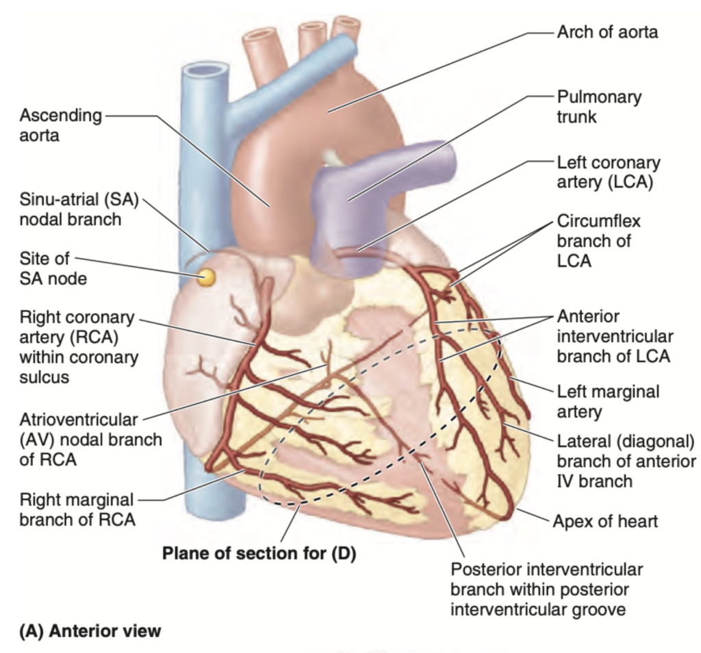
Question 29
問題文: Which vein accompanies the anterior interventricular artery?
a. Great cardiac vein
b. Middle cardiac vein
c. Small cardiac vein
d. Anterior cardiac vein
Answer: a. Great cardiac vein
解説: 大心臓静脈は、前室間枝(または左前下行枝)と共に走り、この動脈からの血液を集めて冠状静脈洞に運びます。この配置は心筋の前部から効率的に静脈血を排出するために重要です。中心臓静脈(選択肢b)、小心臓静脈(選択肢c)、および前心臓静脈(選択肢d)は他の領域や動脈に隣接しています。
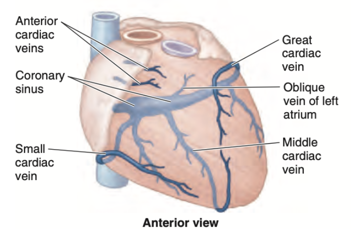
Question 30
問題文: What is the primary function of the pericardium?
a. To pump blood
b. To reduce friction between the heart and surrounding tissues
c. To generate electrical impulses
d. To regulate blood pressure
Answer: b. To reduce friction between the heart and surrounding tissues
解説: 心膜の主な機能は、心臓と周囲の組織間の摩擦を減少させることです。心膜は滑らかな液体で満たされた二重層の膜で、心臓が収縮および拡張する際の摩擦を最小限に抑え、心臓の動きを円滑にします。選択肢aの血液ポンプ、選択肢cの電気インパルスの生成、および選択肢dの血圧調節は心膜の機能ではありません。
Question 31
問題文: Which condition is commonly associated with the thickening of the ventricular walls?
a. Myocardial infarction
b. Ventricular fibrillation
c. Cardiac tamponade
d. Hypertrophic cardiomyopathy
Answer: d. Hypertrophic cardiomyopathy
解説: 心室肥大症(Hypertrophic cardiomyopathy)は、心室壁の厚化が特徴的な状態で、これが心機能に影響を与えることがあります。特に左心室の壁が厚くなることが一般的で、心臓の効率的なポンプ機能を妨げる可能性があります。心筋梗塞(選択肢a)は通常、心筋の壊死によるもので、心室細動(選択肢b)は不整脈の一種で、心膜タンポナーデ(選択肢c)は心膜腔内の液体蓄積によるものです。
Question 32
問題文: What is the primary function of the semilunar valves in the human heart?
a. To prevent backflow of blood into the atria during ventricular contraction
b. To prevent backflow of blood into the ventricles during atrial contraction
c. To prevent backflow of blood from the arteries into the ventricles
d. To regulate the flow of blood between the chambers of the heart
Answer: c. To prevent backflow of blood from the arteries into the ventricles
解説: 半月弁(semilunar valves)の主な機能は、大動脈と肺動脈から心室への血液の逆流を防ぐことです。これにより、心室がリラックスする際に血液が再び心室に戻るのを防ぎ、血液の一方向の流れが確保されます。選択肢aおよびbは房室弁(atrioventricular valves)の機能に関連しており、選択肢dはより広範な心内部の血流調節に関する誤解です。
Question 33
問題文: Which artery is most commonly affected in cases of coronary artery disease?
a. Pulmonary artery
b. Coronary artery
c. Carotid artery
d. Renal artery
Answer: b. Coronary artery
解説: 冠状動脈疾患(coronary artery disease)に最も一般的に影響されるのは冠状動脈です。この疾患は、冠状動脈にプラークが蓄積することにより起こり、心筋への血流が妨げられることで心臓病や心筋梗塞を引き起こすリスクが高まります。選択肢aの肺動脈、選択肢cの頚動脈、および選択肢dの腎動脈は冠状動脈疾患とは直接関係がありません。
Question 34
問題文: The opening of the coronary sinus is located in which chamber of the heart?
a. Right atrium
b. Left atrium
c. Right ventricle
d. Left ventricle
Answer: a. Right atrium
解説: 冠状静脈洞の開口部は右心房に位置しています。この静脈は心筋からの脱酸素血を集め、それを右心房に排出する役割を持ちます。これにより、血液は肺へ送られて酸素化されるプロセスが効率的に進行します。左心房(選択肢b)、右心室(選択肢c)、および左心室(選択肢d)には冠状静脈洞の開口部はありません。
Question 35
問題文: Pulmonary veins return oxygenated blood to which part of the heart?
a. Right atrium
b. Left atrium
c. Right ventricle
d. Left ventricle
Answer: b. Left atrium
解説: 肺静脈は酸素化された血液を左心房に戻します。これにより、血液は左心房から左心室に移動し、その後全身にポンプされます。この酸素豊富な血液の効率的な流れは、生体システム全体の酸素供給と関連して重要です。選択肢aの右心房、選択肢cの右心室、および選択肢dの左心室は、酸素化された血液が直接入る場所ではありません。
Question 36
問題文: Which cardiac structure is crucial for the synchronization of ventricular contraction?
a. Sinoatrial node
b. Atrioventricular node
c. Purkinje fibers
d. Coronary arteries
Answer: c. Purkinje fibers
解説: プルキンエ繊維は、心室収縮を同期させるために不可欠な心臓構造です。これらの繊維は心室壁の内膜下に広がっており、心室全体に迅速に電気的インパルスを伝えることで、均等かつ効率的な心室収縮を促進します。洞房結節(選択肢a)と房室結節(選択肢b)は心拍の発生と心房から心室へのインパルス伝達を調整しますが、心室収縮の同期はプルキンエ繊維の主要な役割です。冠状動脈(選択肢d)は心筋への血液供給を担いますが、収縮同期には関与しません。
Question 37
問題文: Which condition is indicated by the presence of a murmur heard between the second and third intercostal spaces at the left sternal border?
a. Mitral valve prolapse
b. Aortic stenosis
c. Tricuspid regurgitation
d. Pulmonary hypertension
Answer: b. Aortic stenosis
解説: 大動脈狭窄症(Aortic stenosis)は、第二と第三肋間、左胸骨境界で聞こえる雑音(心音)によって示されることが多い状態です。この病態は大動脈弁の開口部が狭くなり、血液が心臓から大動脈へと流れ出る際に異常な流れを生じさせるためです。僧帽弁逸脱症(選択肢a)は異なる音と場所で特定され、三尖弁逆流(選択肢c)は通常右胸部で聞かれ、肺高血圧(選択肢d)は他の特徴的な徴候を伴います。
Question 38
問題文: Which structure is directly involved in regulating the timing of blood flow between the heart chambers?
a. Cardiac veins
b. Cardiac valves
c. Coronary arteries
d. Myocardium
Answer: b. Cardiac valves
解説: 心臓弁(Cardiac valves)は、心房と心室間および心室から大血管への血液の流れのタイミングを調節するために直接的に関与しています。これにより、心臓が効率的に血液を全身にポンプすることが保証されます。心臓静脈(選択肢a)は血液の排出を助け、冠状動脈(選択肢c)は心筋に酸素と栄養を供給し、心筋(選択肢d)は心臓の収縮力を提供しますが、これらは直接的なタイミング調節には関与しません。
Question 39
問題文: What is the primary function of the atrial natriuretic peptide (ANP) secreted by the heart?
a. To increase blood pressure
b. To decrease blood pressure
c. To stimulate red blood cell production
d. To increase blood glucose levels
Answer: b. To decrease blood pressure
解説: 心房性ナトリウム利尿ペプチド(ANP)は、心臓によって分泌され、主に血圧を低下させる機能を持ちます。ANPはナトリウムの排泄を促進し、血管を拡張することで、体内の液体量を減少させ、結果的に血圧を下げます。選択肢aの血圧増加、選択肢cの赤血球産生刺激、および選択肢dの血糖レベル増加は、ANPの機能ではありません。
Question 40
問題文: In which scenario would the endocardium be most directly involved?
a. Regulating heart rate
b. Infective endocarditis
c. Coronary artery bypass grafting
d. Myocardial infarction
Answer: b. Infective endocarditis
解説: 心内膜炎(Infective endocarditis)は、心内膜が直接関与するシナリオです。心内膜は心室の内側を覆う膜であり、感染がこの膜を侵すことで炎症や弁の損傷が生じる可能性があります。選択肢aの心拍数調節、選択肢cの冠状動脈バイパス移植、および選択肢dの心筋梗塞は、心内膜とは直接関連が少ないプロセスです。
Question 45
問題文: Which of the following best describes the role of venae cordis minimae (Thebesian veins) in cardiac physiology?
a. They drain oxygenated blood from the pulmonary veins
b. They supply blood to the myocardium
c. They provide a secondary route for myocardial blood drainage
d. They regulate the contraction of the atria
Answer: c. They provide a secondary route for myocardial blood drainage
解説: 心臓最小静脈(Thebesian veins)は、心筋の血液排出における二次的な経路を提供します。これらの小さな静脈は心臓の壁に散在しており、特に心筋から直接心房に血液を排出する役割を持ちます。選択肢aの肺静脈からの酸素化血液の排出、選択肢bの心筋への血液供給、選択肢dの心房収縮の調節は、心臓最小静脈の機能とは関連しません。
Question 46
問題文: What is the primary consequence of ventricular septal defect (VSD)?
a. Over-oxygenation of blood in the lungs
b. Increased blood flow from the left to the right ventricle
c. Decreased cardiac output
d. Elevated pulmonary vascular resistance
Answer: b. Increased blood flow from the left to the right ventricle
解説: 心室中隔欠損症(VSD)の主な結果は、左心室から右心室への血液流の増加です。この状態では、中隔に異常開口が存在し、血液が高圧の左心室から低圧の右心室へとシャントされます。これにより、右心室および肺動脈への負担が増大し、長期的には肺高血圧のリスクが高まります。選択肢aの肺での血液の過酸素化、選択肢cの心拍出量の低下、選択肢dの肺血管抵抗の上昇は、VSDの直接的な結果ではなく、発展的な合併症です。
Question 48
問題文: The presence of which type of vessel primarily distinguishes the coronary circulation from other vascular beds in the body?
a. Arteries
b. Veins
c. Capillaries
d. Anastomoses
Answer: d. Anastomoses
解説: 冠状循環を他の血管系と区別する主要な特徴は、動脈間の吻合(Anastomoses)の存在です。これらは冠状動脈同士が形成する直接的な接続であり、一部動脈が閉塞した場合に別の血流経路を提供する機能を持ちます。この特性は冠状動脈系が持つユニークな適応機構であり、心筋虚血時に重要な役割を果たします。他の血管系では、このような広範な吻合は一般的ではありません。
Question 49
問題文: What is the primary role of the heart’s fibrous skeleton?
a. To facilitate the contraction of cardiac muscle
b. To prevent the spread of electrical impulses
c. To provide structural support and attachment for cardiac muscle and valves
d. To regulate the exchange of electrolytes across the myocardium
Answer: c. To provide structural support and attachment for cardiac muscle and valves
解説: 心臓の線維性骨格の主な役割は、心筋と心臓弁の構造的支持および付着点を提供することです。この骨格は、心臓の電気的インパルスが適切な経路を通ることを確保するとともに、心臓内の各弁の適切な位置と機能を保持します。選択肢aの心筋収縮の促進、選択肢bの電気的インパルスの拡散防止、選択肢dの電解質交換の調節は、線維性骨格の直接的な機能ではありません。
Question 50
問題文: During a cardiac cycle, the “dub” sound (second heart sound) is produced by the closure of which valves?
a. Mitral and tricuspid valves
b. Pulmonary and aortic valves
c. Pulmonary and mitral valves
d. Aortic and tricuspid valves
Answer: b. Pulmonary and aortic valves
解説: 心拍サイクル中の「ダブ」音(第二心音)は、肺動脈弁と大動脈弁の閉鎖によって生じます。これらの半月弁が閉じるとき、心室の収縮後のリラクゼーションフェーズで逆流を防ぐためにしっかりと閉じます。この音は、血液がこれらの弁に衝突して閉じる際に生じる音です。選択肢aの僧帽弁と三尖弁は第一心音を生成し、選択肢cおよびdは正確な弁の組み合わせを示していません。

コメント